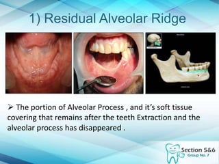
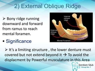
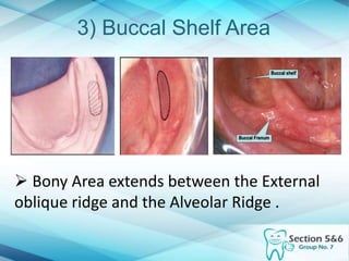
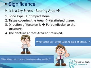
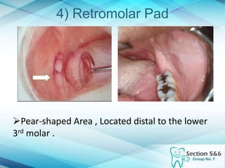
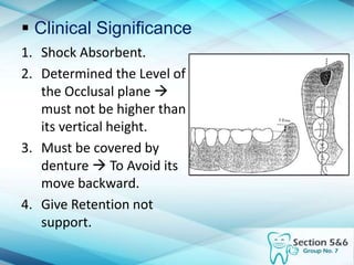
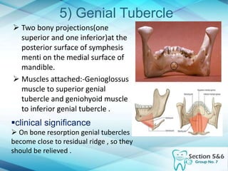
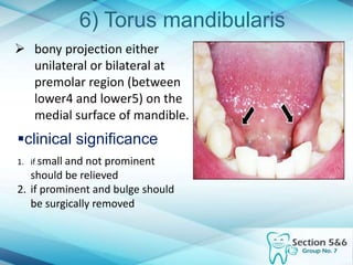
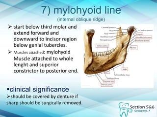
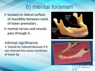
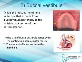
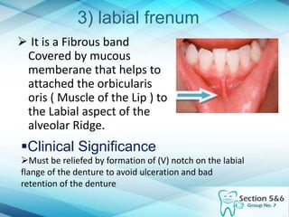
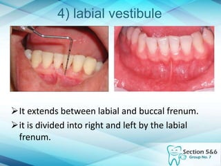
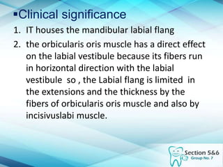
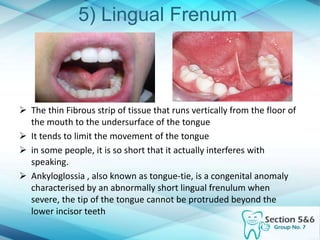
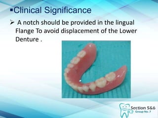
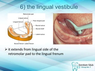
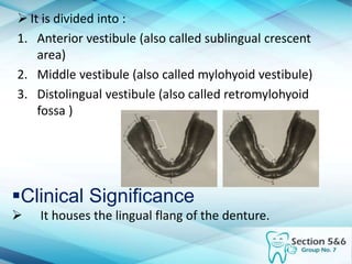
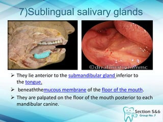
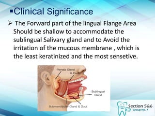
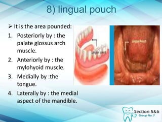
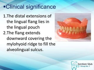
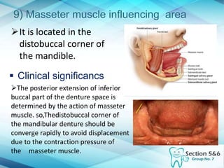
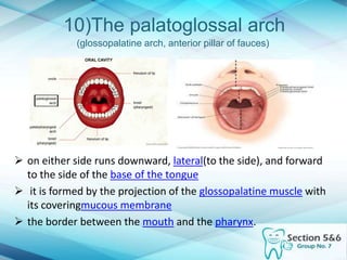
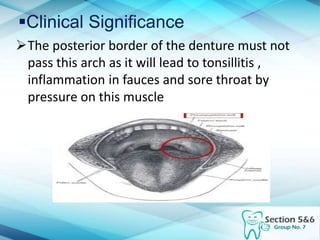
Intra Oral Landmarks
 Supporting Structures
 Limiting Structures
( The Mandible )
 Supporting Structures
1. Residual Alveolar Ridge
2. External Oblique Ridge
3. Buccal Shelf Area
4. Retromolar Pad
5. Genial Tubricle
6. Torus Mandibularis
7. Intrnal Oblique Ridge
8. Mental Foramen
1) Residual Alveolar Ridge
 The portion of Alveolar Process , and it’s soft tissue
covering that remains after the teeth Extraction and the
alveolar process has disappeared .
 Clinical Significance
1. The highest Portion of the ridge is called  Crest of
the ridge ( 2ry Stress-Bearing Area) .
2. The crest is covered By Fibrous C.T with
Keratinized Layer , and bone type is
Cancellous Bone.
3. Sometimes , the crest of the ridge being sharp ( Knife
edge ) so the denture should be relived opposite it .
4. The slopes of the residual ridge Both ( Buccal &
Lingual ) have compact Bone , and can contribute to
resist the horizontal forces .
2) External Oblique Ridge
 Bony ridge running
downward and forward
from ramus to reach
mental foramen.
 Significance
 It’s a limiting structure , the lower denture must
covered but not extend beyond it  To avoid the
displacment by Powerful musculature in this Area
3) Buccal Shelf Area
 Bony Area extends between the External
oblique ridge and the Alveolar Ridge .
 Significance
 It Is a 1ry Stress - Bearing Area 
1. Bone Type  Compact Bone.
2. Tissue covering the Area  Keratinized tissue.
3. Direction of force on it  Perpendicular to the
structure.
4. The denture at that Area not relieved.
What is the 2ry stress Bearing area of Mand. !?
What about the 1ry stress bearing Area For maxilla !?
4) Retromolar Pad
Pear-shaped Area , Located distal to the lower
3rd molar .
 Clinical Significance
1. Shock Absorbent.
2. Determined the Level of
the Occlusal plane 
must not be higher than
its vertical height.
3. Must be covered by
denture  To Avoid its
move backward.
4. Give Retention not
support.
5) Genial Tubercle
 Two bony projections(one
superior and one inferior)at the
posterior surface of symphesis
menti on the medial surface of
mandible.
 Muscles attached:-Genioglossus
muscle to superior genial
tubercle and geniohyoid muscle
to inferior genial tubercle .
clinical significance
 On bone resorption genial tubercles
become close to residual ridge , so they
should be relieved .
6) Torus mandibularis
 bony projection either
unilateral or bilateral at
premolar region (between
lower4 and lower5) on the
medial surface of mandible.
clinical significance
1. if small and not prominent
should be relieved
2. if prominent and bulge should
be surgically removed
7) mylohyoid line
(internal oblique ridge)
 start below third molar and
extend forward and
downward to incisor region
below genial tubercles.
 Musclesattached: mylohyoid
Muscle attached to whole
lenght and superior
constrictor to posterior end.
clinical significance
should be covered by denture if
sharp should be surgically removed.
8) mental foramen
 located on lateral surface
of mandible between roots
of lower premolars .
 mental nerves and vessels
pass through it .
clinical significance
 should be relieved because if it
not relieved this cause numbness
of lower lip
 Limiting Structures
1. Buccal Frenum
2. Buccal Vestibule
3. Labial Frenum
4. Labial Vestibule
5. Lingual Frenum
6. Lingual Vestibule
7. Sublingual Glands
8. Lingual Pouch
9. Platoglossal Arch
10.Masseter muscle influencing area
1) Buccal frenum
Thin band of tissue
extends from alveolar
ridge to cheecks
Form support to
cheecks
Clinical Significance
Must be reliefed by formation of (V)
notch on the buccal flange of the
denture to avoid ulceration and bad
retention of the denture
2) Buccal vestibule
 It is the mucous membrane
reflection that extends from
buccalfrenum posteriorly to the
outside back corner of the
retromolar pad.
The size of buccal vestibule varies with :
1. The contraction of buccinator muscle
2. The amount of bone lost from the
mandible
Clinical significance
1. It houses the buccal flang of the mandibular
denture
2. it determines the length of the flang of the
denture.
3. When it properly filled with the denture
flang,It is greatly enhances the stability and
the retention.
3) labial frenum
 It is a Fibrous band
Covered by mucous
memberane that helps to
attached the orbicularis
oris ( Muscle of the Lip ) to
the Labial aspect of the
alveolar Ridge.
Clinical Significance
Must be reliefed by formation of (V) notch on the labial
flange of the denture to avoid ulceration and bad
retention of the denture
4) labial vestibule
It extends between labial and buccal frenum.
it is divided into right and left by the labial
frenum.
Clinical significance
1. IT houses the mandibular labial flang
2. the orbicularis oris muscle has a direct effect
on the labial vestibule because its fibers run
in horizontal direction with the labial
vestibule so , the Labial flang is limited in
the extensions and the thickness by the
fibers of orbicularis oris muscle and also by
incisivuslabi muscle.
5) Lingual Frenum
 The thin Fibrous strip of tissue that runs vertically from the floor of
the mouth to the undersurface of the tongue
 It tends to limit the movement of the tongue
 in some people, it is so short that it actually interferes with
speaking.
 Ankyloglossia , also known as tongue-tie, is a congenital anomaly
characterised by an abnormally short lingual frenulum when
severe, the tip of the tongue cannot be protruded beyond the
lower incisor teeth
Clinical Significance
 A notch should be provided in the lingual
Flange To avoid displacement of the Lower
Denture .
6) the lingual vestibule
 it extends from lingual side of the
retromolar pad to the lingual frenum
 It is divided into :
1. Anterior vestibule (also called sublingual crescent
area)
2. Middle vestibule (also called mylohyoid vestibule)
3. Distolingual vestibule (also called retromylohyoid
fossa )
Clinical Significance
 It houses the lingual flang of the denture.
7)Sublingual salivary glands
 They lie anterior to the submandibular gland inferior to
the tongue.
 beneaththemucous membrane of the floor of the mouth.
 They are palpated on the floor of the mouth posterior to each
mandibular canine.
Clinical Significance
 The Forward part of the lingual Flange Area
Should be shallow to accommodate the
sublingual Salivary gland and to Avoid the
irritation of the mucous membrane , which is
the least keratinized and the most sensetive.
8) lingual pouch
 It is the area pounded:
1. Posteriorly by : the
palate glossus arch
muscle.
2. Anteriorly by : the
mylohyoid muscle.
3. Medially by :the
tongue.
4. Laterally by : the medial
aspect of the mandible.
Clinical significance
1.The distal extensions of
the lingual flang lies in
the lingual pouch
2.The flang extends
downward covering the
mylohyoid ridge to fill the
alveolingual sulcus.
9) Masseter muscle influencing area
It is located in the
distobuccal corner of
the mandible.
 Clinical significancs
The posterior extension of inferior
buccal part of the denture space is
determined by the action of masseter
muscle. so,Thedistobuccal corner of
the mandibular denture should be
converge rapidly to avoid displacement
due to the contraction pressure of
the masseter muscle.
10)The palatoglossal arch
(glossopalatine arch, anterior pillar of fauces)
 on either side runs downward, lateral(to the side), and forward
to the side of the base of the tongue
 it is formed by the projection of the glossopalatine muscle with
its coveringmucous membrane
 the border between the mouth and the pharynx.
Clinical Significance
The posterior border of the denture must not
pass this arch as it will lead to tonsillitis ,
inflammation in fauces and sore throat by
pressure on this muscle
Before & After …
Made By …
Hesham Sayed
Ahmed Mohsen
Mohamed Essam
MohamedAboGhadier
Mohamedfawzy
Intra oral mandibular landmarks

Intra oral mandibular landmarks

  • 3.
    Intra Oral Landmarks Supporting Structures  Limiting Structures ( The Mandible )
  • 4.
     Supporting Structures 1.Residual Alveolar Ridge 2. External Oblique Ridge 3. Buccal Shelf Area 4. Retromolar Pad 5. Genial Tubricle 6. Torus Mandibularis 7. Intrnal Oblique Ridge 8. Mental Foramen
  • 5.
    1) Residual AlveolarRidge  The portion of Alveolar Process , and it’s soft tissue covering that remains after the teeth Extraction and the alveolar process has disappeared .
  • 6.
     Clinical Significance 1.The highest Portion of the ridge is called  Crest of the ridge ( 2ry Stress-Bearing Area) . 2. The crest is covered By Fibrous C.T with Keratinized Layer , and bone type is Cancellous Bone. 3. Sometimes , the crest of the ridge being sharp ( Knife edge ) so the denture should be relived opposite it . 4. The slopes of the residual ridge Both ( Buccal & Lingual ) have compact Bone , and can contribute to resist the horizontal forces .
  • 7.
    2) External ObliqueRidge  Bony ridge running downward and forward from ramus to reach mental foramen.  Significance  It’s a limiting structure , the lower denture must covered but not extend beyond it  To avoid the displacment by Powerful musculature in this Area
  • 8.
    3) Buccal ShelfArea  Bony Area extends between the External oblique ridge and the Alveolar Ridge .
  • 9.
     Significance  ItIs a 1ry Stress - Bearing Area  1. Bone Type  Compact Bone. 2. Tissue covering the Area  Keratinized tissue. 3. Direction of force on it  Perpendicular to the structure. 4. The denture at that Area not relieved. What is the 2ry stress Bearing area of Mand. !? What about the 1ry stress bearing Area For maxilla !?
  • 10.
    4) Retromolar Pad Pear-shapedArea , Located distal to the lower 3rd molar .
  • 11.
     Clinical Significance 1.Shock Absorbent. 2. Determined the Level of the Occlusal plane  must not be higher than its vertical height. 3. Must be covered by denture  To Avoid its move backward. 4. Give Retention not support.
  • 12.
    5) Genial Tubercle Two bony projections(one superior and one inferior)at the posterior surface of symphesis menti on the medial surface of mandible.  Muscles attached:-Genioglossus muscle to superior genial tubercle and geniohyoid muscle to inferior genial tubercle . clinical significance  On bone resorption genial tubercles become close to residual ridge , so they should be relieved .
  • 13.
    6) Torus mandibularis bony projection either unilateral or bilateral at premolar region (between lower4 and lower5) on the medial surface of mandible. clinical significance 1. if small and not prominent should be relieved 2. if prominent and bulge should be surgically removed
  • 14.
    7) mylohyoid line (internaloblique ridge)  start below third molar and extend forward and downward to incisor region below genial tubercles.  Musclesattached: mylohyoid Muscle attached to whole lenght and superior constrictor to posterior end. clinical significance should be covered by denture if sharp should be surgically removed.
  • 15.
    8) mental foramen located on lateral surface of mandible between roots of lower premolars .  mental nerves and vessels pass through it . clinical significance  should be relieved because if it not relieved this cause numbness of lower lip
  • 16.
     Limiting Structures 1.Buccal Frenum 2. Buccal Vestibule 3. Labial Frenum 4. Labial Vestibule 5. Lingual Frenum 6. Lingual Vestibule 7. Sublingual Glands 8. Lingual Pouch 9. Platoglossal Arch 10.Masseter muscle influencing area
  • 17.
    1) Buccal frenum Thinband of tissue extends from alveolar ridge to cheecks Form support to cheecks Clinical Significance Must be reliefed by formation of (V) notch on the buccal flange of the denture to avoid ulceration and bad retention of the denture
  • 18.
    2) Buccal vestibule It is the mucous membrane reflection that extends from buccalfrenum posteriorly to the outside back corner of the retromolar pad. The size of buccal vestibule varies with : 1. The contraction of buccinator muscle 2. The amount of bone lost from the mandible
  • 19.
    Clinical significance 1. Ithouses the buccal flang of the mandibular denture 2. it determines the length of the flang of the denture. 3. When it properly filled with the denture flang,It is greatly enhances the stability and the retention.
  • 20.
    3) labial frenum It is a Fibrous band Covered by mucous memberane that helps to attached the orbicularis oris ( Muscle of the Lip ) to the Labial aspect of the alveolar Ridge. Clinical Significance Must be reliefed by formation of (V) notch on the labial flange of the denture to avoid ulceration and bad retention of the denture
  • 21.
    4) labial vestibule Itextends between labial and buccal frenum. it is divided into right and left by the labial frenum.
  • 22.
    Clinical significance 1. IThouses the mandibular labial flang 2. the orbicularis oris muscle has a direct effect on the labial vestibule because its fibers run in horizontal direction with the labial vestibule so , the Labial flang is limited in the extensions and the thickness by the fibers of orbicularis oris muscle and also by incisivuslabi muscle.
  • 23.
    5) Lingual Frenum The thin Fibrous strip of tissue that runs vertically from the floor of the mouth to the undersurface of the tongue  It tends to limit the movement of the tongue  in some people, it is so short that it actually interferes with speaking.  Ankyloglossia , also known as tongue-tie, is a congenital anomaly characterised by an abnormally short lingual frenulum when severe, the tip of the tongue cannot be protruded beyond the lower incisor teeth
  • 24.
    Clinical Significance  Anotch should be provided in the lingual Flange To avoid displacement of the Lower Denture .
  • 25.
    6) the lingualvestibule  it extends from lingual side of the retromolar pad to the lingual frenum
  • 26.
     It isdivided into : 1. Anterior vestibule (also called sublingual crescent area) 2. Middle vestibule (also called mylohyoid vestibule) 3. Distolingual vestibule (also called retromylohyoid fossa ) Clinical Significance  It houses the lingual flang of the denture.
  • 27.
    7)Sublingual salivary glands They lie anterior to the submandibular gland inferior to the tongue.  beneaththemucous membrane of the floor of the mouth.  They are palpated on the floor of the mouth posterior to each mandibular canine.
  • 28.
    Clinical Significance  TheForward part of the lingual Flange Area Should be shallow to accommodate the sublingual Salivary gland and to Avoid the irritation of the mucous membrane , which is the least keratinized and the most sensetive.
  • 29.
    8) lingual pouch It is the area pounded: 1. Posteriorly by : the palate glossus arch muscle. 2. Anteriorly by : the mylohyoid muscle. 3. Medially by :the tongue. 4. Laterally by : the medial aspect of the mandible.
  • 30.
    Clinical significance 1.The distalextensions of the lingual flang lies in the lingual pouch 2.The flang extends downward covering the mylohyoid ridge to fill the alveolingual sulcus.
  • 31.
    9) Masseter muscleinfluencing area It is located in the distobuccal corner of the mandible.  Clinical significancs The posterior extension of inferior buccal part of the denture space is determined by the action of masseter muscle. so,Thedistobuccal corner of the mandibular denture should be converge rapidly to avoid displacement due to the contraction pressure of the masseter muscle.
  • 32.
    10)The palatoglossal arch (glossopalatinearch, anterior pillar of fauces)  on either side runs downward, lateral(to the side), and forward to the side of the base of the tongue  it is formed by the projection of the glossopalatine muscle with its coveringmucous membrane  the border between the mouth and the pharynx.
  • 33.
    Clinical Significance The posteriorborder of the denture must not pass this arch as it will lead to tonsillitis , inflammation in fauces and sore throat by pressure on this muscle
  • 34.
  • 35.
    Made By … HeshamSayed Ahmed Mohsen Mohamed Essam MohamedAboGhadier Mohamedfawzy