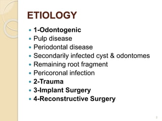
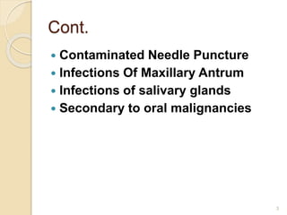
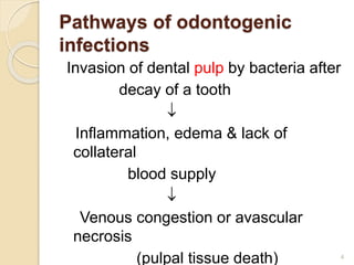
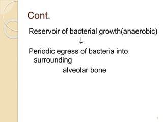
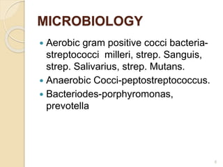
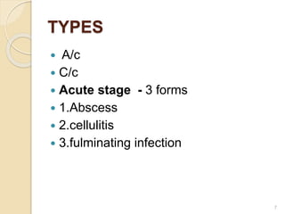
This document provides information on orofacial and neck infections, including their etiology, types, pathways of spread, microbiology, clinical features, treatment, and classifications of fascial spaces. It discusses various types of infections such as acute periapical abscesses, acute dentoalveolar abscesses, acute periodontal abscesses, and infections of specific spaces like the canine space, buccal space, and infratemporal space. Treatment involves both medical approaches like antibiotics and surgical drainage of affected areas.
An oroantral communication is an unnatural perforation between oral cavity and maxillary sinus.
Oroantral fistula is an epithelized, pathological, communication between these two cavities. A fistulous tract present more than 14 days should be considered as chronic fistula.
Dr. Ahmed M. Adawy, Professor Emeritus, Dep. Oral & Maxillofacial Surgery. Former Dean, Faculty of Dental Medicine
Al-Azhar University. The Pathogenesis of infection in oro-facial region due to odontogenic origin is a common clinical issue. bacterial invasion to deeper tissues usually a spread from diseased dental pulp. Recent evidences indicated a multi-microbial nature. The spread of infection is governed by the thickness of the investing bone and the anatomical relation of the tooth root to the attached muscle. Infection could spread from one facial space to another, and the condition may be aggravated to life threatening situations.
Detailed description of diagnosis and management of maxillofacial and neck space infections. Discussion of anatomy of the spaces is also done in details. Drainage of such spaces are also discussed. Medical management is also discussed. Complications are also discussed.
Space infection 2 /certified fixed orthodontic courses by Indian dental academy Indian dental academy
The Indian Dental Academy is the Leader in continuing dental education , training dentists in all aspects of dentistry and offering a wide range of dental certified courses in different formats.
Indian dental academy provides dental crown & Bridge,rotary endodontics,fixed orthodontics,
Dental implants courses.for details pls visit www.indiandentalacademy.com ,or call
0091-9248678078
The Indian Dental Academy is the Leader in continuing dental education , training dentists in all aspects of dentistry and
offering a wide range of dental certified courses in different formats.for more details please visit
www.indiandentalacademy.com
Spaces of head and neck and infections /certified fixed orthodontic courses b...Indian dental academy
Welcome to Indian Dental Academy
The Indian Dental Academy is the Leader in continuing dental education , training dentists in all aspects of dentistry and offering a wide range of dental certified courses in different formats.
Indian dental academy has a unique training program & curriculum that provides students with exceptional clinical skills and enabling them to return to their office with high level confidence and start treating patients
State of the art comprehensive training-Faculty of world wide repute &Very affordable
Introduction
Definition
Pathway of odontogenic infection
Classification
Maxillary space infection
Mandibular space infection
Ludwigs angina
Cavernous sinus thrombophlebitis
Occurrence of infectious disease is determined by interaction of host , the microorganism and the environment
In healthy state there is balance among these factors and when the balance is lost disease occurs
Most odontogenic infections arise as a sequel of pulp necrosis caused by caries, trauma, periodontitis
Definition : the fascial spaces are the potential spaces between the various layers of fascia normally filled with loose connective tissue and bounded by anatomical barriers , usually of bone , muscle or fascial layers.
(Moore – 1975)
Invasion of dental pulp by bacteria after decay of a tooth
inflammation edema and lack of blood supply
Venous congestion ,pulpal tissue death
Reservoir for bacterial growth
Periodic egress of bacteria into surrounding alveolar Acute stage
in acute stage ,infection spreading in the soft tissues can take the following forms of in the clinical situation
Abscess
Cellulitis
Fulminating infections
Neck space infections taken from PL. DHINGRA and other sources to cover all o...lordskywalker7878
This presentation covers the important ENT topics of neck space infections with their management and image illustrations. The source is mainly PL. DHINGRA however other sources have been mentioned in the presentation, especially on the images. It is divided into superficial and deep neck infections for clear distinction between the two categories. It is an extremely important topic especially if your goal is towards surgical side of ENT.
New Directions in Targeted Therapeutic Approaches for Older Adults With Mantl...i3 Health
i3 Health is pleased to make the speaker slides from this activity available for use as a non-accredited self-study or teaching resource.
This slide deck presented by Dr. Kami Maddocks, Professor-Clinical in the Division of Hematology and
Associate Division Director for Ambulatory Operations
The Ohio State University Comprehensive Cancer Center, will provide insight into new directions in targeted therapeutic approaches for older adults with mantle cell lymphoma.
STATEMENT OF NEED
Mantle cell lymphoma (MCL) is a rare, aggressive B-cell non-Hodgkin lymphoma (NHL) accounting for 5% to 7% of all lymphomas. Its prognosis ranges from indolent disease that does not require treatment for years to very aggressive disease, which is associated with poor survival (Silkenstedt et al, 2021). Typically, MCL is diagnosed at advanced stage and in older patients who cannot tolerate intensive therapy (NCCN, 2022). Although recent advances have slightly increased remission rates, recurrence and relapse remain very common, leading to a median overall survival between 3 and 6 years (LLS, 2021). Though there are several effective options, progress is still needed towards establishing an accepted frontline approach for MCL (Castellino et al, 2022). Treatment selection and management of MCL are complicated by the heterogeneity of prognosis, advanced age and comorbidities of patients, and lack of an established standard approach for treatment, making it vital that clinicians be familiar with the latest research and advances in this area. In this activity chaired by Michael Wang, MD, Professor in the Department of Lymphoma & Myeloma at MD Anderson Cancer Center, expert faculty will discuss prognostic factors informing treatment, the promising results of recent trials in new therapeutic approaches, and the implications of treatment resistance in therapeutic selection for MCL.
Target Audience
Hematology/oncology fellows, attending faculty, and other health care professionals involved in the treatment of patients with mantle cell lymphoma (MCL).
Learning Objectives
1.) Identify clinical and biological prognostic factors that can guide treatment decision making for older adults with MCL
2.) Evaluate emerging data on targeted therapeutic approaches for treatment-naive and relapsed/refractory MCL and their applicability to older adults
3.) Assess mechanisms of resistance to targeted therapies for MCL and their implications for treatment selection
Title: Sense of Smell
Presenter: Dr. Faiza, Assistant Professor of Physiology
Qualifications:
MBBS (Best Graduate, AIMC Lahore)
FCPS Physiology
ICMT, CHPE, DHPE (STMU)
MPH (GC University, Faisalabad)
MBA (Virtual University of Pakistan)
Learning Objectives:
Describe the primary categories of smells and the concept of odor blindness.
Explain the structure and location of the olfactory membrane and mucosa, including the types and roles of cells involved in olfaction.
Describe the pathway and mechanisms of olfactory signal transmission from the olfactory receptors to the brain.
Illustrate the biochemical cascade triggered by odorant binding to olfactory receptors, including the role of G-proteins and second messengers in generating an action potential.
Identify different types of olfactory disorders such as anosmia, hyposmia, hyperosmia, and dysosmia, including their potential causes.
Key Topics:
Olfactory Genes:
3% of the human genome accounts for olfactory genes.
400 genes for odorant receptors.
Olfactory Membrane:
Located in the superior part of the nasal cavity.
Medially: Folds downward along the superior septum.
Laterally: Folds over the superior turbinate and upper surface of the middle turbinate.
Total surface area: 5-10 square centimeters.
Olfactory Mucosa:
Olfactory Cells: Bipolar nerve cells derived from the CNS (100 million), with 4-25 olfactory cilia per cell.
Sustentacular Cells: Produce mucus and maintain ionic and molecular environment.
Basal Cells: Replace worn-out olfactory cells with an average lifespan of 1-2 months.
Bowman’s Gland: Secretes mucus.
Stimulation of Olfactory Cells:
Odorant dissolves in mucus and attaches to receptors on olfactory cilia.
Involves a cascade effect through G-proteins and second messengers, leading to depolarization and action potential generation in the olfactory nerve.
Quality of a Good Odorant:
Small (3-20 Carbon atoms), volatile, water-soluble, and lipid-soluble.
Facilitated by odorant-binding proteins in mucus.
Membrane Potential and Action Potential:
Resting membrane potential: -55mV.
Action potential frequency in the olfactory nerve increases with odorant strength.
Adaptation Towards the Sense of Smell:
Rapid adaptation within the first second, with further slow adaptation.
Psychological adaptation greater than receptor adaptation, involving feedback inhibition from the central nervous system.
Primary Sensations of Smell:
Camphoraceous, Musky, Floral, Pepperminty, Ethereal, Pungent, Putrid.
Odor Detection Threshold:
Examples: Hydrogen sulfide (0.0005 ppm), Methyl-mercaptan (0.002 ppm).
Some toxic substances are odorless at lethal concentrations.
Characteristics of Smell:
Odor blindness for single substances due to lack of appropriate receptor protein.
Behavioral and emotional influences of smell.
Transmission of Olfactory Signals:
From olfactory cells to glomeruli in the olfactory bulb, involving lateral inhibition.
Primitive, less old, and new olfactory systems with different path
The prostate is an exocrine gland of the male mammalian reproductive system
It is a walnut-sized gland that forms part of the male reproductive system and is located in front of the rectum and just below the urinary bladder
Function is to store and secrete a clear, slightly alkaline fluid that constitutes 10-30% of the volume of the seminal fluid that along with the spermatozoa, constitutes semen
A healthy human prostate measures (4cm-vertical, by 3cm-horizontal, 2cm ant-post ).
It surrounds the urethra just below the urinary bladder. It has anterior, median, posterior and two lateral lobes
It’s work is regulated by androgens which are responsible for male sex characteristics
Generalised disease of the prostate due to hormonal derangement which leads to non malignant enlargement of the gland (increase in the number of epithelial cells and stromal tissue)to cause compression of the urethra leading to symptoms (LUTS
micro teaching on communication m.sc nursing.pdfAnurag Sharma
Microteaching is a unique model of practice teaching. It is a viable instrument for the. desired change in the teaching behavior or the behavior potential which, in specified types of real. classroom situations, tends to facilitate the achievement of specified types of objectives.
- Video recording of this lecture in English language: https://youtu.be/lK81BzxMqdo
- Video recording of this lecture in Arabic language: https://youtu.be/Ve4P0COk9OI
- Link to download the book free: https://nephrotube.blogspot.com/p/nephrotube-nephrology-books.html
- Link to NephroTube website: www.NephroTube.com
- Link to NephroTube social media accounts: https://nephrotube.blogspot.com/p/join-nephrotube-on-social-media.html
Explore natural remedies for syphilis treatment in Singapore. Discover alternative therapies, herbal remedies, and lifestyle changes that may complement conventional treatments. Learn about holistic approaches to managing syphilis symptoms and supporting overall health.
These lecture slides, by Dr Sidra Arshad, offer a quick overview of physiological basis of a normal electrocardiogram.
Learning objectives:
1. Define an electrocardiogram (ECG) and electrocardiography
2. Describe how dipoles generated by the heart produce the waveforms of the ECG
3. Describe the components of a normal electrocardiogram of a typical bipolar leads (limb II)
4. Differentiate between intervals and segments
5. Enlist some common indications for obtaining an ECG
Study Resources:
1. Chapter 11, Guyton and Hall Textbook of Medical Physiology, 14th edition
2. Chapter 9, Human Physiology - From Cells to Systems, Lauralee Sherwood, 9th edition
3. Chapter 29, Ganong’s Review of Medical Physiology, 26th edition
4. Electrocardiogram, StatPearls - https://www.ncbi.nlm.nih.gov/books/NBK549803/
5. ECG in Medical Practice by ABM Abdullah, 4th edition
6. ECG Basics, http://www.nataliescasebook.com/tag/e-c-g-basics
ARTIFICIAL INTELLIGENCE IN HEALTHCARE.pdfAnujkumaranit
Artificial intelligence (AI) refers to the simulation of human intelligence processes by machines, especially computer systems. It encompasses tasks such as learning, reasoning, problem-solving, perception, and language understanding. AI technologies are revolutionizing various fields, from healthcare to finance, by enabling machines to perform tasks that typically require human intelligence.
Pulmonary Thromboembolism - etilogy, types, medical- Surgical and nursing man...VarunMahajani
Disruption of blood supply to lung alveoli due to blockage of one or more pulmonary blood vessels is called as Pulmonary thromboembolism. In this presentation we will discuss its causes, types and its management in depth.
3. Cont.
Contaminated Needle Puncture
Infections Of Maxillary Antrum
Infections of salivary glands
Secondary to oral malignancies
3
4. Pathways of odontogenic
infections
Invasion of dental pulp by bacteria after
decay of a tooth
Inflammation, edema & lack of
collateral
blood supply
Venous congestion or avascular
necrosis
(pulpal tissue death) 4
8. Abscess
It is a circumscribed collection of pus
in a pathologic tissue space.
Infections are characterised by
sphylococci.
8
9. CELLULITIS
It is spreading infection of loose
connective tissues.
It is a diffuse, erythematous, mucosal
or cutaneous infection.
It is the result of streptococcal
infection.
It does not result in accumulation of
large amount of pus.
9
10. Cont.
Streptococcus produces
streptokinase, hyaluronidase &
streptodornase which break down
fibrin, connective tissue ground
substance & lyse cellular debris, which
facilitate rapid spread of bacteria.
10
12. Chronic stage
C/c fistulous tract or sinus
formation
Abscesses neglected for a long time
may discharge intraorally or extra
orally
12
13. Treatment
Medical treatment
Soft or liquid diet
Adequate hydration
Diet rich in protein
Analgesics
Antiseptic mouthwash
Antibiotics
13
14. Cont.
In a non compromised patient, with
well localized abscess, surgical
drainage with dental therapy will
resolve the infection.
In poorly localized, extensive abscess
& cellulitis antibiotic therapy is
needed.
In compromised patients & patients
with trismus, airway obstruction &
fever antibiotic therapy is must.
14
15. Cont.
Penicillin is the drug of choice.
Penicillin+metronidazole Can Also Be
Used.
Clindamycin
Amoxycillin+clavulanic Acid
First & Second Generation
Cephalosporins
15
16. Cont.
Surgical treatment
It involves blunt exploration of the
anatomic space or abscess.
Abscess cavity is then irrigated with
betadine & saline.
A drain is inserted into the space.
Hilton`s method of incision & drainage
◦ No blood vessel or nerve is damaged.
◦ Topical anaesthesia is obtained.
16
17. Cont.
◦ Stab incision is made over the point of
maximum fluctuation in the most
dependent area along the skin creases,
through skin & subcutaneous tissue.
◦ If pus is not encountered deepening of
surgical site is done with artery forceps.
◦ Closed forceps are pushed through deep
fascia & advanced towards the pus
collection.
◦ Abscess cavity is entered & forceps is
opened parallel to vital structures.
17
18. Cont.
◦ Pus flows along the beaks of the forceps.
◦ A rubber drain is inserted into the depth of
cavity & secured to the wound margin with
the help of sutures.
◦ Drain is left for 24 hrs.
◦ Dressing is given without pressure.
◦ Drain allows discharge of tissue fluids &
pus from the wound.
◦ Drain is removed when the drainage is
completely ceased
18
19. ACUTE PERIAPICAL
ABSCESS
Etiology
◦ Caries
◦ Contamination of traumatic exposure of
pulp.
◦ Chemical or thermal damage to pulp.
The entry to periapical tissues is by
◦ Apical foramina,
◦ Accessary canals,
◦ Endodontic perforation,
◦ Opening in the floor of pulp chamber,
19
20. Cont.
Clinical features
◦ Severe throbbing pain in the affected
tooth
◦ The offending tooth may be sensitive to
percussion.
◦ Mobility may or may not be present.
Radiographic features
◦ Tooth has caries with periapical pathology,
root # or erosion.
◦ There may be periapical radiolucency.
20
22. Acute dentoalveolar
abscess
Etiology
Continuation of periapical abscess.
Clinical features
Pain
Submucosal swelling in the sulcus on
the outer aspect of alveolar process.
If left untreated, swelling bursts &
produces a sinus.
22
23. Cont.
Radiologic features
More marked radiolucency than
periapical abscess.
Treatment
Same as periapical abscess.
Extraoral incision & drainage may be
required.
23
24. Acute periodontal abscess
Etiology
Periodontitis with periodontal pockets.
Clinical features
Dull pain
Pus discharge via gingival pocket
Sinus either on the outer or inner
aspect of alveolar process.
24
25. Spread of oral infection
Routes of spread
Direct continuity through tissues
By lymphatics to the lymph
nodes.From lymph nodes to tissues
results in secondary areas of cellulitis
or tissue space abscess.
By blood stream-local
thrombophlebitis may spread via the
veins entering the cranial cavity
producing cavernous sinus 25
26. Cont.
Factors influencing spread
◦ General factors
Host resistance
Virulance of micro organism
Combination of both
◦ Local factors
Anatomic barriers-
Alveolar bone
Periosteum
Adjacent muscles & fascia
26
27. General clinical features in
patient with orofacial infection
Redness due to vasodialtation
Swelling due to accumulation of
exudate or pus
Temperature over the infected area
due to increased blood flow &
increased metabolism
Pain due to pressure in nerve endings
& release of mediators of pain.
Fever
27
28. Cont.
Head ache
Lymphadenopathy
◦ Acute infection-soft, tender, enlarged,
surrounding tissues are edematous&
overlying skin is erythematous
◦ Chronic infection-firm, nontender enlarged
lymph nodes.
Presence of draining sinus & fistula
Difficulty in opening mouth
28
31. General principles of
management of a/c orofacial
infections
Immediate hospitalization
Medical treatment
Surgical management
31
32. Medical management
Antibiotics
Hydration of the patient through iv
route
Analgesics
Bed rest
Mouth rinses
Opening of tooth for drainage
32
33. Surgical management
Needle decompression
Done in case of pterigomandibular,
peritonsillar,lateral pharyngeal space
infection that is likely to rupture during
passage of endotracheal tube.
Extraction of tooth
Early extraction leads to early
resolution of infection by eliminating
the source of infection & provides a
portal of drainage 33
34. Cont.
Surgical drainage-
Incision is placed on the most
dependent areas.
Incision should be parallel to skin
creases
Incision should lie in aesthetically
acceptable site as far as possible.
Incision should be supported by
healthy underlying dermis &
subcutaneous tissue. 34
35. Cont.
Intraoral incision should not be placed
over frenal attachments, should be
placed parallel to nerve fibers in the
region of mental nerve.
Removal cause such as infected
tooth, segment of necrotic bone,
foreign body, if not already done, then
is done at the time of drainage
procedure
35
38. Canine space infection
Etiology
Infection of maxillary canine, premolar
& mesiobuccal root of 1st molar.
Boundaries
Inferiorly-caninus muscle
Anteriorly-orbicularis oris muscle
Posteriorly-buccinator muscle
Medially-anterolateral surface of
maxilla
38
39. Cont.
Clinical features
Swelling of cheek & upper lip
Obliteration of nasolabial fold
Drooping of angle of mouth
Edema of lower eyelids
Marked Periorbital Edema
Redness & Marked Tenderness Of
Facial Tissues
39
40. Cont.
In c/c stage-fistula near the medial
canthus eye.
Offended tooth is mobile & tender to
percussion
Treatment
Incision & drainage-
Through the mucosa of buccal
vestibule in the region of lateral incisor
& canine.
40
41. Cont.
A curved mosquito artery forceps is
inserted, pus is evacuated & a drain is
inserted & is secured with suture
41
42. Buccal space infection
Etiology
Infection of maxillary & mandibular
premolars & molars
Pericoronitis of lower 3rd molar.
Boundaries
Anteromedially-buccinator muscle
Posteromedially-masseter muscle
Laterally-deep fascia from parotid
capsule & platysma muscle
42
43. Cont.
Inferiorly-deep fascia & depressor
anguli oris
Superiorly-zygomatic process of
maxilla & zygomaticus major & minor
muscles
Contents
Buccal pad of fat
Stenson`s duct
Facial artery
43
44. Cont.
Clinical features
Gum boil in vestibule
Swelling extending from lower border
of mandible to infraorbital margin, from
anterior border of masseter to angle of
mouth
Edema of lower eyelid
44
45. Cont.
Spread
To pterigomandibular space
Infratemporal space
Submasseteric space
Treatment
Incision & drainage through mucosa of
cheek in premolar molar region.
45
46. Infratemporal space infection
Also called retrozygomatic space
because it is situated behind the
zygomatic bone.
Etiology
Infection of buccal roots of maxillary
2nd &3rd molars
LA injection with contaminated
needles in the area of tuberosity
Spread from other spaces
46
47. Cont.
Boundaries
Laterally - by ramus of mandible,
temporalis muscle & its tendon .
Medially - medial pterygoid plate ,
lateral pterygoid muscle , medial
pterygoid muscle ,lower part of
temporal fossa of the skull & lateral
wall of pharynx .
Superiorly - greater wing of sphenoid
& by zygomatic arch . 47
49. Cont.
Middle meningeal artery
Clinical features
Limitation of mouth opening
Swelling in front of ear on the affected
side
Proptosis of eye
Swelling in the area of tuberosity
Elevation of temperature
49
50. Cont.
Incision & drainage
Incision is given in buccal vestibule
opposite the 2nd & 3rd molars
In severe infection incision is made at
the upper posterior edge of temporalis
muscle.
Sinus forceps is directed upwards &
medially.
50
51. Cont.
In case of failure to improve mouth
opening temporalis myotomy or
excision of coronoid process is done.
Spread
To temporal space
Pterigomandibular space
Cavernous sinus
51
52. Abscess of upper lip
Etiology
Infection of upper incisors & canine
Clinical features
Swelling in the base of the upper lip
Swelling in vestibule
Treatment
Antibiotics
Incision & drainage
Extraction of offending tooth or RCT
52
53. Palatal abscess
Etiology
Periodontal abscess from palatal
pockets
Apical abscess from palatal roots of
posterior teeth usually from the lateral
incisor
Boundaries
Inferiorly-hard palate
Superiorly-periosteum & mucosa
Laterally-alveolar process of maxilla &53
54. Cont.
Clinical features
Fluctuant swelling in palate near the
offending tooth
Offending tooth is tender to
percussion
Incision & drainage
Anterioposterior incision is made
through the mucosa down to bone
54
55. Submental space infection
Etiology
Infection from 6 mandibular anterior
teeth
Infection of submental lymph nodes
Boundaries
Laterally-lower border of mandible,
anterior belly of digastric muscle
Superiorly-mylohoid muscle
55
57. Cont.
Skin overlying the swelling is board
like & taut
Fluctuation of swelling
Nonvital, fractured or carious anterior
teeth
Offending tooth is tender on
percussion& sometimes mobile
57
58. Cont.
Incision & drainage
Transverse incision in skin below
symphysis of mandible.
Spread
Submandibular space
58
59. Submandibular space
infection
Etiology
Infection From Mandibular Molars
Infection Of Submandibular Salivary
Gland
Infection From Submental Space
Infection From Submental Lyph Nodes
Infection From Sublingual Space
Infection from middle 1/3 of tongue,
posterior part of floor of mouth,
maxillary teeth, cheek, maxillary sinus59
60. Cont.
Boundaries
Anteromedially-mylohyoid Muscle
Posteromedially-hyoglossusmuscle
Superolaterally-medial Surface Of
Mandible
Anteroposteriorly-anterior belly of
digastric
Posterosuperiorly-posterior belly of
digastric,stylohyoid ,stylopharyngeus
musle 60
62. Cont.
Tenderness of swelling
Redness of overlying skin
Teeth Are Sensitive To Percussion &
Mobile
Dysphagia
Moderate Trismus
62
63. Cont.
Incision & drainage
Incision of 1.5 to 2cm length is made
2cm below the lower border of
mandible in the skin creases.
Skin & subcutaneous tissues are
incised.
Spread
Submental space
Submandibular space of opposite side
Sublingual space 63
64. Sublingual space infection
Etiology
Infection from mandibular incisors,
canines, premolars & molars
Boundaries
Inferiorly-mylohyoid muscle
Laterally-medial side of mandible
Medially-hyoglossus, genioglossus,
geniohyoid muscles
Posteriorly-hyoid bone
64
65. Cont.
Contents
Geniohyoid, genioglossus, mylohyoid
muscle
Deep part of submandibular salivary
gland
Sublingual salivary gland
Lingual nerve
Hypoglossal nerve
65
66. Cont.
Clinical features
Enlarged tender lymph nodes.
Pain & discomfort on deglutition
Speech is affected
Painful swelling in floor of mouth
Tongue may be pushed superiorly
Incision & drainage
Incision made close to lingual cortical
plate.
66
67. Cont.
Spread
Sublingual space of opposite side
Submandibular space
Pterigomandibular space
Parapharyngeal space
Submental & submandibular
lyphnodes
67
68. Temporal space
Etiology
Secondary to the involvement of
infratemporal space
Boundaries
Superficial temporal space-b/w
temporal fascia & temporalis muscle.
Deep temporal space-b/w temporalis
muscle & skull
68
69. Cont.
Clinical features
Pain
Trismus
Swelling over temporal region
Incision & drainage
Incision in temporal region in hairline
45 to zygomatic arch
69
70. Parotid space
Etiology
Infection through stenson`s duct
Blood borne infection
Infection from
submasseteric,pterigomandibular &
lateral pharyngeal space
Boundaries
Inferiorly-stylomandibular ligament
Anteriorly-masseteric space
70
71. Cont.
Space formed by splitting deep
cervical fascia around the parotid
gland
Contents
Parotid gland
Parotid lymph nodes
Facial nerve
Retromandibular vein
External carotid artery
71
72. Cont.
Clinical features
Severe pain referring to ear
accentuated by eating
Swelling extending from zygomatic
arch to lower border of mandible.
Ear lobe may be lifted up
Pus escapes from stenson`s duct
when gland is milked
72
73. Cont.
Incision & drainage
Incision is made on skin behind the
posterior border of mandible extending
from inferior aspect of lobule of ear to
just above mandible
Spread
Submasseteric space
Pterigomandibular space
Lateral pharyngeal space
73
74. Submasseteric space infection
Etiology
Infection Of Lower 3rd Molar
Boundaries
Anterior-anterior border of masseter &
buccinator muscle
Posterior-parotid gland,posterior part
of masseter
Inferior- attachment of masseter to
lower border of mandible
74
75. Cont.
Medial-lateral surface of ramus of
mandible
Lateral-medial surface of masseter
muscle
Contents
Masseteric Nerve
Superficial Temporal Artery
Transverse Facial Artery
75
76. Cont.
Clinical Features
Moderate swelling extending from
lower border of mandible to
zygomatic arch, anteriorly to anterior
border of masseter, posteriorly to
posterior border of mandible
Tenderness over angle of mandible
Complete Limitation Of Mouth
Opening
Pyrexia & Malaise 76
77. Cont.
Incision & drainage
Intraoral-incision is made vertically
over the lower part of anterior border
of ramus of mandible, deep to bone
Extraoral-incision is placed in skin
behind the angle of mandible
77
78. Pterigo - mandibular space
infection
Etiology
Pericoronitis related to the mandibular
third molar .
Inferior alveolar nerve block using
contaminated needle .
Infection form maxillary third molar .
Boundaries .
Posterior - parotid gland .
78
80. Cont.
Inferior alveolar artery .
Mylohyoid muscle
Clinical features .
Limitation of mouth opening .
Tenderness & swelling medial to
anterior border of ramus of the
mandible .
Dysphagia .
Difficulty in breathing
80
81. Cont.
Incision & drainage .
Intraoral – a vertical incision;
approximately 1.5 cm in length , is
made on the anterior & medial aspect
of the ramus of mandible .
Extraoral - an incision is taken in the
skin below the angle of the mandible .
Spread .
Infra temporal space
81
85. Cont.
Clinical Features .
Respiratory Embarrassment Due To
Edema Of The Larynx .
Malaise .
Pyrexia .
Brawny Induration Of The Face .
Trismus .
Severe pain
Dysphagia
85
86. Cont.
Incision & drainage
Extraoral - an incision is made along
the anterior border of
sternocleidomastoid muscle ,
extending from below the angle of the
mandible , to the middle third of
submandibular gland .
Intraoral - a vertical incision is placed
over the pterygomandibular raphe .
86
87. Retropharyngeal space
(prevertebral space )
Etiology
Infection from the iateral pharyngeal
space
Boundaries .
Laterally - carotid sheath
Inferiorly-6th thoracic vertebra
Clinical features .
Painful deglutition .
Snoring .
87
88. Cont.
Choking .
Stertorous breathing .
Incision & drainage .
Same as lateral pharyngeal space
88
89. Pericoronitis
Definition
An inflammatory process involving the
soft tissue covering the crown of
partially erupted or unerupted teeth
Etiology
Impacted teeth .
Trauma to the overlying gingivae from
the cusps of an opposing tooth .
89
90. Cont.
Clinical features
Dull pain
Swollen ,red,tender gingival pad
Pus discharge from the gingival pad
Foetor oris
Indentations of cusps of upper teeth
Discomfort on swallowing
Restriction of oral opening
90
91. Cont.
Enlarged tender submandibular lymph
nodes
Pyrexia/fever
Malaise
Anorexia
Spread
Buccal space
Submandibular space
Pterigomandibular space
91
93. Cont.
◦ Pericoronal abscess
◦ Infected mandibular cyst
Iatrogenic
◦ La using contaminated needles
Trauma in orofacial region
Osteomyelitis
Submandibular & sublingual
sialadenitis
Secondary infections of oral
malignancies 93
94. Cont.
Tonsillitis
Foreign bodies like fish bone
Oral soft tissue lacerations
Clinical features
Pyrexia .
Anorexia
Chills .
Malaise .
Dysphagia .
94
95. Cont.
Impaired speech .
Hoarseness of voice .
Firm or hard brawny swelling in
bilateral submandibular & submental
regions extending to the clavicles .
Swelling is non pitting , non fluctuant
,tender with ill defined borders .
Restricted mouth opening .
Air way obstruction .
95
96. Cont.
Mouth remains open due to edema of
sublingual tissues
Reduced tongue movements .
Increased respiratory rate .
Cyanosis .
Raised floor of mouth .
Tongue is raised against palate .
Increased salivation .
Drooling of saliva .
96
97. Cont.
Spread
Submasseteric space .
Pterygomandibular space .
Parapharyngeal space .
Paratonsillar space .
Mediastinum .
Cavernous sinus thrombosis .
97
98. Cont.
Treatment
Maintenance of air way .
◦ Nasotracheal intubation
Surgical decompression.
◦ Bilateral submandibular incision s & a
midline submental incision 1cm below
inferior border of mandible for drainage
.
Extraction of offending tooth .
98
99. Cont.
Antibiotic therapy .
◦ Aqueous penicillin G 2 - 4 million units , i
v 4-6 hourly or 500mg 6 hourly orally
◦ Ampicillin or amoxycillin 500mg 6 & 8
hourly i v & orally respectively .
◦ Cloxacillin 500mg orally 8 hourly .
◦ Erythromycin 600mg 6- 8 hourly .
◦ Gentamycin 80mg i m bd .
◦ Clindamycin i v 300mg 600mg 8 hourly .
or orally
99
100. Cont.
◦ Metronidazole 400mg 8 hourly orally or i
v .
Hydration of the pt .
Hydro therapy
◦ Cold application decreases inflammation ,
exudates , edema .
Complications
Osteomyelitis .
Maxillary Sinusitis .
Septicaemia . 100