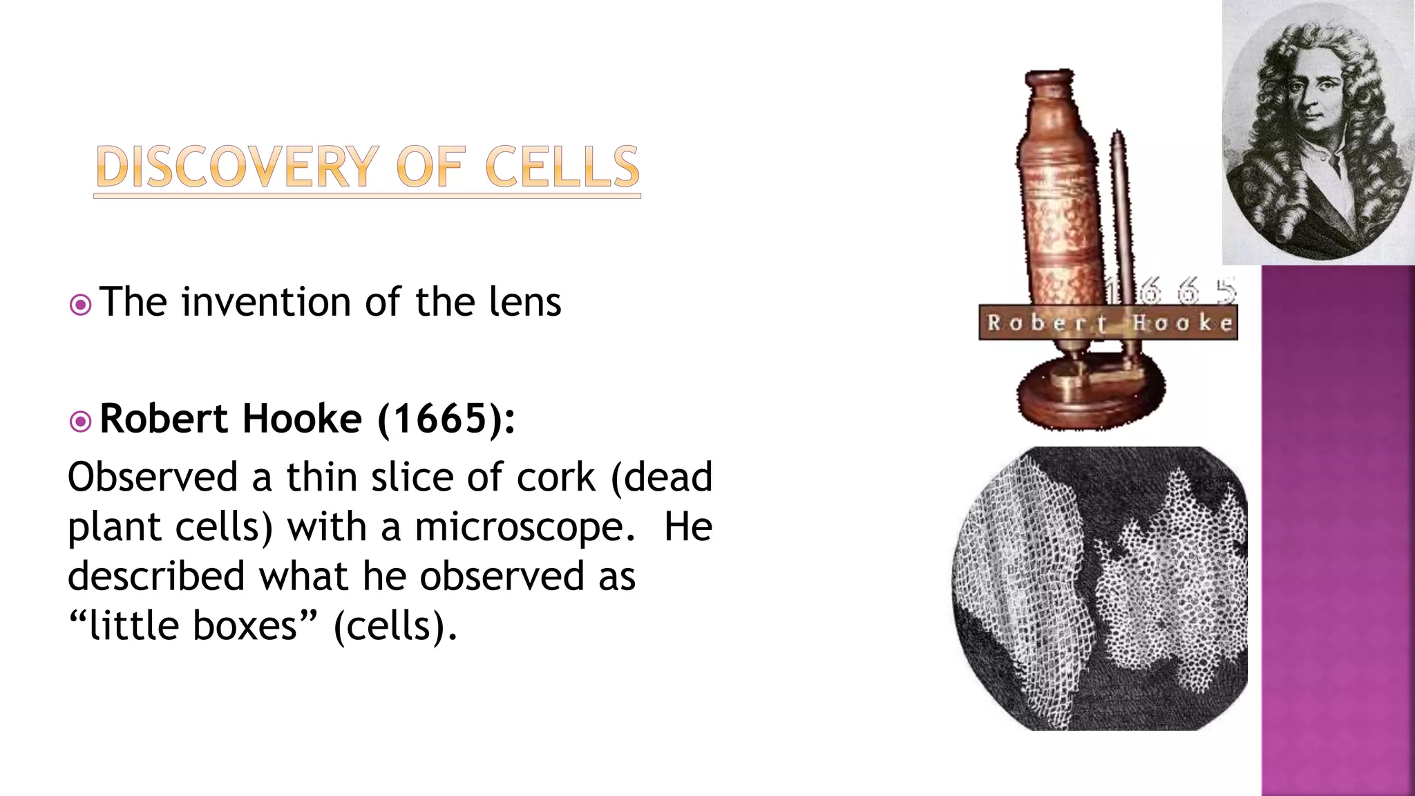
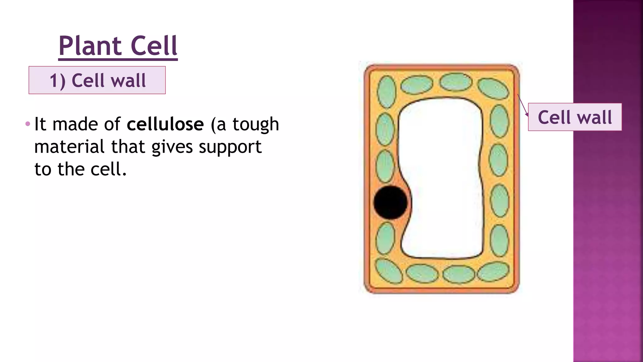
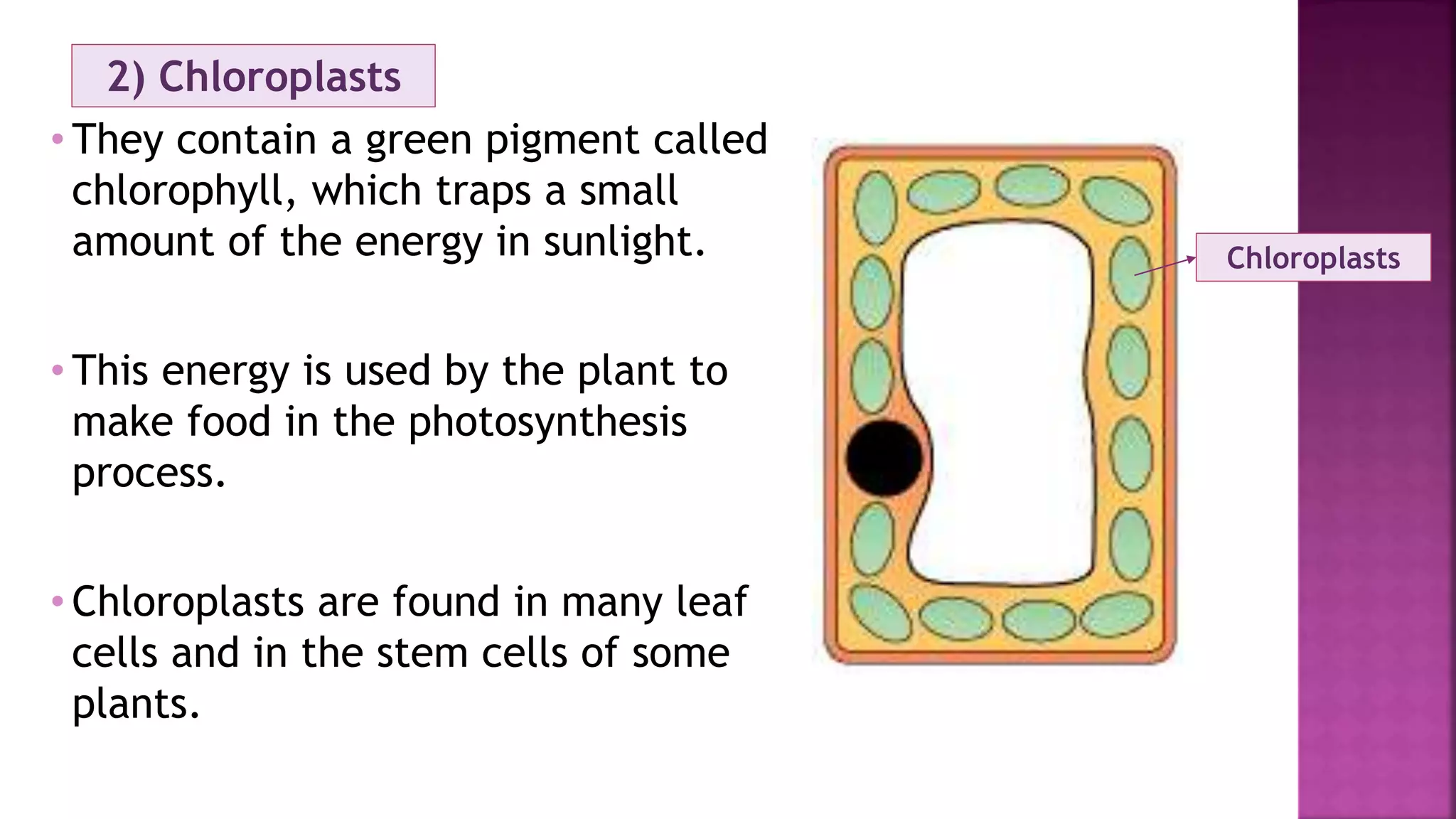
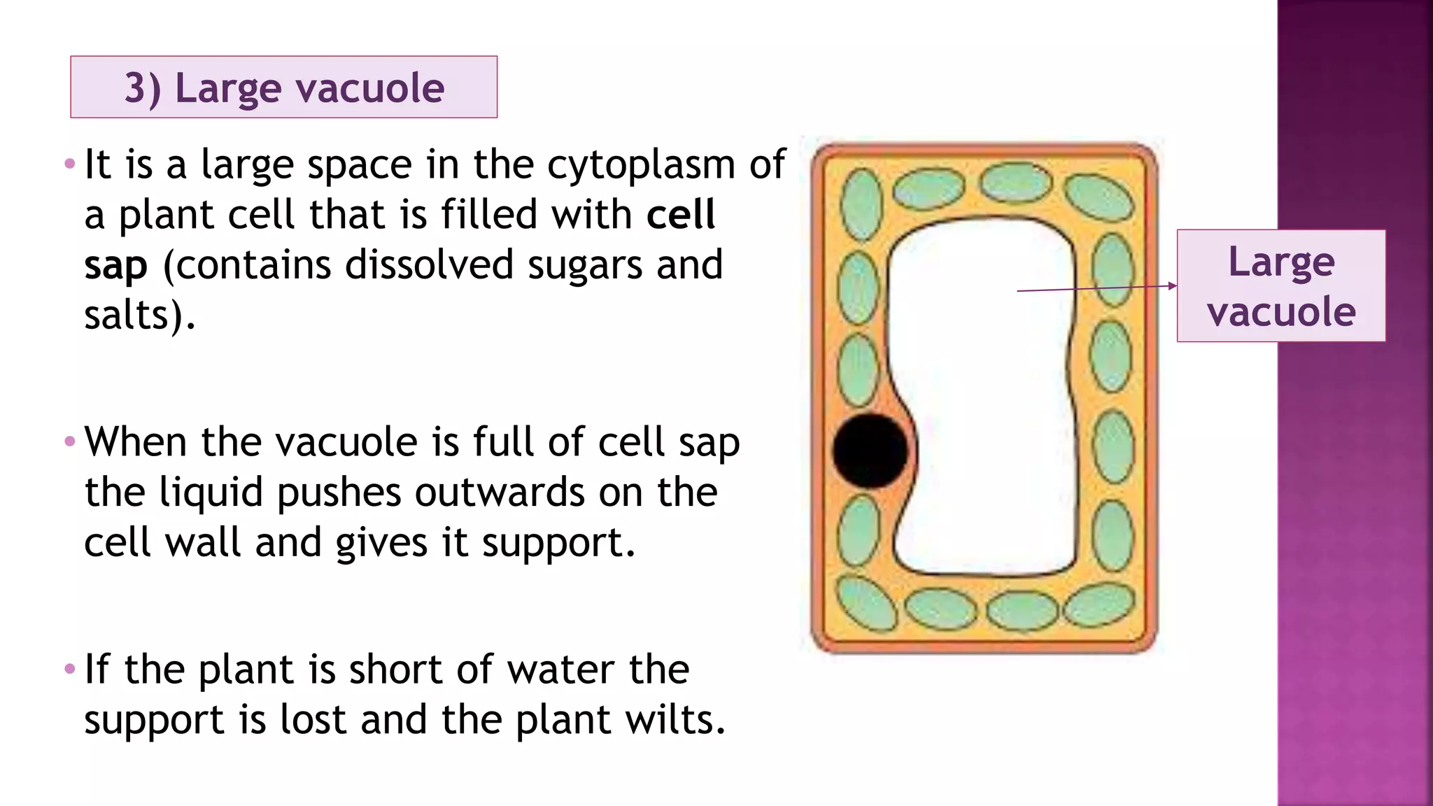
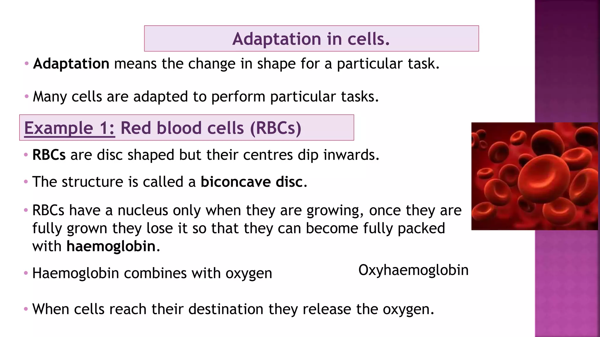
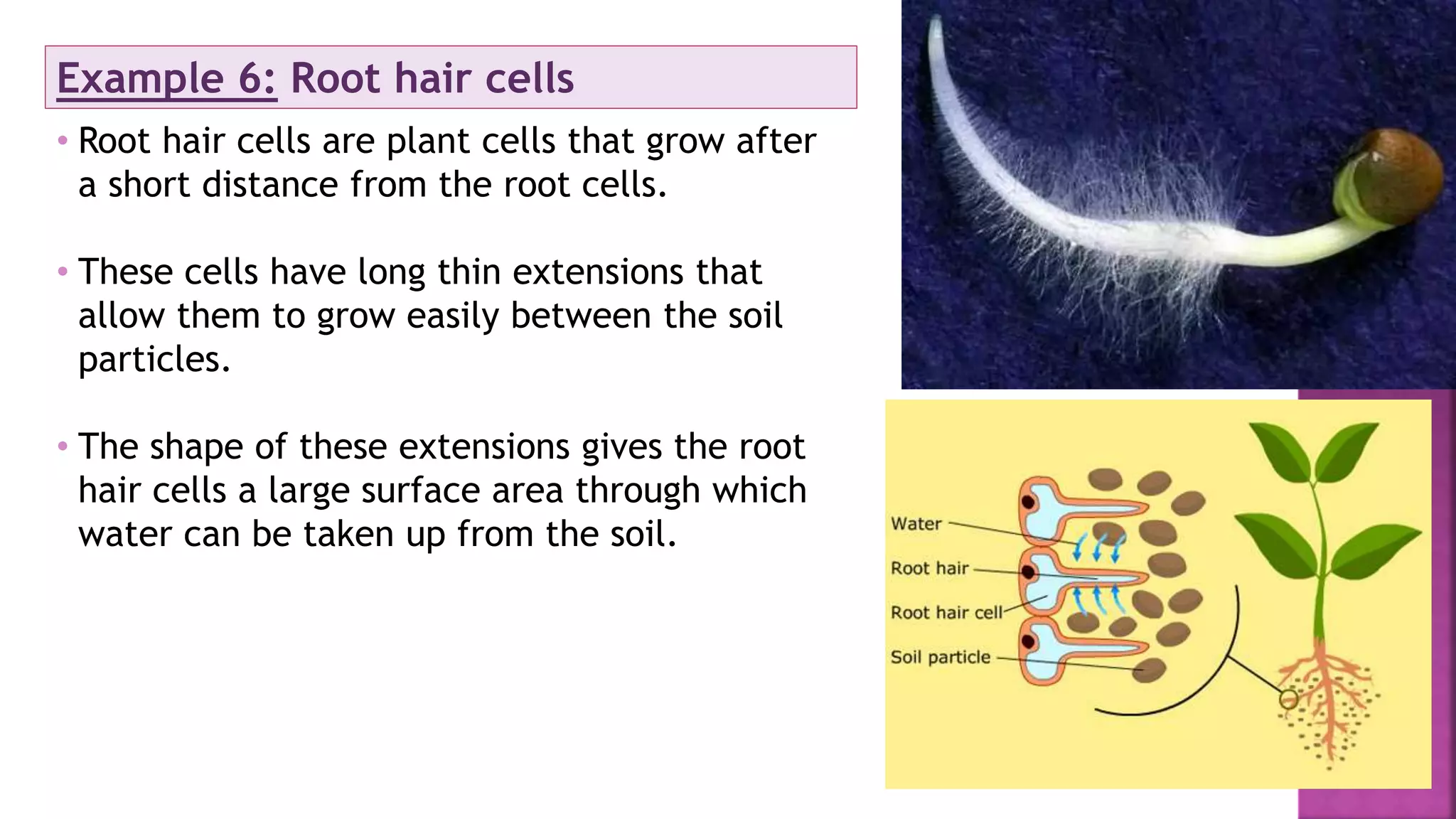
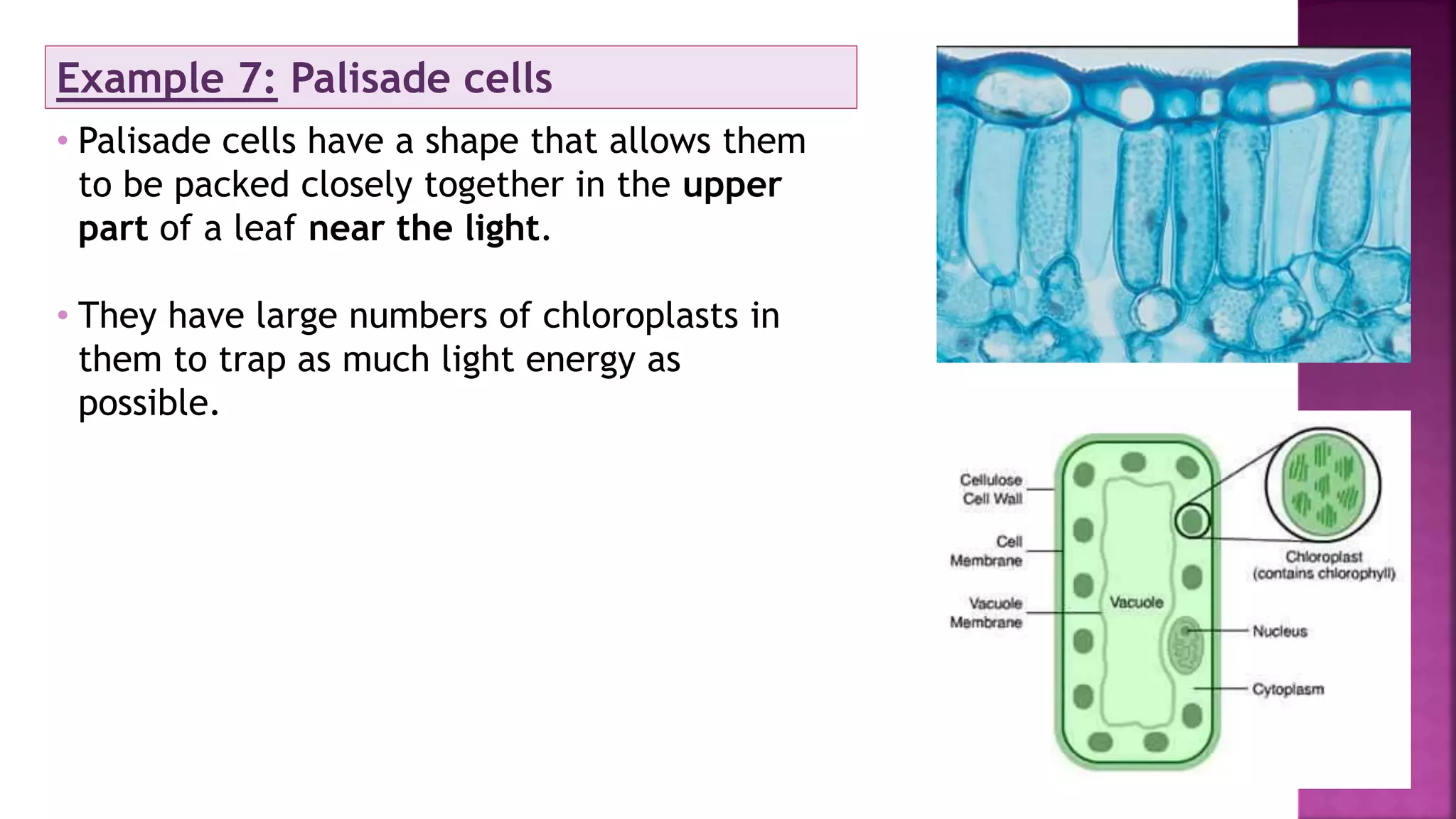
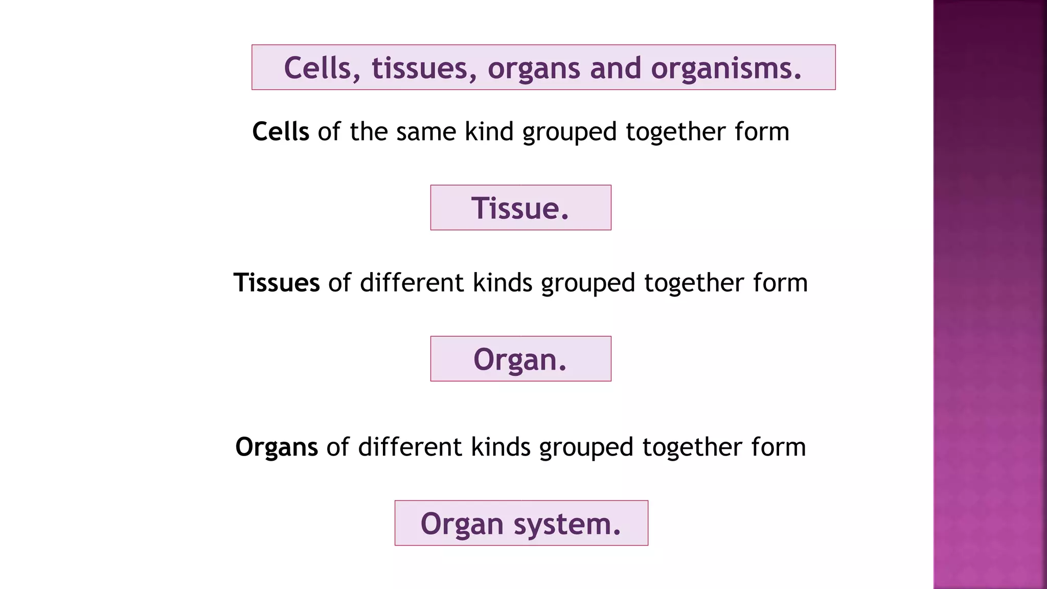
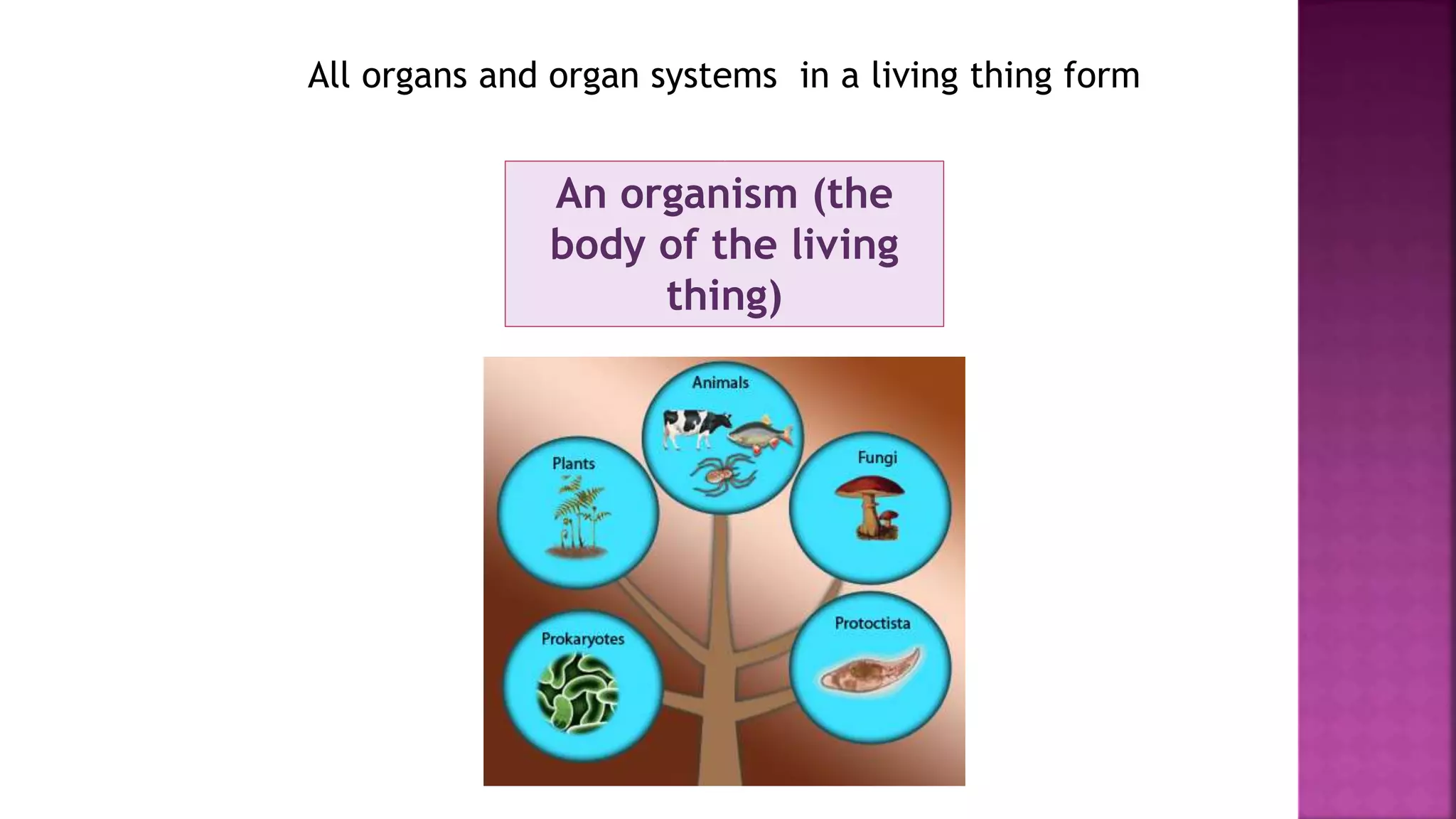
1. Robert Hooke observed plant cells under a microscope in 1665 and described them as "little boxes", introducing the concept of the cell.
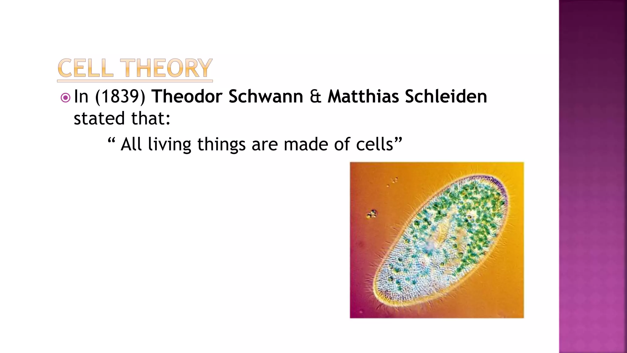
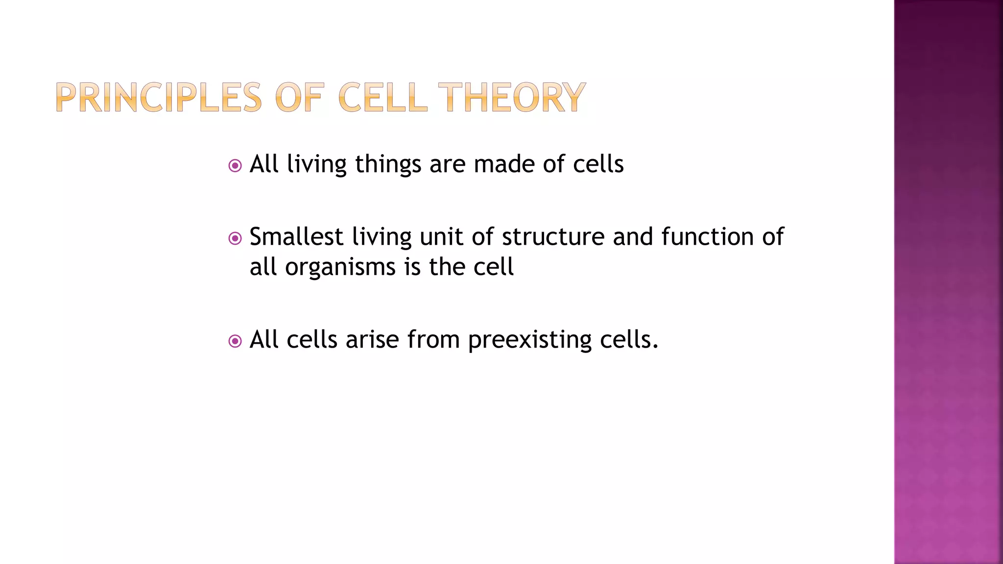
2. In 1839, Schwann and Schleiden stated that all living things are made of cells, establishing the cell theory.
3. The cell is the smallest unit of structure and function in living organisms, and all cells arise from preexisting cells.