162 calcium imaging
•Download as PPT, PDF•
0 likes•241 views
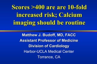
Scores >400 are associated with a 10-fold increased risk of future events. Calcium imaging should be used routinely to assess risk. Matthew Budoff is an assistant professor of cardiology who discloses a relationship with Imatron, Inc as a speaker.
Report
Share
Report
Share

Recommended
250 patients with very abnormal test results
1) Clinical data confirms that coronary calcium detected by electron beam computed tomography (EBT) is found in 96% of patients presenting with or developing acute coronary syndromes, and that these patients have significantly greater coronary artery calcium (CAC) at baseline compared to asymptomatic matched individuals.
2) Histologic studies show that CAC occurs in all types of coronary plaques, including stable plaques (43%), ruptured plaques (77%), and eroded plaques (54%), indicating that calcium is not a marker of plaque stability or instability.
3) Because all plaque types exhibit considerable overlap in the presence of calcium, CAC detected by EBT is not a marker of either stable or unstable plaque.
Mi or scd in patients with very abnormal cardiac test results
Mi or scd in patients with very abnormal cardiac test resultsSociety for Heart Attack Prevention and Eradication
1) Clinical data confirms that coronary calcium detected by electron beam computed tomography (EBT) is found in 96% of patients presenting with or developing acute coronary syndromes, and that these patients have significantly greater coronary artery calcium (CAC) at baseline compared to asymptomatic matched individuals.
2) Histologic studies show that CAC occurs in all types of coronary plaques, including stable plaques (43%), ruptured plaques (77%), and eroded plaques (54%), indicating that calcium is not a marker of plaque stability or instability.
3) Because all plaque types exhibit considerable overlap in CAC, calcium alone cannot be used to characterize plaques as stable or unstable.3rd vulnerable plaque rumberger 3 16-02 3
1) Clinical data confirms that coronary calcium detected by electron beam computed tomography (EBT) is found in 96% of patients presenting with or developing acute coronary syndromes, and that these patients have significantly greater coronary artery calcium (CAC) at baseline compared to asymptomatic matched individuals.
2) Histologic studies show that CAC occurs in all types of coronary plaques, including stable plaques (43%), ruptured plaques (77%), and eroded plaques (54%), indicating that calcium is not a marker of plaque stability or instability.
3) Because all plaque types exhibit considerable overlap in the presence of calcium, CAC detected by EBT is not a marker of either stable or unstable plaque.
Rumberg3
1) Clinical data confirms that coronary calcium detected by electron beam computed tomography (EBT) is found in 96% of patients presenting with or developing acute coronary syndromes, and that these patients have significantly greater coronary artery calcium scores at baseline compared to asymptomatic matched individuals.
2) Histologic studies show that coronary artery calcium occurs in all types of plaques, including stable (43%), rupture-prone (54%), and erosion-prone (77%) plaques.
3) Therefore, coronary artery calcium detected by imaging is a marker of atherosclerotic plaque burden but not necessarily a marker of stable or unstable plaque, as there is considerable overlap in calcium presence between different plaque types.
Ebt rumberger- ohio state u- part 3
1) Clinical data confirms that coronary calcium detected by electron beam computed tomography (EBT) is found in 96% of patients presenting with or developing acute coronary syndromes, and that these patients have significantly greater coronary artery calcium (CAC) at baseline compared to asymptomatic matched individuals.
2) Histologic studies show that CAC occurs in all types of coronary plaques, including stable plaques (43%), ruptured plaques (77%), and eroded plaques (54%), indicating that calcium is not a marker of plaque stability or instability.
3) Because all plaque types exhibit considerable overlap in the presence of calcium, CAC detected by EBT is not a marker of either stable or unstable plaque.
3rd vulnerable plaque rumberger 3 16-02 3
1) Clinical data confirms that coronary calcium detected by electron beam computed tomography (EBT) is found in 96% of patients presenting with or developing acute coronary syndromes, and that these patients have significantly greater coronary artery calcium scores at baseline compared to asymptomatic matched individuals.
2) Histologic studies show that coronary artery calcium occurs in all types of plaques, including stable (43%), rupture-prone (54%), and erosion-prone (77%) plaques.
3) Therefore, coronary artery calcium detected by imaging is a marker of atherosclerotic plaque burden but not necessarily a marker of stable or unstable plaque, as there is considerable overlap in calcium presence between different plaque types.
Cardio respiratory nuclear imaging ihab - copy
Nuclear cardiology imaging uses radiotracers and gamma cameras to image cardiac physiology and function. It is useful for diagnosing coronary artery disease, assessing risk, guiding treatment decisions, and evaluating outcomes. The presentation covered the basics of nuclear tracers, instrumentation, stress testing, image interpretation, and provided examples of clinical applications including assessing viability and guiding management of heart disease.
Cardiology lecture toIternal Medicine 21/10/2013
This document provides an outline and content for a cardiology lecture for internal medicine board exam preparation. It includes multiple choice questions (MCQs), a picture quiz, and explanatory notes on topics like myocardial infarction, heart failure, atrial fibrillation, and hypertension. The lecture was given by Dr. Ihab Suliman on October 20, 2013 and covered diagnostic tests, treatments, medications, and pathophysiology related to cardiology.
Cardiology lecture to i moct2013final
This document provides an outline and questions for a cardiology lecture for internal medicine board exam preparation. It includes multiple choice questions on topics like medications for lowering triglycerides, treatments for arrhythmias, and causes of petechial rashes and leg pain in post-MI patients. Explanatory notes are provided for some of the questions.
Cardiac amyloidosis
one of the form of restrictive cardiomyopathy usually missed in diagnosis. correct diagnosis can make N different outcome.
Out flow tract ventricular tachycardia
A woman in her late 40s with a history of hypertension presented to the emergency department after multiple episodes of palpitations with near syncope. While in the
emergency department, she developed monomorphic ventricular tachycardia (VT) with hemodynamic instability and was successfully cardioverted. She continued to have nonsustained monomorphic VT, so intravenous amiodarone and oral metoprolol were initiated. She was admitted for further evaluation. Results of tests of electrolyte levels and coronary angiography were normal. Cardiac magnetic resonance imaging with
gadolinium contrast revealed normal-sized cardiac chambers and normal biventricular
function without delayed enhancement. The presenting electrocardiogram (ECG)
is shown in Figure 1.
Early repolarisation syndrome
Early repolarization (ER) is an ECG pattern characterized by J-point elevation. While historically considered a benign variant, recent studies have linked ER to an increased risk of arrhythmia. ER syndrome describes those with both ER on ECG and symptomatic arrhythmias like ventricular fibrillation. Diagnosis requires excluding other causes through testing of survivors of sudden cardiac death. While the ER pattern itself usually requires no treatment, an implantable cardioverter-defibrillator is recommended for secondary prevention in ER syndrome patients with a history of resuscitated sudden cardiac death.
arrhythmogenic right ventricular dysplasia/Cardiomyopathy
This case discusses arrhythmogenic right ventricular cardiomyopathy (ARVC) in a 19-year-old female whose sister recently passed away from the condition. ARVC is characterized by replacement of the RV myocardium with fibrofatty tissue and electrical instability. The patient's sister's autopsy confirmed ARVC. The doctor discusses the pathology, diagnosis, treatment including ICDs and screening of relatives of ARVC to help inform the patient of her risk.
Bayes syndrome and atrial fibrillation and stroke
1) Advanced interatrial block (aIAB) is associated with delayed and retrograde activation of the left atrium due to impairment of the Bachmann's bundle, which connects the left and right atria. This conduction delay between atria is linked to a high risk of atrial arrhythmias like atypical atrial flutter or fibrillation.
2) aIAB can be diagnosed on electrocardiogram by a prolonged P wave over 120ms with biphasic morphology in leads II, III, and aVF, indicating retrograde left atrial activation. aIAB prevalence increases with age and is a risk factor for atrial fibrillation and stroke.
3) "Bayes syndrome" refers
ARVD
ARVC is a heritable heart muscle disorder that predominantly affects the right ventricle. It is caused by genetic defects in cardiac desmosomes, which are important for cell-to-cell adhesion. This leads to progressive loss of right ventricular myocardium and replacement by fibrofatty tissue. ARVC can cause dangerous ventricular arrhythmias and is a leading cause of sudden cardiac death in young people. Diagnosis involves imaging tests and electrocardiography to detect right ventricular structural abnormalities and arrhythmias.
Arrhythmogenic right ventricular 2003
The document summarizes arrhythmogenic right ventricular dysplasia (ARVD), a condition where the right ventricle of the heart is replaced by fat and fibrous tissue. It affects mostly young males and can cause sudden cardiac death. Genetic factors are involved in many cases. The condition starts with fatty infiltration of the right ventricle and progresses to include fibrosis, thinning of the ventricular wall, and later involvement of the left ventricle. Diagnosis involves criteria related to structural changes, electrocardiogram abnormalities, arrhythmias, and family history.
Arrhythmogenic right ventricular dysplasia
Arrhythmogenic right ventricular dysplasia (ARVD) is a genetic heart condition characterized by replacement of right ventricular myocardium with fat and fibrous tissue. This causes ventricular arrhythmias which can lead to sudden cardiac death. ARVD is diagnosed based on criteria involving cardiac imaging, biopsy, electrocardiogram findings and genetic testing. A combination of major and minor criteria must be met, including ventricular dysfunction, aneurysms, conduction abnormalities and evidence of fibrofatty tissue replacement on biopsy. ARVD has an autosomal dominant inheritance pattern and is a leading cause of sudden cardiac death in young people.
Acute Pyelonephritis of Crossed Right Fused Renal Ectopia- Crimson Publishers
Acute Pyelonephritis of Crossed Right Fused Renal
Ectopia by Batsaikhan Bat Erdene* in Surgical Medicine Open Access Journal
ARRHYTHMOGENIC RIGHT VENTRICULAR DYSPLASIA/CARDIOMYOPATHY
ARVD is one of important coardiomyopathy in our clinical practice,early diagnosis, risk stratification and early diagnosis of CHF, management of VT will make big difference in patient life
Arrhythmogenic right ventricular dysplasia
Arrhythmogenic right ventricular dysplasia/cardiomyopathy (ARVD/C) is an inherited heart muscle disease characterized by structural abnormalities and fatty replacement of the right ventricle muscle leading to ventricular arrhythmias. It is an important cause of sudden cardiac death in young adults. The disease results from genetic mutations that cause programmed cell death and fibrosis of the right ventricle muscle. Diagnosis is based on ECG findings like inverted T-waves in the right ventricle leads and epsilon waves, along with imaging showing right ventricle structural changes. Treatment involves medications like beta-blockers and implantable defibrillators to prevent arrhythmias.
Arvd vs uhls anomaly
This document discusses Uhl's anomaly and arrhythmogenic right ventricular dysplasia/cardiomyopathy (ARVD/C). It presents a case study of a 50-year-old man with palpitations and ventricular ectopic beats. Key differences between Uhl's anomaly and ARVD/C are described. Uhl's anomaly is a rare congenital disorder characterized by a thin-walled right ventricle, while ARVD/C is an inherited cardiomyopathy characterized by structural abnormalities and fatty infiltration of the right ventricle myocardium. Diagnostic criteria for ARVD/C include family history, electrocardiogram abnormalities, arrhythmias on monitoring, and structural changes seen on imaging like echocard
Early repolarization
12-lead electrocardiogram features of arrhythmic risk: A focus on early repolarization
Caterina Rizzo, Francesco Monitillo, Massimo Iacoviello
Caterina Rizzo, Francesco Monitillo, School of Cardiology, Department of Emergency and Organ Transplantation, University of Bari, 70124 Bari, Italy
Massimo Iacoviello, Cardiology Unit, Department of Cardiothoracic, Policlinic University Hospital, 70124 Bari, Italy
Why should we measure endothelial function
This document discusses the importance of measuring endothelial function for cardiovascular risk assessment. It begins with background on cardiovascular disease being the leading cause of death globally and the problems with traditional risk assessment based only on risk factors. It then discusses how endothelial dysfunction underlies many disease states and can serve as an integrated measure of risk. The document reviews different techniques for measuring endothelial function, including flow-mediated dilation of the brachial artery. It argues that a comprehensive cardiovascular risk assessment should include measures of both subclinical disease and endothelial function.
Remote Ischemic Conditioning - Dr. Robert Kloner
This document summarizes a presentation on ischemic conditioning and myocardial infarction. It discusses how brief periods of ischemia can protect the heart from subsequent longer periods of ischemia, known as preconditioning. Studies in animal models and clinical trials show remote ischemic conditioning, using brief ischemia in another part of the body like a limb, can protect the heart. Remote ischemic conditioning reduced infarct size and improved outcomes in patients having a heart attack or undergoing procedures like bypass surgery. Ongoing clinical trials are investigating remote ischemic conditioning for other conditions involving ischemia in organs like the brain and kidneys.
ARVD (Arrythmogenic right ventricular cardiomyopathy) - updated task force cr...
This document discusses arrythmogenic right ventricular cardiomyopathy (ARVC). It begins by explaining the genetics of ARVC, noting that mutations can be either dominant or recessive. It then describes the natural history, clinical presentation, diagnosis, and criteria used to diagnose ARVC based on the revised Task Force Criteria. This includes major and minor criteria in categories such as imaging, electrocardiography findings, biopsy results, and family history. The document concludes by discussing management strategies for ARVC including ICD therapy, antiarrhythmic drugs, ablation, heart failure treatment, and transplantation.
Braunwald
This document outlines an approach to identifying and managing coronary risk. It recommends that prevention must be the primary goal through intensive global risk factor reduction for all patients with clinically apparent heart disease. It also recommends identifying asymptomatic high-risk individuals through testing like the Framingham Risk Score so they can receive prevention. It proposes a risk stratification approach from low to very high risk based on annual risk levels and corresponding testing and treatments, with very high risk patients receiving the most intensive treatments like invasive detection of unstable plaques and procedures like CABG or multiple drug-eluting stents.
083 non invasive imaging of the vulnerable plaque
This document discusses non-invasive imaging techniques for coronary artery plaque and vulnerable patients. It notes that while lipid-rich plaque makes up about 20% of total plaque volume, it is often not detectable without invasive techniques. Newer CT and electron beam tomography techniques aim to non-invasively detect plaque composition through measurements of density and heterogeneity. The document raises questions about which patients should be imaged, what parameters need to be measured, and how imaging results could potentially change patient outcomes. It also discusses technical considerations for these non-invasive imaging modalities.
129 inflammation and culprit lesions
This document discusses inflammation in coronary arteries and culprit lesions. It finds that:
1. Inflammation is present in many non-culprit lesions, not just culprit lesions, based on a study of 74 coronary arteries.
2. Inflammation therefore has a very low positive predictive value (<3%) for identifying the actual culprit lesion responsible for a clinical event like a heart attack.
3. Inflammation appears to be a local arterial phenomenon, as cross-sectional sampling of inflammation in one part of an artery did not strongly correlate with levels in other parts or in paired left/right arteries.
086 unstable angina, lad stenosis
A patient presented with unstable angina and stenosis of the left anterior descending artery. Angiography and thermography were performed, which identified the site of stenosis. A stent was implanted in the left anterior descending artery, after which thermography and angiography were repeated and showed improvement. Additional thermography and angiography were done for a circumflex lesion, which also identified the site of stenosis.
160 5th vulnerable plaque symposium
This document announces an industry roundtable event on vulnerable plaque to be held on March 29, 2003 in Chicago. The roundtable aims to bring together cardiologists, researchers, and industry executives to discuss regulatory challenges, reimbursement issues, and identifying areas of common interest in developing and regulating new products for detecting and treating vulnerable plaque. The program will highlight emerging trends in treating heart disease caused by vulnerable plaque and the current regulatory climate for alternative clinical strategies. A panel of experts from academia, industry and government agencies will discuss these issues.
More Related Content
What's hot
Cardiology lecture to i moct2013final
This document provides an outline and questions for a cardiology lecture for internal medicine board exam preparation. It includes multiple choice questions on topics like medications for lowering triglycerides, treatments for arrhythmias, and causes of petechial rashes and leg pain in post-MI patients. Explanatory notes are provided for some of the questions.
Cardiac amyloidosis
one of the form of restrictive cardiomyopathy usually missed in diagnosis. correct diagnosis can make N different outcome.
Out flow tract ventricular tachycardia
A woman in her late 40s with a history of hypertension presented to the emergency department after multiple episodes of palpitations with near syncope. While in the
emergency department, she developed monomorphic ventricular tachycardia (VT) with hemodynamic instability and was successfully cardioverted. She continued to have nonsustained monomorphic VT, so intravenous amiodarone and oral metoprolol were initiated. She was admitted for further evaluation. Results of tests of electrolyte levels and coronary angiography were normal. Cardiac magnetic resonance imaging with
gadolinium contrast revealed normal-sized cardiac chambers and normal biventricular
function without delayed enhancement. The presenting electrocardiogram (ECG)
is shown in Figure 1.
Early repolarisation syndrome
Early repolarization (ER) is an ECG pattern characterized by J-point elevation. While historically considered a benign variant, recent studies have linked ER to an increased risk of arrhythmia. ER syndrome describes those with both ER on ECG and symptomatic arrhythmias like ventricular fibrillation. Diagnosis requires excluding other causes through testing of survivors of sudden cardiac death. While the ER pattern itself usually requires no treatment, an implantable cardioverter-defibrillator is recommended for secondary prevention in ER syndrome patients with a history of resuscitated sudden cardiac death.
arrhythmogenic right ventricular dysplasia/Cardiomyopathy
This case discusses arrhythmogenic right ventricular cardiomyopathy (ARVC) in a 19-year-old female whose sister recently passed away from the condition. ARVC is characterized by replacement of the RV myocardium with fibrofatty tissue and electrical instability. The patient's sister's autopsy confirmed ARVC. The doctor discusses the pathology, diagnosis, treatment including ICDs and screening of relatives of ARVC to help inform the patient of her risk.
Bayes syndrome and atrial fibrillation and stroke
1) Advanced interatrial block (aIAB) is associated with delayed and retrograde activation of the left atrium due to impairment of the Bachmann's bundle, which connects the left and right atria. This conduction delay between atria is linked to a high risk of atrial arrhythmias like atypical atrial flutter or fibrillation.
2) aIAB can be diagnosed on electrocardiogram by a prolonged P wave over 120ms with biphasic morphology in leads II, III, and aVF, indicating retrograde left atrial activation. aIAB prevalence increases with age and is a risk factor for atrial fibrillation and stroke.
3) "Bayes syndrome" refers
ARVD
ARVC is a heritable heart muscle disorder that predominantly affects the right ventricle. It is caused by genetic defects in cardiac desmosomes, which are important for cell-to-cell adhesion. This leads to progressive loss of right ventricular myocardium and replacement by fibrofatty tissue. ARVC can cause dangerous ventricular arrhythmias and is a leading cause of sudden cardiac death in young people. Diagnosis involves imaging tests and electrocardiography to detect right ventricular structural abnormalities and arrhythmias.
Arrhythmogenic right ventricular 2003
The document summarizes arrhythmogenic right ventricular dysplasia (ARVD), a condition where the right ventricle of the heart is replaced by fat and fibrous tissue. It affects mostly young males and can cause sudden cardiac death. Genetic factors are involved in many cases. The condition starts with fatty infiltration of the right ventricle and progresses to include fibrosis, thinning of the ventricular wall, and later involvement of the left ventricle. Diagnosis involves criteria related to structural changes, electrocardiogram abnormalities, arrhythmias, and family history.
Arrhythmogenic right ventricular dysplasia
Arrhythmogenic right ventricular dysplasia (ARVD) is a genetic heart condition characterized by replacement of right ventricular myocardium with fat and fibrous tissue. This causes ventricular arrhythmias which can lead to sudden cardiac death. ARVD is diagnosed based on criteria involving cardiac imaging, biopsy, electrocardiogram findings and genetic testing. A combination of major and minor criteria must be met, including ventricular dysfunction, aneurysms, conduction abnormalities and evidence of fibrofatty tissue replacement on biopsy. ARVD has an autosomal dominant inheritance pattern and is a leading cause of sudden cardiac death in young people.
Acute Pyelonephritis of Crossed Right Fused Renal Ectopia- Crimson Publishers
Acute Pyelonephritis of Crossed Right Fused Renal
Ectopia by Batsaikhan Bat Erdene* in Surgical Medicine Open Access Journal
ARRHYTHMOGENIC RIGHT VENTRICULAR DYSPLASIA/CARDIOMYOPATHY
ARVD is one of important coardiomyopathy in our clinical practice,early diagnosis, risk stratification and early diagnosis of CHF, management of VT will make big difference in patient life
Arrhythmogenic right ventricular dysplasia
Arrhythmogenic right ventricular dysplasia/cardiomyopathy (ARVD/C) is an inherited heart muscle disease characterized by structural abnormalities and fatty replacement of the right ventricle muscle leading to ventricular arrhythmias. It is an important cause of sudden cardiac death in young adults. The disease results from genetic mutations that cause programmed cell death and fibrosis of the right ventricle muscle. Diagnosis is based on ECG findings like inverted T-waves in the right ventricle leads and epsilon waves, along with imaging showing right ventricle structural changes. Treatment involves medications like beta-blockers and implantable defibrillators to prevent arrhythmias.
Arvd vs uhls anomaly
This document discusses Uhl's anomaly and arrhythmogenic right ventricular dysplasia/cardiomyopathy (ARVD/C). It presents a case study of a 50-year-old man with palpitations and ventricular ectopic beats. Key differences between Uhl's anomaly and ARVD/C are described. Uhl's anomaly is a rare congenital disorder characterized by a thin-walled right ventricle, while ARVD/C is an inherited cardiomyopathy characterized by structural abnormalities and fatty infiltration of the right ventricle myocardium. Diagnostic criteria for ARVD/C include family history, electrocardiogram abnormalities, arrhythmias on monitoring, and structural changes seen on imaging like echocard
Early repolarization
12-lead electrocardiogram features of arrhythmic risk: A focus on early repolarization
Caterina Rizzo, Francesco Monitillo, Massimo Iacoviello
Caterina Rizzo, Francesco Monitillo, School of Cardiology, Department of Emergency and Organ Transplantation, University of Bari, 70124 Bari, Italy
Massimo Iacoviello, Cardiology Unit, Department of Cardiothoracic, Policlinic University Hospital, 70124 Bari, Italy
Why should we measure endothelial function
This document discusses the importance of measuring endothelial function for cardiovascular risk assessment. It begins with background on cardiovascular disease being the leading cause of death globally and the problems with traditional risk assessment based only on risk factors. It then discusses how endothelial dysfunction underlies many disease states and can serve as an integrated measure of risk. The document reviews different techniques for measuring endothelial function, including flow-mediated dilation of the brachial artery. It argues that a comprehensive cardiovascular risk assessment should include measures of both subclinical disease and endothelial function.
Remote Ischemic Conditioning - Dr. Robert Kloner
This document summarizes a presentation on ischemic conditioning and myocardial infarction. It discusses how brief periods of ischemia can protect the heart from subsequent longer periods of ischemia, known as preconditioning. Studies in animal models and clinical trials show remote ischemic conditioning, using brief ischemia in another part of the body like a limb, can protect the heart. Remote ischemic conditioning reduced infarct size and improved outcomes in patients having a heart attack or undergoing procedures like bypass surgery. Ongoing clinical trials are investigating remote ischemic conditioning for other conditions involving ischemia in organs like the brain and kidneys.
ARVD (Arrythmogenic right ventricular cardiomyopathy) - updated task force cr...
This document discusses arrythmogenic right ventricular cardiomyopathy (ARVC). It begins by explaining the genetics of ARVC, noting that mutations can be either dominant or recessive. It then describes the natural history, clinical presentation, diagnosis, and criteria used to diagnose ARVC based on the revised Task Force Criteria. This includes major and minor criteria in categories such as imaging, electrocardiography findings, biopsy results, and family history. The document concludes by discussing management strategies for ARVC including ICD therapy, antiarrhythmic drugs, ablation, heart failure treatment, and transplantation.
What's hot (17)
arrhythmogenic right ventricular dysplasia/Cardiomyopathy
arrhythmogenic right ventricular dysplasia/Cardiomyopathy
Acute Pyelonephritis of Crossed Right Fused Renal Ectopia- Crimson Publishers
Acute Pyelonephritis of Crossed Right Fused Renal Ectopia- Crimson Publishers
ARRHYTHMOGENIC RIGHT VENTRICULAR DYSPLASIA/CARDIOMYOPATHY
ARRHYTHMOGENIC RIGHT VENTRICULAR DYSPLASIA/CARDIOMYOPATHY
ARVD (Arrythmogenic right ventricular cardiomyopathy) - updated task force cr...
ARVD (Arrythmogenic right ventricular cardiomyopathy) - updated task force cr...
Viewers also liked
Braunwald
This document outlines an approach to identifying and managing coronary risk. It recommends that prevention must be the primary goal through intensive global risk factor reduction for all patients with clinically apparent heart disease. It also recommends identifying asymptomatic high-risk individuals through testing like the Framingham Risk Score so they can receive prevention. It proposes a risk stratification approach from low to very high risk based on annual risk levels and corresponding testing and treatments, with very high risk patients receiving the most intensive treatments like invasive detection of unstable plaques and procedures like CABG or multiple drug-eluting stents.
083 non invasive imaging of the vulnerable plaque
This document discusses non-invasive imaging techniques for coronary artery plaque and vulnerable patients. It notes that while lipid-rich plaque makes up about 20% of total plaque volume, it is often not detectable without invasive techniques. Newer CT and electron beam tomography techniques aim to non-invasively detect plaque composition through measurements of density and heterogeneity. The document raises questions about which patients should be imaged, what parameters need to be measured, and how imaging results could potentially change patient outcomes. It also discusses technical considerations for these non-invasive imaging modalities.
129 inflammation and culprit lesions
This document discusses inflammation in coronary arteries and culprit lesions. It finds that:
1. Inflammation is present in many non-culprit lesions, not just culprit lesions, based on a study of 74 coronary arteries.
2. Inflammation therefore has a very low positive predictive value (<3%) for identifying the actual culprit lesion responsible for a clinical event like a heart attack.
3. Inflammation appears to be a local arterial phenomenon, as cross-sectional sampling of inflammation in one part of an artery did not strongly correlate with levels in other parts or in paired left/right arteries.
086 unstable angina, lad stenosis
A patient presented with unstable angina and stenosis of the left anterior descending artery. Angiography and thermography were performed, which identified the site of stenosis. A stent was implanted in the left anterior descending artery, after which thermography and angiography were repeated and showed improvement. Additional thermography and angiography were done for a circumflex lesion, which also identified the site of stenosis.
160 5th vulnerable plaque symposium
This document announces an industry roundtable event on vulnerable plaque to be held on March 29, 2003 in Chicago. The roundtable aims to bring together cardiologists, researchers, and industry executives to discuss regulatory challenges, reimbursement issues, and identifying areas of common interest in developing and regulating new products for detecting and treating vulnerable plaque. The program will highlight emerging trends in treating heart disease caused by vulnerable plaque and the current regulatory climate for alternative clinical strategies. A panel of experts from academia, industry and government agencies will discuss these issues.
073 collagenases vs collagen
This document discusses research on the roles of collagen and collagenases in atherosclerotic plaque vulnerability. It presents several hypotheses: 1) that human and experimental plaques contain collagenases, 2) that lipid lowering improves features associated with vulnerability by reducing macrophages and collagenase expression while increasing collagen, and 3) that collagenolysis regulates collagen accumulation in plaques of genetically altered mice. Several studies are described that provide evidence supporting these hypotheses, finding that collagenases are expressed in human and animal plaques, lipid lowering reduces vulnerability features in rabbits by altering collagenase and collagen levels, and collagenase-resistant mice have altered collagen in plaques. The research suggests collagenases critically regulate collagen metabolism in plaques and their clinical complications.
257 inhibition of atherosclerosis in mice
This document discusses research into identifying immune responses against oxidized LDL in humans and developing an immunization therapy to inhibit atherosclerosis. The research found that immunization of apoE null mice with specific MDA-modified apoB-100 peptide sequences activated athero-protective immune responses and significantly reduced atherosclerotic plaque development compared to controls. This suggests vaccination with these peptide sequences may be a potential therapy for atherosclerosis by stimulating protective immune responses against oxidized LDL.
The Impact of carotid plaque screening on motivation for smoking cessation a...
Presented by:
Nicolas Rodondi, MD, MAS, Reto Auer, MD, Patrick J. Devine, MD, Patrick G. O’Malley, MD, MPH, Daniel Hayoz, MD, Jacques Cornuz, MD, MPH
149 revisiting the basics
The document discusses several factors that are considered predictors of plaque vulnerability, including luminal narrowing, plaque volume and composition, fibrous cap thickness, and plaque inflammation. It reviews studies that show myocardial infarction can develop from previously non-severe lesions and that lipid content, cap thickness, inflammation, and stress factors like circumferential stress are correlated with plaque stability or rupture. In conclusion, the size and composition of the lipid core, thickness and composition of the fibrous cap, and inflammation are well-established predictors of plaque rupture.
Acc 2002 mehran for print
This document discusses the use of DNA microarrays in studying vulnerable atherosclerotic plaques. It provides background on atherosclerosis and plaque rupture. DNA microarrays allow high-throughput analysis of gene and protein expression, which can provide insights into molecular mechanisms underlying plaque vulnerability. One study used microarrays to analyze gene expression differences between ruptured and stable plaques, identifying perilipin as upregulated in ruptured plaques. However, microarray analysis of atherosclerosis is still in its early stages with many technical challenges to address.
No correlation and low agreement of imaging and inflammatory atherosclerosis’...
Presented by:
Lilton R.C. Martinez MD, Marcio H. Miname MD, Luiz A. Bortolotto MD, Ana P.M. Chacra MD, Carlos E. Rochitte MD, Andrei C. Sposito MD, Raul D. Santos MD,PhD.
140 mr imaging of fibrin
The document discusses research into using MRI with fibrin-targeted gadolinium contrast agents to detect vulnerable plaques associated with heart attacks, presenting findings that this approach can successfully identify fibrin deposits in thrombi in animal studies and has potential for sensitive detection of injured plaques in humans; it also notes the need for more studies to develop highly sensitive targeted contrast agents for multiple imaging modalities to identify vulnerable plaque features beyond structure.
154 plaque rupture definition
The document defines plaque rupture as the structural failure of the fibrocellular cap separating an atheromatous core from the lumen of an atherosclerotic artery. It then discusses factors that increase the propensity of plaque rupture like a lipid-rich core and lack of a fibrous cap. Several studies are summarized that show plaque rupture is the cause of fatal thrombosis in 75-100% of cases of acute MI and sudden cardiac death when examined post-mortem. The role of traditional risk factors like age, sex, cholesterol levels, and smoking in rupture vs. non-rupture cases is then assessed based on multiple studies.
054 vulnerable plaques and vulnerable patients
This document outlines guidelines for defining vulnerable plaques and vulnerable patients from the Association for Eradication of Heart Attack. It discusses how atherosclerotic diseases are not limited to developed countries and are a major cause of death worldwide. Vulnerable plaques, myocardium, and hypercoagulable blood can lead to sudden cardiac death and heart attacks. The document proposes histological and clinical criteria for defining vulnerable plaques and screening methods. It also discusses diagnosing plaque inflammation, thin caps, endothelial dysfunction, and other factors at both the plaque and systemic level.
076 cardiac magnetic resonance imaging
MRI can effectively evaluate patients presenting to the emergency department with possible acute coronary syndrome. In a study of 161 such patients, MRI had higher sensitivity and specificity than electrocardiography, troponin levels, or clinical risk scores for detecting acute coronary syndrome, non-ST elevation myocardial infarction, and ischemic heart disease. Regional wall motion abnormalities detected by MRI had 89% sensitivity and 99% specificity for detecting ischemic heart disease compared to 67% sensitivity and 99% specificity for detecting ischemic heart disease using delayed hyperenhancement. MRI provides accurate information to guide patient management in the emergency department.
258 dr renu virmani
Dr. Renu Virmani is the Chairperson of the Department of Cardiovascular Pathology at the Armed Forces Institute of Pathology in Washington, DC. She received her medical degrees in India and has held positions at multiple universities in the United States. Dr. Virmani has established an independent research program focused on cardiovascular pathology, publishing over 300 papers. She leads a department of 23 researchers studying devices, sudden death, and cardiovascular diseases.
146 animal models of vulnerable plaque
This study examined plaque rupture in the brachiocephalic arteries of 98 fat-fed apolipoprotein E knockout mice over time. Ruptured plaques were significantly larger, more occlusive, had thinner fibrous caps, more previous ruptures, and a greater lipid burden than intact plaques. The earliest ruptures occurred after 7 weeks on a high-fat diet. A pravastatin treatment significantly reduced sudden deaths, suggesting statins may prevent plaque rupture.
1 spio-mri studies by dr naghavi - amersham oct 2003
This document discusses non-invasive imaging of macrophages for detection of vulnerable plaque using magnetic resonance imaging (MRI) with ultrasmall particles of iron oxide (USPIO). USPIOs are taken up by macrophages and shorten MRI relaxation time, allowing visualization of macrophage activity. Studies show USPIO uptake correlates with macrophage density and plaque inflammation in animal models. While MRI can identify plaque inflammation, limitations include poor resolution for coronary imaging and high cost compared to alternatives.
164 calcium in atherosclerosis
Calcium can be both good and bad in atherosclerosis. While calcium burden is a significant predictor of future cardiovascular events, papers on plaque rupture do not always discuss calcification. The document discusses several past studies that have shown calcium can be present deep within plaques, near the lipid core and necrotic areas. This suggests calcium is sometimes located in areas prone to rupture rather than only being present in stable, fibrotic regions. The location and extent of calcium deposits may help determine whether it indicates a more stable or high-risk plaque.
123 coronary dialsysis
Atherosclerosis is a leading cause of death worldwide. It is a systemic inflammatory disease involving the buildup of plaque in artery walls. Inflammatory markers like C-reactive protein are associated with future coronary events. The authors describe a Cardiac Dialysis System designed to specifically target and remove inflammatory markers from the bloodstream to stabilize vulnerable plaques and prevent acute cardiac events like heart attack.
Viewers also liked (20)
The Impact of carotid plaque screening on motivation for smoking cessation a...
The Impact of carotid plaque screening on motivation for smoking cessation a...
No correlation and low agreement of imaging and inflammatory atherosclerosis’...
No correlation and low agreement of imaging and inflammatory atherosclerosis’...
1 spio-mri studies by dr naghavi - amersham oct 2003
1 spio-mri studies by dr naghavi - amersham oct 2003
Similar to 162 calcium imaging
Cardiogenicshock by Dr.Afroza Prioty -140123092109-phpapp02
1) Cardiogenic shock is a clinical condition caused by the heart's inability to pump an adequate amount of blood to vital organs, resulting in inadequate tissue perfusion.
2) The document discusses the causes, pathophysiology, clinical manifestations, diagnosis, and management of cardiogenic shock, with a focus on cardiogenic shock caused by acute myocardial infarction.
3) Early revascularization through percutaneous coronary intervention or coronary artery bypass grafting is recommended for suitable patients with cardiogenic shock due to acute myocardial infarction, along with supportive therapies like intra-aortic balloon pump counterpulsation or ventricular assist devices.
Carotid artery stenosis
This document provides an overview of carotid artery stenosis. It discusses the anatomy of the carotid arteries and how stenosis can increase the risk of stroke by reducing blood flow to the brain. Symptoms of stenosis range from transient ischemic attacks to full strokes, depending on the location and severity of the blockage. Imaging plays a key role in detecting and evaluating carotid artery stenosis. Treatment may involve medications, lifestyle changes, or carotid endarterectomy surgery to remove plaque buildup.
Stable ischemic heart disease bv tam duc
Bài Học Sưu Tầm nên muốn chia sẻ cùng mọi người, Nếu có vấn đề bản quyền tôi sẽ gỡ xuống liền Cám ơn đã theo dõi bài đăng.
ECG in young
A 33-year-old man presented to the emergency department after collapsing. His ECG showed Brugada pattern, which is characterized by ST-segment elevation in leads V1-V3 and increased risk of ventricular arrhythmias and sudden cardiac death. Brugada syndrome is a genetic condition caused by sodium channel mutations and commonly presents with syncope or cardiac arrest in young males. The diagnosis can be confirmed with ajmaline/flecainide provocation test showing transient Brugada pattern. Treatment involves lifestyle modifications and implantable cardioverter-defibrillator for high-risk patients.
Pfo
1. A patent foramen ovale (PFO) is present in 25% of the population but is more common in younger people. 2. Stroke and PFO as well as migraine and PFO have a proven or suspected causal relationship. 3. Catheter based PFO closure is considered the safest therapeutic intervention for cardiology but randomized studies comparing it to drug therapy are still ongoing and results are not expected until at least 2010.
06 the anesthesia patient with acute coronary syndrome copia
This document discusses the management of patients with acute coronary syndrome (ACS) in the perioperative period. It begins with an overview of ACS, distinguishing between unstable angina, myocardial infarction without ST-segment elevation (NSTEMI), and myocardial infarction with ST-segment elevation (STEMI). It then reviews the diagnosis, pathophysiology, and standard treatment of ACS outside of surgery, including antiplatelet therapy, antithrombin therapy, and beta-blockade. The document indicates that the anesthesiologist must understand how ACS is typically treated to properly manage patients who present for surgery with ACS.
Aortic dissection
This document presents the case of a 52-year-old male who presented with sudden onset severe back pain. Examination and tests revealed type B aortic dissection with possible acute kidney injury and polycystic kidney disease. The patient was managed medically but required endovascular intervention which was delayed, and he collapsed on the fourth day after referral. A review of literature discusses aortic dissection including causes, classification, clinical manifestations, physical findings, and management considerations.
Ventricular fibrillation eMedicine Article
Ventricular fibrillation (VF) is a life-threatening heart rhythm disorder that usually results in cardiac arrest if not treated promptly. It occurs when the lower chambers of the heart beat in an uncoordinated, chaotic fashion, preventing the heart from pumping blood effectively. VF accounts for about 300,000 deaths per year in the United States, making it the leading cause of sudden cardiac death. It is commonly caused by coronary artery disease and often presents as the first sign of a heart attack. Prompt treatment with cardiopulmonary resuscitation and defibrillation can help restore a normal heart rhythm and prevent death from VF in some cases.
2014session5 3
The document discusses the lack of consensus around treating asymptomatic carotid artery disease. While surgery reduces the risk of stroke, the absolute benefit is small given the already low risk with medical management alone. More recent data favors a purely medical approach for most asymptomatic patients. The evidence for invasive treatments like carotid endarterectomy (CEA) or stenting is considered weak, with no clear benefits shown for subgroups like women, those over 75, or different degrees of stenosis. Treating asymptomatic disease at a population level may lead to many unnecessary interventions that provide little benefit given evolving medical therapies.
Xaheer shah...pulmonary embolism
This document discusses pulmonary embolism (PE). It notes that PE is a common disorder that can be deadly if left untreated. The presentation of PE is often vague and nonspecific. While the classic triad of symptoms is hemoptysis, dyspnea, and pleuritic pain, this occurs in less than 20% of patients. The document reviews risk factors, clinical features, diagnostic testing options including Wells criteria, imaging studies, D-dimer testing and their limitations. Treatment involves anticoagulation with heparin or warfarin to prevent clot extension and recurrence while educating patients.
Acute pulmonary thromboembolism
The document describes the case of a 26-year-old female who presented with shortness of breath and was initially diagnosed with anxiety but later diagnosed with acute pulmonary thromboembolism. It then reviews the epidemiology, pathophysiology, risk factors, clinical features, diagnosis, natural history, and management of acute pulmonary thromboembolism, with a focus on topics relevant to critically ill patients.
Heart failure syndrome1
This document discusses heart failure with preserved systolic function, also known as heart failure with normal ejection fraction. Some key points:
- This condition represents 20-50% of all heart failure cases. However, relatively little is known about its morbidity and mortality.
- Median prevalence in studies was 36%, with a range from 13-74%. Rates of readmission were similar to heart failure with low ejection fraction but mortality was lower at 9% versus 18%.
- Causes include inaccurate diagnosis, episodic systolic dysfunction, diastolic dysfunction from various conditions like hypertension, cardiomyopathy, and aging.
- Diastolic dysfunction refers to abnormally elevated ventricular filling pressures despite normal or
Coronary artery perforation complicating percutaneous coronary intervention
This study summarizes the management of 24 patients who experienced coronary artery perforation as a complication of percutaneous coronary intervention (PCI) at a hospital in Iraq from 2009-2016. The majority of perforations involved the left anterior descending artery and were classified as Type II or III, requiring sealing with covered stents. Thirteen patients also required drainage of pericardial effusions. All perforations were immediately diagnosed and treated, with no patients requiring surgery or experiencing mortality. The low rate of coronary artery perforation complications in this study, primarily managed using covered stents, demonstrates the effectiveness of the approaches used at this hospital.
Management of Atrial Fibrillation Science:Myths & Fashion
This document discusses the management of atrial fibrillation. It notes that AF prevalence is increasing with an aging population and is associated with increased risk of stroke and mortality. Treatment involves rate or rhythm control with medications, electrical cardioversion, or newer options like catheter ablation. Risk stratification tools like CHADS2 are used to determine stroke risk and need for anticoagulation. Newer oral anticoagulants offer alternatives to warfarin by avoiding the need for INR monitoring.
Nuclear Imaging In Cardiology Cme
Nuclear imaging techniques have various applications in cardiology, including assessing coronary artery disease, left ventricular function, cardiomyopathy, valvular heart disease, cardiac shunts, pulmonary hypertension, and more. Myocardial perfusion imaging can accurately diagnose and assess the prognosis of coronary artery disease, viability after myocardial infarction, and effectiveness of revascularization procedures. Gated SPECT allows evaluation of both cardiac function and perfusion simultaneously. Other nuclear techniques help evaluate conditions like myocarditis, pulmonary embolism, and secondary causes of hypertension.
Pulmonary embolism radiology imaging
Pulmonary embolism is caused by a blockage in the pulmonary artery from substances traveling through the bloodstream, most commonly from deep vein thrombosis. It is a common and potentially lethal condition. Diagnosis is challenging as symptoms are non-specific and it is often overlooked or missed. Imaging tests like CT pulmonary angiography, ventilation-perfusion scans, and pulmonary angiography are used to diagnose pulmonary embolism, while ultrasound of the legs can identify deep vein thrombosis, a major risk factor. Prompt diagnosis and treatment are important to prevent mortality from this potentially serious condition.
Carotid artery disease
The document describes the anatomy of the carotid arteries and their branches, evaluation and imaging of carotid artery disease, and treatment strategies including lifestyle modifications to reduce risk factors, carotid endarterectomy to remove plaques from significantly stenotic arteries, and outcomes data from clinical trials on endarterectomy for symptomatic and asymptomatic carotid stenosis. Imaging modalities like carotid duplex ultrasound, CTA, and MRA are described for evaluating the degree of carotid stenosis. The benefits of carotid endarterectomy are greater for symptomatic high-grade stenosis while more moderate for asymptomatic disease.
3rd vulnerable plaque rumberger 3 16-02 4
The document discusses that less obstructive plaques pose a greater risk of coronary occlusion than severely obstructed plaques due to their greater numbers. It also states that the aggregate risk of rupture from many non-significant lesions exceeds that of fewer significant lesions, so a myocardial infarction is more likely to originate from a non-significant lesion. Additionally, it explains that while electron beam tomography (EBT) cannot identify vulnerable plaques directly, it can identify vulnerable patients based on their coronary artery calcium (CAC) scores and percentile ranks, as risk increases with higher scores. EBT is also useful for estimating prognosis and tracking changes in plaque in response to treatment over time.
199 plaque severity and coronary occlusion
The document discusses that less obstructive plaques pose a greater risk of coronary occlusion than severely obstructed plaques due to their greater numbers. It also states that the aggregate risk of rupture from many non-significant lesions exceeds that of fewer significant lesions, so a myocardial infarction is more likely to originate from a non-significant lesion. Additionally, while electron beam tomography (EBT) cannot identify vulnerable plaques directly, it can identify vulnerable patients based on their coronary artery calcium (CAC) scores and percentiles, as risk increases with higher scores. EBT is also useful for estimating prognosis and tracking changes in plaque burden in response to treatment over time.
Plaque severity and coronary occlusion
The document discusses that less obstructive plaques pose a greater risk of coronary occlusion than severely obstructed plaques due to their greater numbers. It also states that the aggregate risk of rupture from many non-significant lesions exceeds that of fewer significant lesions, so a myocardial infarction is more likely to originate from a non-significant lesion. Additionally, it explains that while electron beam tomography (EBT) cannot identify vulnerable plaques directly, it can identify vulnerable patients based on their coronary artery calcium (CAC) scores and percentiles, as risk increases with higher scores. EBT is also useful for estimating prognosis and tracking changes in plaque in response to treatment over time.
Similar to 162 calcium imaging (20)
Cardiogenicshock by Dr.Afroza Prioty -140123092109-phpapp02
Cardiogenicshock by Dr.Afroza Prioty -140123092109-phpapp02
06 the anesthesia patient with acute coronary syndrome copia
06 the anesthesia patient with acute coronary syndrome copia
Coronary artery perforation complicating percutaneous coronary intervention
Coronary artery perforation complicating percutaneous coronary intervention
Management of Atrial Fibrillation Science:Myths & Fashion
Management of Atrial Fibrillation Science:Myths & Fashion
More from SHAPE Society
Barth imt
This document provides background on Jacques Barth, an expert in cardiovascular imaging and risk assessment. It discusses the evolution of ultrasound technology for measuring intima-media thickness (IMT) from 1986 to 2005. IMT is an early marker of atherosclerosis and cardiovascular risk. The document also addresses issues around vulnerable plaques, reporting IMT measurements, and assessing cardiovascular risk in children and adolescents.
Acc vp-org 3-02 stone
This document describes a study that uses intravascular ultrasound (IVUS), biplane coronary angiography, and blood flow measurements to characterize endothelial shear stress (ESS) in coronary arteries. It found that over 6 months, areas of low ESS demonstrated plaque progression, areas of normal ESS remained stable, and areas of high ESS developed outward remodeling. The technology allows in vivo determination of intracoronary flow velocity and ESS, which has not previously been possible. This provides a method to predict progression of atherosclerosis and vascular remodeling. A pilot study applied this technique in 8 patients at baseline and 6 months to analyze changes in native coronary artery disease and in-stent restenosis while taking candesartan vs fel
101205 shape aha 2005 f
Zahi A. Fayad is an Associate Professor who studies molecular imaging of atherosclerosis using MRI. His research focuses on developing targeted contrast agents to noninvasively detect unstable plaque. Some agents under investigation include annexin A5 labeled with a radioisotope to detect apoptosis, FDG-PET to assess plaque activity, and fibrin-targeted and MMP-targeting Gd-based contrasts. Additional work involves lipid-based particulate agents using reconstituted HDL or iron oxide nanoparticles. The goal is to improve MRI detection sensitivity and specificity for high-risk plaque characterization.
Acc v porg becker
This document discusses the use of coronary CT angiography (CTA) to detect and characterize coronary atherosclerosis beyond just detecting coronary stenoses. CTA can identify calcified plaques, non-calcified plaques, mixed plaques, atheromas, thrombi, and myocardial infarction scars. CTA provides information on plaque composition and distribution that can help understand coronary artery disease and be used to follow patients under therapy. The limitations of CTA include artifacts from cardiac motion, breathing, blooming effect, and poor contrast opacification of small vessels. Advances in multislice CT technology are helping to address some of these limitations.
Acc 2005-1, v pl-vp
This document discusses approaches to cardiovascular disease (CVD) and the need for new approaches. It summarizes that the emphasis is shifting from high risk plaques to high risk symptomatic patients, and from high risk asymptomatic to intermediate and low risk patients. It discusses diagnostic tools like magnetic resonance imaging to identify high risk asymptomatic patients and computed tomography to identify intermediate risk patients using coronary artery calcium scoring and CRP biomarkers. It also discusses prevention and treatment strategies like a polypill for acute coronary syndrome patients and those with chronic atherothrombosis.
Acc presentation macrophage (1)
This document proposes a non-invasive method using SPIO (super paramagnetic iron oxide) nanoparticles to image macrophage infiltration and inflammation in vulnerable atherosclerotic plaques. Rabbits and mice were injected with SPIO, which accumulated in inflamed plaque areas correlated with macrophage density. SPIO-enhanced MRI then successfully identified these inflamed plaques non-invasively in vivo. This technique could provide a way to detect rupture-prone plaques and better understand plaque vulnerability.
Acc v porg
This document discusses the use of coronary CT angiography (CTA) to detect and characterize coronary atherosclerosis beyond just detecting coronary stenoses. CTA can identify calcified plaques, non-calcified plaques, and mixed plaques. It can detect atheromas and characterize plaque density. CTA can also identify intracoronary thrombi and myocardial infarction scars. The document outlines the CTA scanning parameters and techniques used to minimize motion artifacts and optimize image quality for plaque detection and characterization.
Acc presentation (1)
This document describes a study that introduces a non-invasive method for imaging macrophage infiltration in inflamed atherosclerotic plaques using superparamagnetic iron oxide (SPIO) nanoparticles and MRI. The researchers injected SPIO into hypercholesterolemic and normal rabbits and found that SPIO profoundly accumulated in areas of macrophage infiltration in the atherosclerotic plaques, as confirmed by histology. SPIO-enhanced MRI was able to identify these inflamed plaques non-invasively. The results suggest SPIO-enhanced MRI can be a novel method for detecting rupture-prone inflamed plaques associated with heart attacks and strokes.
Ac cculprit
The document discusses several factors that are considered predictors of plaque vulnerability, including luminal narrowing, plaque volume and composition, fibrous cap thickness, and plaque inflammation. It reviews studies that show myocardial infarction can develop from previously non-severe lesions and that lipid content, cap thickness, inflammation, and stress factors like circumferential stress are correlated with plaque stability and vulnerability. In conclusion, the size and composition of the lipid core, thickness and composition of the fibrous cap, and inflammation are well-established predictors of plaque rupture.
Acc presentation lipid coated(2)
This document discusses a novel tracer for MRI imaging of macrophage infiltration in atherosclerotic plaque. It summarizes research into lipid-coated superparamagnetic iron oxide nanoparticles (SPIOs) that are phagocytosed by macrophages. The researchers tested various SPIO coatings and sizes to maximize macrophage uptake while minimizing oxidative stress. Lipid-coated SPIOs combined with certain aminoglycans showed the highest uptake and lowest induction of reactive oxygen species. The goal is to develop an MRI contrast agent that can noninvasively image vulnerable, inflamed plaques by detecting macrophage presence.
Acc 2002 microarray mehran for print
This document discusses the use of DNA microarrays in vulnerable plaque research. It provides background on atherosclerosis and identifies DNA microarrays as a tool that can be used to investigate the molecular mechanisms underlying plaque vulnerability. The document outlines the basic steps of a DNA microarray experiment and discusses considerations for experimental design, data analysis, and validation of results. It also summarizes several studies that have used DNA microarrays or related techniques to examine gene expression in atherosclerosis.
Acc booth inventory
The document provides details about AEHA's booth at the ACC/AEHA Exhibition from March 30 to April 1, 2003. It lists the booth equipment and inventory, proposed activities like an Ecode on Friday morning and a VP Symposium. Giveaways include Magellan GPS devices in a raffle and Dove chocolates. It also discusses the need for an immediate membership sign-up page on the AEHA website and a vision for the future of AEHA.
Acc 04 vuln pt
This document discusses approaches to identifying and managing coronary risk. It states that the primary goal should be preventing acute cardiac events through intensive risk factor reduction for all patients with clinically apparent heart disease. Additionally, it notes that one third of sudden cardiac deaths and heart attacks occur in previously asymptomatic individuals with undiagnosed risk factors or pre-clinical disease. The document proposes identifying high-risk asymptomatic individuals through testing to provide prevention. It presents a risk stratification approach using testing like CRP, cholesterol, glucose and imaging to guide different levels of risk factor reduction and management.
Acc2002 thermography
Vulnerable plaques are prone to rupture and cause heart attacks. This document proposes criteria for defining vulnerable plaques based on histopathology and clinical factors. It also explores using infrared thermography to identify vulnerable plaques by detecting heat from macrophage inflammation. Studies in animal models and humans found temperature heterogeneity in atherosclerotic arteries that correlated with plaque vulnerability features. Further research aims to develop non-invasive thermography techniques to accurately detect vulnerable plaques and help predict heart attack risk.
A20 gene
The document presents findings on the A20 gene, which encodes a zinc finger protein that inhibits NF-kB activity and TNF-induced apoptosis. The study found that C57 and FVB mouse strains have a coding difference in A20 that generates a phosphorylation site in C57 mice. C57-A20 was less effective at shutting down TNF-induced NF-kB activity and C57 cells were less susceptible to TNF-induced apoptosis compared to FVB cells. This suggests less active A20 in C57 mice leads to increased inflammation and reduced apoptosis, while more active A20 in FVB mice decreases inflammation and increases apoptosis, contributing to differences in atherosclerosis susceptibility between the strains.
#1 killer of human beings in the 21st century
Vulnerable plaque refers to dangerous forms of atherosclerotic plaques that can rupture or induce thrombosis, disrupting blood flow. The document discusses the history and research around vulnerable plaque, including pioneers in the field and emerging techniques to detect vulnerable plaque such as intravascular ultrasound, optical coherence tomography, and magnetic resonance imaging. It summarizes that vulnerable plaques are typically characterized by a thin fibrous cap, large lipid core, and presence of macrophages.
A20 is an atherosclerosis
The document presents findings on the A20 gene, which encodes a zinc finger protein that inhibits NF-kB activity and TNF-induced apoptosis. The study found that C57 and FVB mouse strains have a coding difference in A20 that generates a putative phosphorylation site in C57 mice. Experiments showed the C57 version of A20 is less effective at shutting down NF-kB activity and C57 cells are less susceptible to TNF-induced apoptosis. This suggests the A20 variation could contribute to differences in atherosclerosis susceptibility between C57 and FVB strains by affecting inflammation and apoptosis.
103100
This progress report discusses ongoing near-infrared (NIR) spectroscopy studies. It notes that a new probe design is being developed to improve signal detection in the NIR range. Characterization of a new light source and additional tissue phantom studies are needed. The report identifies ongoing difficulties with depth penetration studies and experimental setup issues. Priorities include analyzing existing data to inform probe redesign, characterizing the new light source, conducting depth penetration and tissue phantom studies, and addressing experimental setup challenges.
259 crp as a risk factor
This document discusses the use of C-reactive protein (CRP) and low-density lipoprotein (LDL) cholesterol levels to predict cardiovascular risk. It summarizes a study that found CRP to be a stronger predictor of future cardiovascular events than LDL. The study measured CRP and LDL levels in 27,939 healthy women and followed them for 8 years, finding that most cardiovascular events occurred in women with normal or low LDL (<160 mg/dl) but elevated CRP. The document concludes that combining CRP and LDL measurements provides better risk assessment than either marker alone.
511 first steps towards understanding
This study investigated genetic differences in vascular remodeling and shear stress regulation in response to altered blood flow in four inbred rat strains. The results showed significant differences among strains in their ability to maintain normal endothelial shear stress levels through outward arterial remodeling when flow was increased or decreased. Specifically, the GH strain was better able to regulate shear stress through remodeling compared to the SHR-SP strain. These genetic differences in vascular responses to changes in blood flow have important implications for understanding the variable manifestations of atherosclerosis and susceptibility to cardiovascular disease in individuals and populations. Future studies are needed to investigate whether similar genetic differences exist in humans and their role in clinical outcomes.
More from SHAPE Society (20)
Recently uploaded
Identifying Major Symptoms of Slip Disc.
Our backs are like superheroes, holding us up and helping us move around. But sometimes, even superheroes can get hurt. That’s where slip discs come in.
All info about Diabetes and how to control it.
One health condition that is becoming more common day by day is diabetes.
According to research conducted by the National Family Health Survey of India, diabetic cases show a projection which might increase to 10.4% by 2030.
Cell Therapy Expansion and Challenges in Autoimmune Disease
There is increasing confidence that cell therapies will soon play a role in the treatment of autoimmune disorders, but the extent of this impact remains to be seen. Early readouts on autologous CAR-Ts in lupus are encouraging, but manufacturing and cost limitations are likely to restrict access to highly refractory patients. Allogeneic CAR-Ts have the potential to broaden access to earlier lines of treatment due to their inherent cost benefits, however they will need to demonstrate comparable or improved efficacy to established modalities.
In addition to infrastructure and capacity constraints, CAR-Ts face a very different risk-benefit dynamic in autoimmune compared to oncology, highlighting the need for tolerable therapies with low adverse event risk. CAR-NK and Treg-based therapies are also being developed in certain autoimmune disorders and may demonstrate favorable safety profiles. Several novel non-cell therapies such as bispecific antibodies, nanobodies, and RNAi drugs, may also offer future alternative competitive solutions with variable value propositions.
Widespread adoption of cell therapies will not only require strong efficacy and safety data, but also adapted pricing and access strategies. At oncology-based price points, CAR-Ts are unlikely to achieve broad market access in autoimmune disorders, with eligible patient populations that are potentially orders of magnitude greater than the number of currently addressable cancer patients. Developers have made strides towards reducing cell therapy COGS while improving manufacturing efficiency, but payors will inevitably restrict access until more sustainable pricing is achieved.
Despite these headwinds, industry leaders and investors remain confident that cell therapies are poised to address significant unmet need in patients suffering from autoimmune disorders. However, the extent of this impact on the treatment landscape remains to be seen, as the industry rapidly approaches an inflection point.
share - Lions, tigers, AI and health misinformation, oh my!.pptx
• Pitfalls and pivots needed to use AI effectively in public health
• Evidence-based strategies to address health misinformation effectively
• Building trust with communities online and offline
• Equipping health professionals to address questions, concerns and health misinformation
• Assessing risk and mitigating harm from adverse health narratives in communities, health workforce and health system
Journal Article Review on Rasamanikya
Rasamanikya is a excellent preparation in the field of Rasashastra, it is used in various Kushtha Roga, Shwasa, Vicharchika, Bhagandara, Vatarakta, and Phiranga Roga. In this article Preparation& Comparative analytical profile for both Formulationon i.e Rasamanikya prepared by Kushmanda swarasa & Churnodhaka Shodita Haratala. The study aims to provide insights into the comparative efficacy and analytical aspects of these formulations for enhanced therapeutic outcomes.
Promoting Wellbeing - Applied Social Psychology - Psychology SuperNotes
A proprietary approach developed by bringing together the best of learning theories from Psychology, design principles from the world of visualization, and pedagogical methods from over a decade of training experience, that enables you to: Learn better, faster!
Top Effective Soaps for Fungal Skin Infections in India
Swisschem Dermacare has mentioned the List of The Best Antifungal Soap In India 2022. All of these soaps are trusted by various Dermatology Experts.
REGULATION FOR COMBINATION PRODUCTS AND MEDICAL DEVICES.pptx
It includes regulation of combination products and medical devices. FDA and industry liaisons.
Efficacy of Avartana Sneha in Ayurveda
Avartana Sneha is a unique method of Preparation of Sneha Kalpana in Ayurveda, mainly it is indicated for the Vataja rogas.
LOOPS in orthodontics t loop bull loop vertical loop mushroom loop stop loop
vertical loop
horizontal loop
Artificial Intelligence Symposium (THAIS)
Artificial Intelligence Symposium (THAIS). Parc Taulí. Sabadell
The Nervous and Chemical Regulation of Respiration
These lecture slides, by Dr Sidra Arshad, offer a simplified look into the mechanisms involved in the regulation of respiration:
Learning objectives:
1. Describe the organisation of respiratory center
2. Describe the nervous control of inspiration and respiratory rhythm
3. Describe the functions of the dorsal and respiratory groups of neurons
4. Describe the influences of the Pneumotaxic and Apneustic centers
5. Explain the role of Hering-Breur inflation reflex in regulation of inspiration
6. Explain the role of central chemoreceptors in regulation of respiration
7. Explain the role of peripheral chemoreceptors in regulation of respiration
8. Explain the regulation of respiration during exercise
9. Integrate the respiratory regulatory mechanisms
10. Describe the Cheyne-Stokes breathing
Study Resources:
1. Chapter 42, Guyton and Hall Textbook of Medical Physiology, 14th edition
2. Chapter 36, Ganong’s Review of Medical Physiology, 26th edition
3. Chapter 13, Human Physiology by Lauralee Sherwood, 9th edition
Does Over-Masturbation Contribute to Chronic Prostatitis.pptx
In some case, your chronic prostatitis may be related to over-masturbation. Generally, natural medicine Diuretic and Anti-inflammatory Pill can help mee get a cure.
Hemodialysis: Chapter 4, Dialysate Circuit - Dr.Gawad
- Video recording of this lecture in English language: https://youtu.be/kqbnxVAZs-0
- Video recording of this lecture in Arabic language: https://youtu.be/SINlygW1Mpc
- Link to download the book free: https://nephrotube.blogspot.com/p/nephrotube-nephrology-books.html
- Link to NephroTube website: www.NephroTube.com
- Link to NephroTube social media accounts: https://nephrotube.blogspot.com/p/join-nephrotube-on-social-media.html
TEST BANK For Community and Public Health Nursing: Evidence for Practice, 3rd...
TEST BANK For Community and Public Health Nursing: Evidence for Practice, 3rd Edition by DeMarco, Walsh, Verified Chapters 1 - 25, Complete Newest Version TEST BANK For Community and Public Health Nursing: Evidence for Practice, 3rd Edition by DeMarco, Walsh, Verified Chapters 1 - 25, Complete Newest Version TEST BANK For Community and Public Health Nursing: Evidence for Practice, 3rd Edition by DeMarco, Walsh, Verified Chapters 1 - 25, Complete Newest Version Test Bank For Community and Public Health Nursing: Evidence for Practice 3rd Edition Pdf Chapters Download Test Bank For Community and Public Health Nursing: Evidence for Practice 3rd Edition Pdf Download Stuvia Test Bank For Community and Public Health Nursing: Evidence for Practice 3rd Edition Study Guide Test Bank For Community and Public Health Nursing: Evidence for Practice 3rd Edition Ebook Download Stuvia Test Bank For Community and Public Health Nursing: Evidence for Practice 3rd Edition Questions and Answers Quizlet Test Bank For Community and Public Health Nursing: Evidence for Practice 3rd Edition Studocu Test Bank For Community and Public Health Nursing: Evidence for Practice 3rd Edition Quizlet Test Bank For Community and Public Health Nursing: Evidence for Practice 3rd Edition Stuvia Community and Public Health Nursing: Evidence for Practice 3rd Edition Pdf Chapters Download Community and Public Health Nursing: Evidence for Practice 3rd Edition Pdf Download Course Hero Community and Public Health Nursing: Evidence for Practice 3rd Edition Answers Quizlet Community and Public Health Nursing: Evidence for Practice 3rd Edition Ebook Download Course hero Community and Public Health Nursing: Evidence for Practice 3rd Edition Questions and Answers Community and Public Health Nursing: Evidence for Practice 3rd Edition Studocu Community and Public Health Nursing: Evidence for Practice 3rd Edition Quizlet Community and Public Health Nursing: Evidence for Practice 3rd Edition Stuvia Community and Public Health Nursing: Evidence for Practice 3rd Edition Test Bank Pdf Chapters Download Community and Public Health Nursing: Evidence for Practice 3rd Edition Test Bank Pdf Download Stuvia Community and Public Health Nursing: Evidence for Practice 3rd Edition Test Bank Study Guide Questions and Answers Community and Public Health Nursing: Evidence for Practice 3rd Edition Test Bank Ebook Download Stuvia Community and Public Health Nursing: Evidence for Practice 3rd Edition Test Bank Questions Quizlet Community and Public Health Nursing: Evidence for Practice 3rd Edition Test Bank Studocu Community and Public Health Nursing: Evidence for Practice 3rd Edition Test Bank Quizlet Community and Public Health Nursing: Evidence for Practice 3rd Edition Test Bank Stuvia
Recently uploaded (20)
Ear and its clinical correlations By Dr. Rabia Inam Gandapore.pptx
Ear and its clinical correlations By Dr. Rabia Inam Gandapore.pptx
Cell Therapy Expansion and Challenges in Autoimmune Disease
Cell Therapy Expansion and Challenges in Autoimmune Disease
share - Lions, tigers, AI and health misinformation, oh my!.pptx
share - Lions, tigers, AI and health misinformation, oh my!.pptx
Promoting Wellbeing - Applied Social Psychology - Psychology SuperNotes
Promoting Wellbeing - Applied Social Psychology - Psychology SuperNotes
Top Effective Soaps for Fungal Skin Infections in India
Top Effective Soaps for Fungal Skin Infections in India
REGULATION FOR COMBINATION PRODUCTS AND MEDICAL DEVICES.pptx
REGULATION FOR COMBINATION PRODUCTS AND MEDICAL DEVICES.pptx
LOOPS in orthodontics t loop bull loop vertical loop mushroom loop stop loop
LOOPS in orthodontics t loop bull loop vertical loop mushroom loop stop loop
Tests for analysis of different pharmaceutical.pptx
Tests for analysis of different pharmaceutical.pptx
The Nervous and Chemical Regulation of Respiration
The Nervous and Chemical Regulation of Respiration
Does Over-Masturbation Contribute to Chronic Prostatitis.pptx
Does Over-Masturbation Contribute to Chronic Prostatitis.pptx
Hemodialysis: Chapter 4, Dialysate Circuit - Dr.Gawad
Hemodialysis: Chapter 4, Dialysate Circuit - Dr.Gawad
TEST BANK For Community and Public Health Nursing: Evidence for Practice, 3rd...
TEST BANK For Community and Public Health Nursing: Evidence for Practice, 3rd...
162 calcium imaging
- 1. Scores >400 are are 10-foldScores >400 are are 10-fold increased risk; Calciumincreased risk; Calcium imaging should be routineimaging should be routine Matthew J. Budoff, MD, FACCMatthew J. Budoff, MD, FACC Assistant Professor of MedicineAssistant Professor of Medicine Division of CardiologyDivision of Cardiology Harbor-UCLA Medical Center Torrance, CA
- 2. Presenter Disclosure Information DISCLOSURE INFORMATION: The following relationships exist related to this presentation: Dr. Budoff is on the Speaker’s Bureau for Imatron, Inc. Matthew Budoff, MD, FACC High Speed CT
- 4. 20%20% 80%80% Total Coronary Artery PlaqueTotal Coronary Artery Plaque and EBCT Coronary Calciumand EBCT Coronary Calcium 80%80% PlaquePlaque DetectableDetectable by IVUS,by IVUS, PathologyPathology Lipid RichLipid Rich FibroticFibrotic CalcifiedCalcified 20%20% 80%80%
- 5. 926 Asymptomatic Patients followed for over 3 years All patients who suffered hard events had coronary calcium scores >16 at baseline (sensitivity = 100%) Increasing scores (plaque burden) associated with increasing event rates Odds ratios of 8.8 for future events Odds ratio included standard risk factors in a multivariate model Prognostic Data WONG and Detrano AJC September 2000
- 6. Predictive Power > 75th Percentile 4.9 21 20 8.8 9.67 10.8 13.4 0 5 10 15 20 25 RelativeRisk Detrano Arad Kondos Georgiou Raggi Wong Detrano
- 7. Event Rates Based upon Scores 0.8 21 41 480204060 Estimated 10 Year Event Rate Zero 1 99 100-400 >400 EBT Calcium Score Raggi, AHJ 2001
- 8. Rotterdam Heart Study 2,013 patients Scores of 101-500 were associated with more than a two-fold increased risk of stroke. Scores above 500 were over three times more likely to suffer a stroke over the next three years. Age-adjusted odds ratio for MI in subjects with the highest calcium scores was 7.7 for men, and 6.7 for women Vliegenthart R et al. Stroke. 2002 and Eur Heart J 2002
- 9. 10 Year All-Cause Mortality 0102030 %Mortality 0-79 80-159 160-599 >600 Scores Agatston 2000: AHA Abstract
- 11. CONVENTIONALCONVENTIONAL (Population based) RISK FACTORS(Population based) RISK FACTORS Family History Diabetes Mellitus Elevated LDL Cholesterol Low HDL Cholesterol Tobacco Use Hypertension Obesity/Physical Inactivity These risk factors only explain 50-66%50-66% of cardiac events
- 12. Over 50% of cardiac events occur in ‘intermediate risk’ patients, as classified by NCEP or Framingham risk analysis 70% of all events occur at mild stenosis (<50%) Compliance with anti-atherosclerotic therapy is less than 50% at one year The Challenge in Diagnosis ofThe Challenge in Diagnosis of CORONARY HEART DISEASECORONARY HEART DISEASE
- 13. Stress Testing is not enough ““The majority of people destined to dieThe majority of people destined to die suddenly will not have a positive exercise test.suddenly will not have a positive exercise test. The likely reason that they will die suddenly isThe likely reason that they will die suddenly is that only a mild, non-flow -limiting coronarythat only a mild, non-flow -limiting coronary plaque will have been present before theplaque will have been present before the sudden development of an occlusivesudden development of an occlusive thrombus.”thrombus.” - Stephen Epstein- Stephen Epstein New England Medical Journal 1989New England Medical Journal 1989
- 14. Asymptomatic Patient Algorithm forAsymptomatic Patient Algorithm for Intermediate Risk PatientsIntermediate Risk Patients Greenland P, et al. Circulation Oct 9, 2001
- 15. Refining Framingham Risk Score EBT derived “Arterial Age” a man is as old as his coronaries… Syndenham 1689
- 16. % of Cases with Calcium Present prior to Cardiovascular Events 0% 10% 20% 30% 40% 50% 60% 70% 80% 90% 100% Risk Factors Arad Agatston Georgio
- 17. ““the finding of advancedthe finding of advanced subclinicalsubclinical atherosclerosis by noninvasive testingatherosclerosis by noninvasive testing can be helpful for confirming thecan be helpful for confirming the presence of high risk persons... andpresence of high risk persons... and have utility in selected persons to guidehave utility in selected persons to guide intensity of risk-reduction therapy”intensity of risk-reduction therapy” NCEP ATP-III : Noninvasive TestingNCEP ATP-III : Noninvasive Testing