Out flow tract ventricular tachycardia

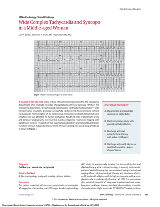
A woman in her late 40s with a history of hypertension presented to the emergency department after multiple episodes of palpitations with near syncope. While in the emergency department, she developed monomorphic ventricular tachycardia (VT) with hemodynamic instability and was successfully cardioverted. She continued to have nonsustained monomorphic VT, so intravenous amiodarone and oral metoprolol were initiated. She was admitted for further evaluation. Results of tests of electrolyte levels and coronary angiography were normal. Cardiac magnetic resonance imaging with gadolinium contrast revealed normal-sized cardiac chambers and normal biventricular function without delayed enhancement. The presenting electrocardiogram (ECG) is shown in Figure 1.

Recommended
Recommended
More Related Content
What's hot
What's hot (18)
Similar to Out flow tract ventricular tachycardia
Similar to Out flow tract ventricular tachycardia (20)
More from Ramachandra Barik
More from Ramachandra Barik (20)
Recently uploaded
Recently uploaded (20)
Out flow tract ventricular tachycardia
- 1. Wide Complex Tachycardia and Syncope in a Middle-aged Woman Laith A. Derbas, MD; Omair K. Yousuf, MD; Faraz Kureshi, MD, MSc A woman in her late 40s with a history of hypertension presented to the emergency department after multiple episodes of palpitations with near syncope. While in the emergency department, she developed monomorphic ventricular tachycardia (VT) with hemodynamic instability and was successfully cardioverted. She continued to have nonsustained monomorphic VT, so intravenous amiodarone and oral metoprolol were initiated. She was admitted for further evaluation. Results of tests of electrolyte levels and coronary angiography were normal. Cardiac magnetic resonance imaging with gadolinium contrast revealed normal-sized cardiac chambers and normal biventricular function without delayed enhancement. The presenting electrocardiogram (ECG) is shown in Figure 1. Diagnosis Outflow tract ventricular tachycardia What to Do Next B. Electrophysiology study with possible catheter ablation Discussion This patient presented with recurrent symptomatic monomorphic VT suggestive of an outflow tract (OT) origin. An electrophysiology (EP) study to anatomically localize the abnormal rhythm and ablation therapy is the preferred strategy in severely symptomatic patients. Medical therapy may be considered; owing to limited and varying efficacy of pharmacologic therapy and its adverse effects, an EP study with ablation, with its high success rate and low com- plication rate, is preferred. Outflow tract VT (OTVT) is a monomor- phic subset of idiopathic VT diagnosed in patients without under- lying structural heart disease, metabolic abnormalities, or cardiac channelopathies. Right ventricular OT (RVOT) VT makes up about I II II III aVR aVL aVF V1 V2 V3 V4 V5 V6 Figure 1. Patient electrocardiogram at presentation. WHAT WOULD YOU DO NEXT? A. Placement of an implantable cardioverter-defibrillator B. Electrophysiology study with possible catheter ablation C. Discharge with oral antiarrhythmic therapy with a class I or III agent D. Discharge with oral β-blocker or nondihydropyridine calcium channel blocker Clinical Review & Education JAMA Cardiology Clinical Challenge jamacardiology.com (Reprinted) JAMA Cardiology March 2019 Volume 4, Number 3 295 © 2018 American Medical Association. All rights reserved. Downloaded From: https://jamanetwork.com/ AIIMS – Bhubaneswar by Ramachandra Barik on 03/23/2019
- 2. 80% of OTVT cases, with most originating just inferior to the pulmonic valve from the anterior and superior septal aspects of the RVOT. The other 20% are left ventricular OT (LVOT) VT, which commonly arises from the region of the aortic cusps in the aortic root.1 A surface ECG with an inferior axis, left bundle branch block (BBB) morphology, and precordial R-wave transition at V4 or after suggests RVOT VT, while one with an inferior axis, right or left BBB morphology, and precordial R-wave transition at V2 or earlier sug- gests LVOT VT.2 Despite surface ECG findings that may differenti- ate LVOT from RVOT, definitive localization is via electroanatomic mapping in an EP study. Presenting ECGs (Figure 1) showed runs of a monomorphic wide complex tachycardia interceded by normal sinus rhythm (with a normal axis and narrow QRS complexes). As the wide com- plex monomorphic tachycardia rhythm developed, a new left BBB morphology with an inferior axis (positive QRS deflections in infe- rior leads II, III, and aVF) and a precordial R-wave transition (a ratio R-wave to S-wave amplitude of Ն1) at lead V3 were observed (Figure 2). Management of OTVT is dictated by symptom frequency and severity and can include medical therapy and/or catheter ablation. Acute termination can be achieved by vagal maneuvers and abor- tive drugs (eg, adenosine, β-blockers, verapamil). Preventive medications include class IA, IC, and III antiarrhythmic drugs; β-blockers; and verapamil. Radiofrequency catheter ablation can be used early in management (with success rates >90% and recurrence of 5% in the first year in RVOT3,4 ); indications include severe symptoms or ones refractory to medical therapy, cardiomyopathy, or younger age and desire to avoid long-term medical therapy.5 Idiopathic VT has a favorable prognosis,6 but symptoms can decrease quality of life, and patients may be at risk for cardiomyopathy if premature ventricular contraction (PVC) burden is high.7 Patient Outcome ThepatienthadsufficientPVCsforactivationmappingwithaCARTO 3advanced3-dimensionalmappingsystem(Biosense-Webster).The origin was localized to the LVOT at the junction between the right and noncoronary cusps of the aortic root. Catheter ablation with radiofrequency energy was performed successfully; VT was nonin- ducible at the end of the EP study, and the patient had no recur- rence of VT or PVCs in the next 24 hours per telemetry. ARTICLE INFORMATION Author Affiliations: Division of Medicine, University of Missouri, Kansas City (Derbas); Division of Cardiology, Saint Luke’s Mid America Heart Institute, Kansas City, Missouri (Yousuf); Advanced Cardiovascular Imaging Laboratory, Cardiovascular and Pulmonary Branch, National Heart, Lung, and Blood Institute, National Institutes of Health, Bethesda, Maryland (Kureshi). Corresponding Author: Laith A. Derbas, MD, Division of Medicine, University of Missouri, Kansas City, 2301 Holmes St, Kansas City, MO 64111 (derbasl@umkc.edu). Published Online: November 7, 2018. doi:10.1001/jamacardio.2018.3687 Conflict of Interest Disclosures: All authors have completed and submitted the ICMJE Form for Disclosure of Potential Conflicts of Interest. Dr Yousuf reports receiving honoraria from Medtronic and Boston Scientific as a part of their speakers’ bureaus and Abbott and Biosense Webster as a consultant. No other disclosures were reported. Additional Contributions: We thank Alan P. Wimmer, MD, Saint Luke’s Health System, for his manuscript review. He was not compensated. REFERENCES 1. Lerman BB, Stein KM, Markowitz SM. Idiopathic right ventricular outflow tract tachycardia: a clinical approach. Pacing Clin Electrophysiol. 1996; 19(12 pt 1):2120-2137. doi:10.1111/j.1540-8159.1996. tb03287.x 2. Park KM, Kim YH, Marchlinski FE. Using the surface electrocardiogram to localize the origin of idiopathic ventricular tachycardia. Pacing Clin Electrophysiol. 2012;35(12):1516-1527. doi:10.1111/ j.1540-8159.2012.03488.x 3. Prystowsky EN, Padanilam BJ, Joshi S, Fogel RI. Ventricular arrhythmias in the absence of structural heart disease. J Am Coll Cardiol. 2012;59(20):1733- 1744. doi:10.1016/j.jacc.2012.01.036 4. Joshi S, Wilber DJ. Ablation of idiopathic right ventricular outflow tract tachycardia: current perspectives. J Cardiovasc Electrophysiol. 2005;16 (suppl 1):S52-S58. doi:10.1111/j.1540-8167.2005. 50163.x 5. Aliot EM, Stevenson WG, Almendral-Garrote JM, et al; European Heart Rhythm Association (EHRA); Registered Branch of the European Society of Cardiology (ESC); Heart Rhythm Society (HRS); American College of Cardiology (ACC); American Heart Association (AHA). EHRA/HRS expert consensus on catheter ablation of ventricular arrhythmias: developed in a partnership with the European Heart Rhythm Association (EHRA), a Registered Branch of the European Society of Cardiology (ESC), and the Heart Rhythm Society (HRS); in collaboration with the American College of Cardiology (ACC) and the American Heart Association (AHA). Heart Rhythm. 2009;6(6):886- 933. doi:10.1016/j.hrthm.2009.04.030 6. Belhassen B, Viskin S. Idiopathic ventricular tachycardia and fibrillation. J Cardiovasc Electrophysiol. 1993;4(3):356-368. doi:10.1111/ j.1540-8167.1993.tb01236.x 7. Mora G, Romero N, van Rendon. Tachycardiomyopathy a rare manifestation of left ventricular outflow tract tachycardia: treatment with radiofrequency catheter ablation. Indian Pacing Electrophysiol J. 2013;13(1):38-42. doi:10. 1016/S0972-6292(16)30587-3 I II II III aVR aVL aVF V1 V2 V3 V4 V5 V6 Figure 2. Patient electrocardiogram on development of a wide complex monomorphic tachycardia rhythm. Note positive QRS deflections in the inferior leads II, III, and aVF, indicating a new left branch bundle block morphology with an inferior axis and a precordial R-wave transition at lead V3. Clinical Review & Education JAMA Cardiology Clinical Challenge 296 JAMA Cardiology March 2019 Volume 4, Number 3 (Reprinted) jamacardiology.com © 2018 American Medical Association. All rights reserved. Downloaded From: https://jamanetwork.com/ AIIMS – Bhubaneswar by Ramachandra Barik on 03/23/2019