Downloaded 136 times


![2D speckle-tracking
• Differentiating cardiac amyloidosis from othercauses of LV
hypertrophy
• Reduced basal strain and regional variations inLS from
base to apex
• A relative ‘apical sparing’ (average apical LS/[average
basal LS + mid LS]) pattern of LS is an easily
recognizable, accurate and reproducible means of
differentiating cardiac amyloidosis from other causes of
LVH
Phelan D, Collier P, Thavendiranathan P, et al.. Heart
2012;98:1442—8.
24](https://image.slidesharecdn.com/cardiacamyloidosis-171208152755/75/Cardiac-amyloidosis-24-2048.jpg)


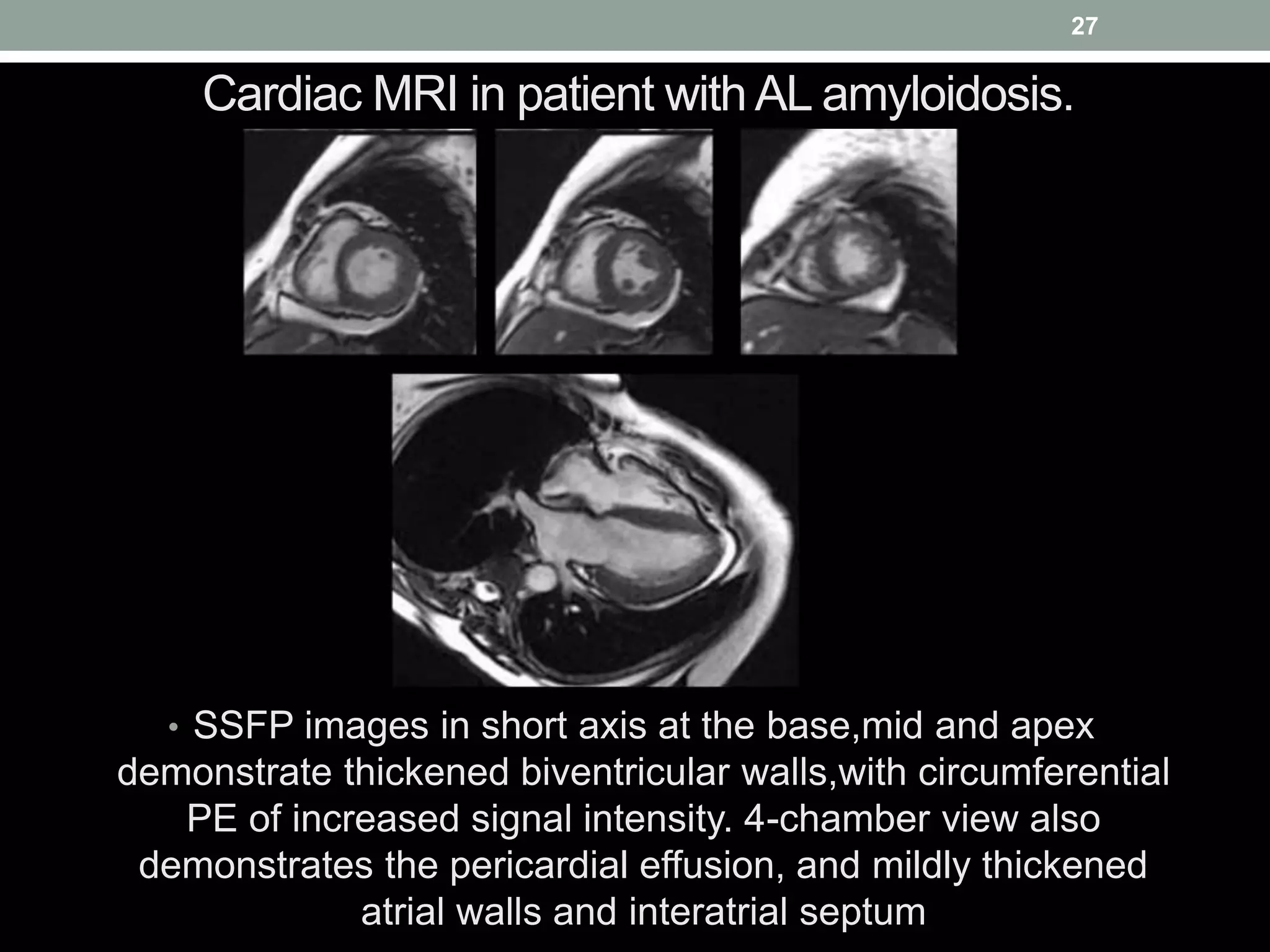


Cardiac amyloidosis is characterized by the extracellular deposition of misfolded protein aggregates, leading to parenchymal stiffness and organ dysfunction, particularly affecting the heart. Diagnosis involves echocardiographic measurements, biomarker assessments, and potentially endomyocardial biopsy, while treatment focuses on chemotherapy and supportive measures to manage heart failure symptoms. Early detection is crucial, as survival rates vary significantly based on the type of amyloidosis and timely intervention.


