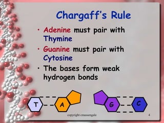
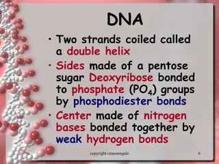
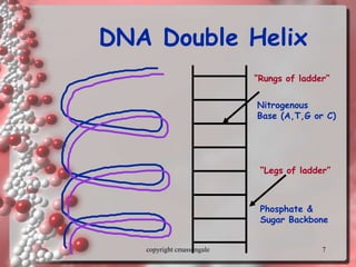
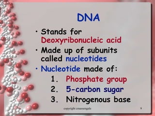
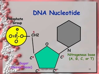
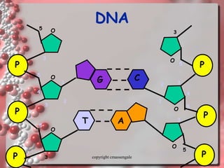
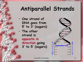
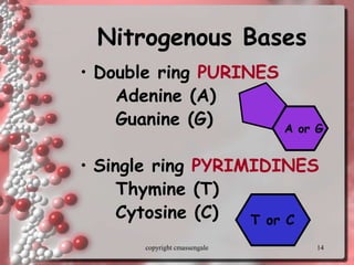
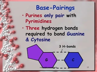
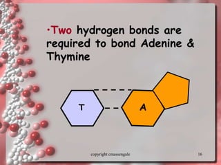
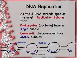
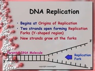
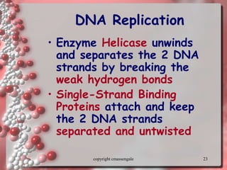
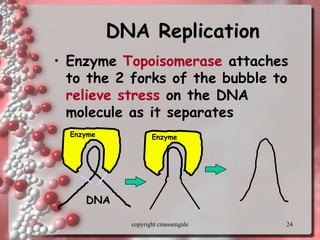
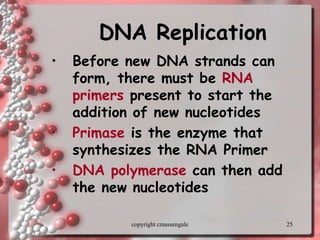
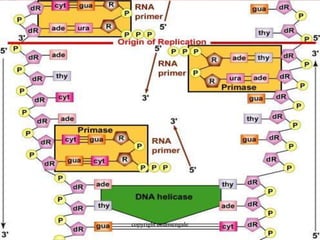
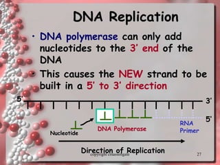
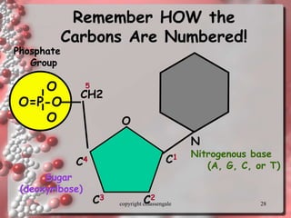
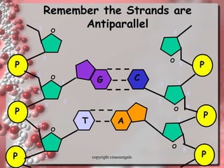
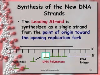
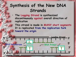
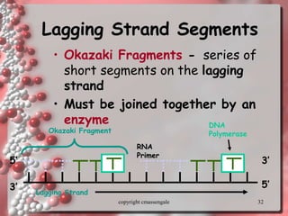
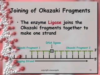
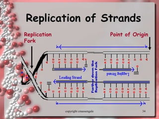
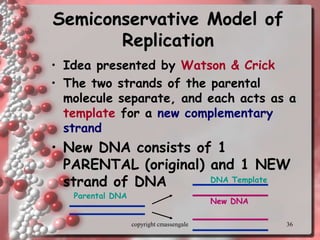
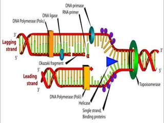
Rosalind Franklin discovered the DNA structure through x-ray crystallography. Watson and Crick then built the first DNA model using Franklin's research. DNA is made of two antiparallel strands coiled into a double helix. Each strand is a backbone of sugar and phosphate groups with nitrogenous bases projecting inward in base pairs - adenine pairs with thymine and cytosine pairs with guanine. DNA replication copies the DNA before cell division, using enzymes like helicase to unwind the strands and DNA polymerase to add complementary nucleotides to each new strand.
DNA and RNA molecules are linear polymers built from individual units called nucleotides connected by bonds called phosphodiester linkages. DNA and RNA are used to store and pass genetic information from one generation to the next.
Nucleic acid play an important role in transmission of hereditary characteristics and biosynthesis of proteins.
DNA and RNA
* CONTENTS Introduction to Nucleic acids History of Nucleic acids Structure of Nucleic acids Description of Nucleic acids Chemical structure of DNA and RNA Classifications of Bases Sites of Nucleic acids Names of Nucleosides and Nucleotides Conclusion References
* Structure of Nucleic acids NA structure is often divided into four different levels: Primary structure Secondary structure Tertiary structure Quaternary structure
* Primary structure: consists of a linear sequence of nucleotides that are linked together by phosphodiester bond. Nucleotides consists of 3 components: Nitrogenous base 5-carbon sugar One or more phosphate groups
* Secondary structure This is the set of interactions between bases. In DNA double helix, the two strands of DNA are held together by hydrogen bonds. The nucleotides on one strand base pairs with the nucleotide on the other strand. The secondary structure is responsible for the shape that the nucleic acid assumes.
* Tertiary structure This is the locations of atoms in three-dimensional space, taking into consideration geometrical and steric constraits. A higher order than the secondary structure in which large scale folding in a linear polymer occurs and the entire chain is folded into a specific 3-dimensional shape.
* Quaternary structure This is similar to that of protein quaternary structure. Although some of the concepts are not exactly the same. QS refers to a higher level of organization of nucleic acids moreover, it refers to the interactions of the nucleic acids with other molecules.
* NNuucclleeiicc AAcciiddss Nucleic acids are molecules that store information for cellular growth and reproduction
* There are two types of nucleic acids: - deoxyribonucleic acid (DNA) and ribonucleic acid (RNA)
* These are polymers consisting of long chains of monomers called nucleotides A nucleotide consists of a nitrogenous base, pentose sugar and a phosphate group.
* DNA and RNA are nucleic acids, long, thread-like polymers made up of a linear array of monomers called nucleotides All nucleotides contain three components: 1. A nitrogen heterocyclic base 2. A pentose sugar 3. A phosphate residue
* Ribonucleotides have a 2’-OH Deoxyribonucleotides have a 2’-H
* Bases are classified as Pyrimidines or Purines
* Nucleus Cytoplasm replication DNA transcription RNA (mRNA) translation Proteins
* reverse transcription messenger RNA (mRNA) transfer RNA (tRNA) ribosomal RNA (rRNA)
* Names of Nucleosides and Nucleotides
* X-ray diffraction patterns produced by DNA fibers Rosalind Franklin and Maurice Wilkins
* 1962 Nobel Prize in Physiology or Medicine for their discoveries concerning the molecular structure of nucleic acids and its significance for information transfer in living material" James Watson Francis Crick Maurice Wilkins
This presentation explains DNA transcription and RNA Processing.
It gives details about prokaryotic DNA transcription and eukaryotic DNA transcription. it also explains post-transcriptional modification both in prokaryotes and eukaryotes.
DNA is maintained in a compressed, supercoiled state.
But basis of replication is the formation of strands based on specific bases pairing with their complementary bases
DNA = deoxyribonucleic acid.
DNA carries the genetic information in the cell – i.e. it carries the instructions for making all the structures and materials the body needs to function.
DNA is capable of self-replication.
Most of the cell’s DNA is carried in the nucleus – a small amount is contained in the mitochondria.
Ozempic: Preoperative Management of Patients on GLP-1 Receptor Agonists Saeid Safari
Preoperative Management of Patients on GLP-1 Receptor Agonists like Ozempic and Semiglutide
ASA GUIDELINE
NYSORA Guideline
2 Case Reports of Gastric Ultrasound
Report Back from SGO 2024: What’s the Latest in Cervical Cancer?bkling
Are you curious about what’s new in cervical cancer research or unsure what the findings mean? Join Dr. Emily Ko, a gynecologic oncologist at Penn Medicine, to learn about the latest updates from the Society of Gynecologic Oncology (SGO) 2024 Annual Meeting on Women’s Cancer. Dr. Ko will discuss what the research presented at the conference means for you and answer your questions about the new developments.
TEST BANK for Operations Management, 14th Edition by William J. Stevenson, Ve...kevinkariuki227
TEST BANK for Operations Management, 14th Edition by William J. Stevenson, Verified Chapters 1 - 19, Complete Newest Version.pdf
Lung Cancer: Artificial Intelligence, Synergetics, Complex System Analysis, S...Oleg Kshivets
RESULTS: Overall life span (LS) was 2252.1±1742.5 days and cumulative 5-year survival (5YS) reached 73.2%, 10 years – 64.8%, 20 years – 42.5%. 513 LCP lived more than 5 years (LS=3124.6±1525.6 days), 148 LCP – more than 10 years (LS=5054.4±1504.1 days).199 LCP died because of LC (LS=562.7±374.5 days). 5YS of LCP after bi/lobectomies was significantly superior in comparison with LCP after pneumonectomies (78.1% vs.63.7%, P=0.00001 by log-rank test). AT significantly improved 5YS (66.3% vs. 34.8%) (P=0.00000 by log-rank test) only for LCP with N1-2. Cox modeling displayed that 5YS of LCP significantly depended on: phase transition (PT) early-invasive LC in terms of synergetics, PT N0—N12, cell ratio factors (ratio between cancer cells- CC and blood cells subpopulations), G1-3, histology, glucose, AT, blood cell circuit, prothrombin index, heparin tolerance, recalcification time (P=0.000-0.038). Neural networks, genetic algorithm selection and bootstrap simulation revealed relationships between 5YS and PT early-invasive LC (rank=1), PT N0—N12 (rank=2), thrombocytes/CC (3), erythrocytes/CC (4), eosinophils/CC (5), healthy cells/CC (6), lymphocytes/CC (7), segmented neutrophils/CC (8), stick neutrophils/CC (9), monocytes/CC (10); leucocytes/CC (11). Correct prediction of 5YS was 100% by neural networks computing (area under ROC curve=1.0; error=0.0).
CONCLUSIONS: 5YS of LCP after radical procedures significantly depended on: 1) PT early-invasive cancer; 2) PT N0--N12; 3) cell ratio factors; 4) blood cell circuit; 5) biochemical factors; 6) hemostasis system; 7) AT; 8) LC characteristics; 9) LC cell dynamics; 10) surgery type: lobectomy/pneumonectomy; 11) anthropometric data. Optimal diagnosis and treatment strategies for LC are: 1) screening and early detection of LC; 2) availability of experienced thoracic surgeons because of complexity of radical procedures; 3) aggressive en block surgery and adequate lymph node dissection for completeness; 4) precise prediction; 5) adjuvant chemoimmunoradiotherapy for LCP with unfavorable prognosis.
New Directions in Targeted Therapeutic Approaches for Older Adults With Mantl...i3 Health
i3 Health is pleased to make the speaker slides from this activity available for use as a non-accredited self-study or teaching resource.
This slide deck presented by Dr. Kami Maddocks, Professor-Clinical in the Division of Hematology and
Associate Division Director for Ambulatory Operations
The Ohio State University Comprehensive Cancer Center, will provide insight into new directions in targeted therapeutic approaches for older adults with mantle cell lymphoma.
STATEMENT OF NEED
Mantle cell lymphoma (MCL) is a rare, aggressive B-cell non-Hodgkin lymphoma (NHL) accounting for 5% to 7% of all lymphomas. Its prognosis ranges from indolent disease that does not require treatment for years to very aggressive disease, which is associated with poor survival (Silkenstedt et al, 2021). Typically, MCL is diagnosed at advanced stage and in older patients who cannot tolerate intensive therapy (NCCN, 2022). Although recent advances have slightly increased remission rates, recurrence and relapse remain very common, leading to a median overall survival between 3 and 6 years (LLS, 2021). Though there are several effective options, progress is still needed towards establishing an accepted frontline approach for MCL (Castellino et al, 2022). Treatment selection and management of MCL are complicated by the heterogeneity of prognosis, advanced age and comorbidities of patients, and lack of an established standard approach for treatment, making it vital that clinicians be familiar with the latest research and advances in this area. In this activity chaired by Michael Wang, MD, Professor in the Department of Lymphoma & Myeloma at MD Anderson Cancer Center, expert faculty will discuss prognostic factors informing treatment, the promising results of recent trials in new therapeutic approaches, and the implications of treatment resistance in therapeutic selection for MCL.
Target Audience
Hematology/oncology fellows, attending faculty, and other health care professionals involved in the treatment of patients with mantle cell lymphoma (MCL).
Learning Objectives
1.) Identify clinical and biological prognostic factors that can guide treatment decision making for older adults with MCL
2.) Evaluate emerging data on targeted therapeutic approaches for treatment-naive and relapsed/refractory MCL and their applicability to older adults
3.) Assess mechanisms of resistance to targeted therapies for MCL and their implications for treatment selection
- Video recording of this lecture in English language: https://youtu.be/lK81BzxMqdo
- Video recording of this lecture in Arabic language: https://youtu.be/Ve4P0COk9OI
- Link to download the book free: https://nephrotube.blogspot.com/p/nephrotube-nephrology-books.html
- Link to NephroTube website: www.NephroTube.com
- Link to NephroTube social media accounts: https://nephrotube.blogspot.com/p/join-nephrotube-on-social-media.html
The prostate is an exocrine gland of the male mammalian reproductive system
It is a walnut-sized gland that forms part of the male reproductive system and is located in front of the rectum and just below the urinary bladder
Function is to store and secrete a clear, slightly alkaline fluid that constitutes 10-30% of the volume of the seminal fluid that along with the spermatozoa, constitutes semen
A healthy human prostate measures (4cm-vertical, by 3cm-horizontal, 2cm ant-post ).
It surrounds the urethra just below the urinary bladder. It has anterior, median, posterior and two lateral lobes
It’s work is regulated by androgens which are responsible for male sex characteristics
Generalised disease of the prostate due to hormonal derangement which leads to non malignant enlargement of the gland (increase in the number of epithelial cells and stromal tissue)to cause compression of the urethra leading to symptoms (LUTS
These simplified slides by Dr. Sidra Arshad present an overview of the non-respiratory functions of the respiratory tract.
Learning objectives:
1. Enlist the non-respiratory functions of the respiratory tract
2. Briefly explain how these functions are carried out
3. Discuss the significance of dead space
4. Differentiate between minute ventilation and alveolar ventilation
5. Describe the cough and sneeze reflexes
Study Resources:
1. Chapter 39, Guyton and Hall Textbook of Medical Physiology, 14th edition
2. Chapter 34, Ganong’s Review of Medical Physiology, 26th edition
3. Chapter 17, Human Physiology by Lauralee Sherwood, 9th edition
4. Non-respiratory functions of the lungs https://academic.oup.com/bjaed/article/13/3/98/278874
MANAGEMENT OF ATRIOVENTRICULAR CONDUCTION BLOCK.pdfJim Jacob Roy
Cardiac conduction defects can occur due to various causes.
Atrioventricular conduction blocks ( AV blocks ) are classified into 3 types.
This document describes the acute management of AV block.
Title: Sense of Smell
Presenter: Dr. Faiza, Assistant Professor of Physiology
Qualifications:
MBBS (Best Graduate, AIMC Lahore)
FCPS Physiology
ICMT, CHPE, DHPE (STMU)
MPH (GC University, Faisalabad)
MBA (Virtual University of Pakistan)
Learning Objectives:
Describe the primary categories of smells and the concept of odor blindness.
Explain the structure and location of the olfactory membrane and mucosa, including the types and roles of cells involved in olfaction.
Describe the pathway and mechanisms of olfactory signal transmission from the olfactory receptors to the brain.
Illustrate the biochemical cascade triggered by odorant binding to olfactory receptors, including the role of G-proteins and second messengers in generating an action potential.
Identify different types of olfactory disorders such as anosmia, hyposmia, hyperosmia, and dysosmia, including their potential causes.
Key Topics:
Olfactory Genes:
3% of the human genome accounts for olfactory genes.
400 genes for odorant receptors.
Olfactory Membrane:
Located in the superior part of the nasal cavity.
Medially: Folds downward along the superior septum.
Laterally: Folds over the superior turbinate and upper surface of the middle turbinate.
Total surface area: 5-10 square centimeters.
Olfactory Mucosa:
Olfactory Cells: Bipolar nerve cells derived from the CNS (100 million), with 4-25 olfactory cilia per cell.
Sustentacular Cells: Produce mucus and maintain ionic and molecular environment.
Basal Cells: Replace worn-out olfactory cells with an average lifespan of 1-2 months.
Bowman’s Gland: Secretes mucus.
Stimulation of Olfactory Cells:
Odorant dissolves in mucus and attaches to receptors on olfactory cilia.
Involves a cascade effect through G-proteins and second messengers, leading to depolarization and action potential generation in the olfactory nerve.
Quality of a Good Odorant:
Small (3-20 Carbon atoms), volatile, water-soluble, and lipid-soluble.
Facilitated by odorant-binding proteins in mucus.
Membrane Potential and Action Potential:
Resting membrane potential: -55mV.
Action potential frequency in the olfactory nerve increases with odorant strength.
Adaptation Towards the Sense of Smell:
Rapid adaptation within the first second, with further slow adaptation.
Psychological adaptation greater than receptor adaptation, involving feedback inhibition from the central nervous system.
Primary Sensations of Smell:
Camphoraceous, Musky, Floral, Pepperminty, Ethereal, Pungent, Putrid.
Odor Detection Threshold:
Examples: Hydrogen sulfide (0.0005 ppm), Methyl-mercaptan (0.002 ppm).
Some toxic substances are odorless at lethal concentrations.
Characteristics of Smell:
Odor blindness for single substances due to lack of appropriate receptor protein.
Behavioral and emotional influences of smell.
Transmission of Olfactory Signals:
From olfactory cells to glomeruli in the olfactory bulb, involving lateral inhibition.
Primitive, less old, and new olfactory systems with different path
These lecture slides, by Dr Sidra Arshad, offer a quick overview of physiological basis of a normal electrocardiogram.
Learning objectives:
1. Define an electrocardiogram (ECG) and electrocardiography
2. Describe how dipoles generated by the heart produce the waveforms of the ECG
3. Describe the components of a normal electrocardiogram of a typical bipolar leads (limb II)
4. Differentiate between intervals and segments
5. Enlist some common indications for obtaining an ECG
Study Resources:
1. Chapter 11, Guyton and Hall Textbook of Medical Physiology, 14th edition
2. Chapter 9, Human Physiology - From Cells to Systems, Lauralee Sherwood, 9th edition
3. Chapter 29, Ganong’s Review of Medical Physiology, 26th edition
4. Electrocardiogram, StatPearls - https://www.ncbi.nlm.nih.gov/books/NBK549803/
5. ECG in Medical Practice by ABM Abdullah, 4th edition
6. ECG Basics, http://www.nataliescasebook.com/tag/e-c-g-basics
2. 2
DNA Structure
• Rosalind Franklin took
diffraction x-ray
photographs of DNA
crystals
• In the 1950’s, Watson &
Crick built the first model
of DNA using Franklin’s
x-rays
copyright cmassengale
3. 3
Discovery of DNA
Structure
• Erwin Chargaff showed the
amounts of the four bases on
DNA ( A,T,C,G)
• In a body or somatic cell:
A = 30.3%
T = 30.3%
G = 19.5%
C = 19.9%
copyright cmassengale
4. 4
Chargaff’s Rule
• Adenine must pair with
Thymine
• Guanine must pair with
Cytosine
• The bases form weak
hydrogen bonds
G C
T A
copyright cmassengale
6. 6
DNA
• Two strands coiled called
a double helix
• Sides made of a pentose
sugar Deoxyribose bonded
to phosphate (PO4) groups
by phosphodiester bonds
• Center made of nitrogen
bases bonded together by
weak hydrogen bonds
copyright cmassengale
8. 8
Helix
• Most DNA has a right-hand
twist with 10 base pairs in a
complete turn
• Left twisted DNA is called
Z-DNA or southpaw DNA
• Hot spots occur where right
and left twisted DNA meet
producing mutations
copyright cmassengale
9. 9
DNA
• Stands for
Deoxyribonucleic acid
• Made up of subunits
called nucleotides
• Nucleotide made of:
1. Phosphate group
2. 5-carbon sugar
3. Nitrogenous base
copyright cmassengale
13. 13
Antiparallel Strands
• One strand of
DNA goes from
5’ to 3’ (sugars)
• The other
strand is
opposite in
direction going
3’ to 5’ (sugars)
copyright cmassengale
14. 14
Nitrogenous Bases
• Double ring PURINES
Adenine (A)
Guanine (G)
• Single ring PYRIMIDINES
Thymine (T)
Cytosine (C) T or C
A or G
copyright cmassengale
15. 15
Base-Pairings
• Purines only pair with
Pyrimidines
• Three hydrogen bonds
required to bond Guanine
& Cytosine
C
G
3 H-bonds
copyright cmassengale
16. 16
T A
•Two hydrogen bonds are
required to bond Adenine &
Thymine
copyright cmassengale
20. 20
Replication Facts
• DNA has to be copied
before a cell divides
• DNA is copied during the S
or synthesis phase
• New cells will need identical
DNA strands
copyright cmassengale
21. 21
DNA Replication
• As the 2 DNA strands open at
the origin, Replication Bubbles
form
• Prokaryotes (bacteria) have a
single bubble
• Eukaryotic chromosomes have
MANY bubbles
Bubbles Bubbles
copyright cmassengale
22. 22
DNA Replication
• Begins at Origins of Replication
• Two strands open forming Replication
Forks (Y-shaped region)
• New strands grow at the forks
Replication
Fork
Parental DNA Molecule
3’
5’
3’
5’
copyright cmassengale
23. 23
DNA Replication
• Enzyme Helicase unwinds
and separates the 2 DNA
strands by breaking the
weak hydrogen bonds
• Single-Strand Binding
Proteins attach and keep
the 2 DNA strands
separated and untwisted
copyright cmassengale
24. 24
DNA Replication
• Enzyme Topoisomerase attaches
to the 2 forks of the bubble to
relieve stress on the DNA
molecule as it separates
Enzyme
DNA
Enzyme
copyright cmassengale
25. 25
DNA Replication
• Before new DNA strands can
form, there must be RNA
primers present to start the
addition of new nucleotides
• Primase is the enzyme that
synthesizes the RNA Primer
• DNA polymerase can then add
the new nucleotides
copyright cmassengale
27. 27
DNA Replication
• DNA polymerase can only add
nucleotides to the 3’ end of the
DNA
• This causes the NEW strand to be
built in a 5’ to 3’ direction
RNA
Primer
DNA Polymerase
Nucleotide
5’
5’ 3’
Direction of Replication
copyright cmassengale
28. 28
Remember HOW the
Carbons Are Numbered!
O
O=P-O
O
Phosphate
Group
N
Nitrogenous base
(A, G, C, or T)
CH2
O
C1
C4
C3 C2
5
Sugar
(deoxyribose)
copyright cmassengale
29. 29
Remember the Strands are
Antiparallel
P
P
P
O
O
O
1
2
3
4
5
5
3
3
5
P
P
P
O
O
O
1
2 3
4
5
5
3
5
3
G C
T A
copyright cmassengale
30. 30
Synthesis of the New DNA
Strands
• The Leading Strand is
synthesized as a single strand
from the point of origin toward
the opening replication fork
RNA
Primer
DNA Polymerase
Nucleotides
3’
5’
5’
copyright cmassengale
31. 31
Synthesis of the New DNA
Strands
• The Lagging Strand is synthesized
discontinuously against overall direction of
replication
• This strand is made in MANY short segments
It is replicated from the replication fork
toward the origin
RNA Primer
Leading Strand
DNA Polymerase
5
’
5’
3’
3’
Lagging Strand
5’
5’
3’
3’ copyright cmassengale
32. 32
Lagging Strand Segments
• Okazaki Fragments - series of
short segments on the lagging
strand
• Must be joined together by an
enzyme
Lagging Strand
RNA
Primer
DNA
Polymerase
3’
3’
5’
5’
Okazaki Fragment
copyright cmassengale
33. 33
Joining of Okazaki Fragments
• The enzyme Ligase joins the
Okazaki fragments together to
make one strand
Lagging Strand
Okazaki Fragment 2
DNA ligase
Okazaki Fragment 1
5’
5’
3’
3’
copyright cmassengale
35. 35
Proofreading New DNA
• DNA polymerase initially makes
about 1 in 10,000 base pairing
errors
• Enzymes proofread and correct
these mistakes
• The new error rate for DNA that
has been proofread is 1 in 1 billion
base pairing errors
copyright cmassengale
36. 36
Semiconservative Model of
Replication
• Idea presented by Watson & Crick
• The two strands of the parental
molecule separate, and each acts as a
template for a new complementary
strand
• New DNA consists of 1
PARENTAL (original) and 1 NEW
strand of DNA
Parental DNA
DNA Template
New DNA
copyright cmassengale
37. 37
DNA Damage & Repair
• Chemicals & ultraviolet radiation
damage the DNA in our body cells
• Cells must continuously repair
DAMAGED DNA
• Excision repair occurs when any of
over 50 repair enzymes remove
damaged parts of DNA
• DNA polymerase and DNA ligase
replace and bond the new nucleotides
together
copyright cmassengale