
More Related Content
What's hot
What's hot (20)
Viewers also liked
Viewers also liked (20)
Similar to 15 16-maxillary-premolars-20
Similar to 15 16-maxillary-premolars-20 (20)
Recently uploaded
Recently uploaded (20)
15 16-maxillary-premolars-20
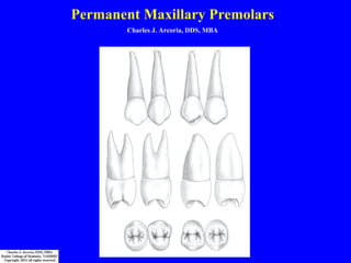
- 1. Permanent Maxillary Premolars Charles J. Arcoria, DDS, MBA
- 2. Permanent Maxillary Premolars Class Traits & Key Considerations: 1. Maxillary premolars have two cusps (facial & lingual) 2. Mandibular premolars have a single facial cusp with one or more lingual cusps The preferred nomenclature for these teeth is “premolar” rather than “bicuspid” which implies the tooth has two and only two cusps 3. There are two major cusps, facial and lingual, that are approximately equal in size and prominence 4. The maxillary first premolar presents a lingual cusp that is approximately 1.0 mm shorter than the facial cusp 5. The crowns, from the occlusal aspect, are relatively much wider (2.0 mm) faciolingually than they are mesiodistally 6. Facial profiles viewed from the proximal aspect, are only slightly inclined lingually from height of contour to cusp apex 7. Lingual height of contour is situated approximately midway between cervical line and the cusp tip in the middle 1/3 of the crown 8. There is a much greater morphological similarity between the two maxillary premolars than between the two mandibular premolars
- 3. Permanent Maxillary Premolars – Type Traits Aspect First Premolar Second Premolar Facial > Prominent, broad shoulders > Facial cusp tip distal of center > Prominent facial lobes > Narrow shoulders > Facial cusp tip mesial of center > Facial lobes not prominent Lingual > Entire facial profile of crown visible > Little or none of the facial profile visible Mesial > Mesial marginal ridge developmental groove interrupts mesial marginal ridge. > Usually two roots, facial and lingual. > Mesial developmental depression extends from bifurcation of root across the cervical line and well onto crown almost to contact area. > No mesial marginal ridge developmental groove. > Single root. > Mesial depression restricted to root surface.
- 4. Permanent Maxillary Premolars – Type Traits Aspect First Premolar Second Premolar Occlusal > Crown profile hexagonal. > Mesiofacial and distofacial line angles are sharp. > Mesial and distal profiles converge lingually. > Occlusal table outline trapezoidal. > Facial cusp ridge has a slight mesial cant, giving the crown a twisted appearance. > Facial cusp wider than lingual cusp. > Central groove long. > Supplemental grooves rare. > Facial ridge, developmental depressions and lobes visible. > Crown profile ovoid. > Mesiofacial and distofacial line angles more rounded. > Little lingual convergence; mesial and distal marginal ridges parallel. > Occlusal table outline rectangular. > Crown not twisted in appearance. > Short central groove & wide marginal ridges > Many supplemental grooves giving a “wrinkled” appearance Cross Section of Root > Outline is kidney shaped with concavity on mesial. > One root canal at cervix, two root canals as root bifurcates > Outline is ovoid. > One root with one or two root canals with varied pulp morphology
- 5. Permanent Maxillary First Premolar – Introductory Points •The maxillary first premolar typically has two roots, one facial and one lingual. It always has (at least) two canals. If a third canal is present, it will be in the facial root. •Rarely, the maxillary 1st premolar will present with 3 roots. These teeth will have two roots situated facially (MF & DF) and one root situated lingually. •The lingual cusp is typically 1.0 mm shorter than the facial cusp. •The mesial marginal ridge is taller than the distal marginal ridge (the DMR is more cervical than the MMR). •The middle facial lobe is the most developed of the facial lobes and gives rise to a prominent facial ridge. •The maxillary first premolar emerges into the oral cavity after the permanent maxillary lateral incisor but before the permanent maxillary canine.
- 6. Permanent Maxillary First Premolar - Facial M D · Facial outline of the crown is a trapezoid with the shorter parallel side at the cervix. · The tooth is broad shouldered when viewed from this aspect with markedly convex mesial and distal contours. · The cusp tip is relatively pointed and will be slightly distal to the mesiodistal long axis bisector. (This is a distinctive characteristic since this is the only permanent tooth where the mesial cuspal ridge of the facial cusp is longer than the distal cuspal ridge). · The mesial proximal contact is slightly more cervically placed than the distal proximal contact. (This is a characteristic of both maxillary and mandibular first premolars and would be expected since the mesial contact meets the distal contact of a canine and both maxillary and mandibular canines have their distal contact areas in the middle 1/3 of the crown incisogingivally). · The mesial outline of the crown between the mesial contact area and the cervical line is slightly concave. This is principally due to the developmental root depression on the mesial surface that extends over the CEJ onto the crown surface better described from the mesial aspect. Although not visible from the facial aspect, this outline is said by one text to be a mirror image of the distal contour of the adjacent canine. · The distal outline of the crown between the contact area and the cervical line is slightly convex. · The apical crest of curvature of the cervical line on premolar crowns will usually be centered but may be slightly distal to the mesiodistal long axis bisector. · The root of this tooth is usually bifurcated into a facial and a lingual root. Since the facial root is usually wider than the lingual root, the lingual root will not always be visible from the facial aspect. However, dental charts depicting a drawing of this tooth from a facial view will usually show the apices of both roots on the drawing. · The apices of both the facial and lingual roots will usually be slightly distal to the mesiodistal long axis bisector. · There is a tendency for the facial surface of the crown to show evidence of lobe formation by having a prominent facial ridge running axially from the facial cusp that is bordered by noticeable developmental depressions mesial and distal to it.
- 7. Permanent Maxillary First Premolar - Facial M D · Facial outline of the crown is a trapezoid with the shorter parallel side at the cervix. · The tooth is broad shouldered when viewed from this aspect with markedly convex mesial and distal contours. · The cusp tip is relatively pointed and will be slightly distal to the mesiodistal long axis bisector. (This is a distinctive characteristic since this is the only permanent tooth where the mesial cuspal ridge of the facial cusp is longer than the distal cuspal ridge). · The mesial proximal contact is slightly more cervically placed than the distal proximal contact. (This is a characteristic of both maxillary and mandibular first premolars and would be expected since the mesial contact meets the distal contact of a canine and both maxillary and mandibular canines have their distal contact areas in the middle 1/3 of the crown incisogingivally). · The mesial outline of the crown between the mesial contact area and the cervical line is slightly concave. This is principally due to the developmental root depression on the mesial surface that extends over the CEJ onto the crown surface better described from the mesial aspect. Although not visible from the facial aspect, this outline is said by one text to be a mirror image of the distal contour of the adjacent canine. · The distal outline of the crown between the contact area and the cervical line is slightly convex. · The apical crest of curvature of the cervical line on premolar crowns will usually be centered but may be slightly distal to the mesiodistal long axis bisector. · The root of this tooth is usually bifurcated into a facial and a lingual root. Since the facial root is usually wider than the lingual root, the lingual root will not always be visible from the facial aspect. However, dental charts depicting a drawing of this tooth from a facial view will usually show the apices of both roots on the drawing. · The apices of both the facial and lingual roots will usually be slightly distal to the mesiodistal long axis bisector. · There is a tendency for the facial surface of the crown to show evidence of lobe formation by having a prominent facial ridge running axially from the facial cusp that is bordered by noticeable developmental depressions mesial and distal to it.
- 8. Permanent Maxillary First Premolar - Facial M D · Facial outline of the crown is a trapezoid with the shorter parallel side at the cervix. · The tooth is broad shouldered when viewed from this aspect with markedly convex mesial and distal contours. · The cusp tip is relatively pointed and will be slightly distal to the mesiodistal long axis bisector. (This is a distinctive characteristic since this is the only permanent tooth where the mesial cuspal ridge of the facial cusp is longer than the distal cuspal ridge). · The mesial proximal contact is slightly more cervically placed than the distal proximal contact. (This is a characteristic of both maxillary and mandibular first premolars and would be expected since the mesial contact meets the distal contact of a canine and both maxillary and mandibular canines have their distal contact areas in the middle 1/3 of the crown incisogingivally). · The mesial outline of the crown between the mesial contact area and the cervical line is slightly concave. This is principally due to the developmental root depression on the mesial surface that extends over the CEJ onto the crown surface better described from the mesial aspect. Although not visible from the facial aspect, this outline is said by one text to be a mirror image of the distal contour of the adjacent canine. · The distal outline of the crown between the contact area and the cervical line is slightly convex. · The apical crest of curvature of the cervical line on premolar crowns will usually be centered but may be slightly distal to the mesiodistal long axis bisector. · The root of this tooth is usually bifurcated into a facial and a lingual root. Since the facial root is usually wider than the lingual root, the lingual root will not always be visible from the facial aspect. However, dental charts depicting a drawing of this tooth from a facial view will usually show the apices of both roots on the drawing. · The apices of both the facial and lingual roots will usually be slightly distal to the mesiodistal long axis bisector. · There is a tendency for the facial surface of the crown to show evidence of lobe formation by having a prominent facial ridge running axially from the facial cusp that is bordered by noticeable developmental depressions mesial and distal to it.
- 9. Permanent Maxillary First Premolar - Facial M D · Facial outline of the crown is a trapezoid with the shorter parallel side at the cervix. · The tooth is broad shouldered when viewed from this aspect with markedly convex mesial and distal contours. · The cusp tip is relatively pointed and will be slightly distal to the mesiodistal long axis bisector. (This is a distinctive characteristic since this is the only permanent tooth where the mesial cuspal ridge of the facial cusp is longer than the distal cuspal ridge). · The mesial proximal contact is slightly more cervically placed than the distal proximal contact. (This is a characteristic of both maxillary and mandibular first premolars and would be expected since the mesial contact meets the distal contact of a canine and both maxillary and mandibular canines have their distal contact areas in the middle 1/3 of the crown incisogingivally). · The mesial outline of the crown between the mesial contact area and the cervical line is slightly concave. This is principally due to the developmental root depression on the mesial surface that extends over the CEJ onto the crown surface better described from the mesial aspect. Although not visible from the facial aspect, this outline is said by one text to be a mirror image of the distal contour of the adjacent canine. · The distal outline of the crown between the contact area and the cervical line is slightly convex. · The apical crest of curvature of the cervical line on premolar crowns will usually be centered but may be slightly distal to the mesiodistal long axis bisector. · The root of this tooth is usually bifurcated into a facial and a lingual root. Since the facial root is usually wider than the lingual root, the lingual root will not always be visible from the facial aspect. However, dental charts depicting a drawing of this tooth from a facial view will usually show the apices of both roots on the drawing. · The apices of both the facial and lingual roots will usually be slightly distal to the mesiodistal long axis bisector. · There is a tendency for the facial surface of the crown to show evidence of lobe formation by having a prominent facial ridge running axially from the facial cusp that is bordered by noticeable developmental depressions mesial and distal to it.
- 10. Permanent Maxillary First Premolar - Facial M D · Facial outline of the crown is a trapezoid with the shorter parallel side at the cervix. · The tooth is broad shouldered when viewed from this aspect with markedly convex mesial and distal contours. · The cusp tip is relatively pointed and will be slightly distal to the mesiodistal long axis bisector. (This is a distinctive characteristic since this is the only permanent tooth where the mesial cuspal ridge of the facial cusp is longer than the distal cuspal ridge). · The mesial proximal contact is slightly more cervically placed than the distal proximal contact. (This is a characteristic of both maxillary and mandibular first premolars and would be expected since the mesial contact meets the distal contact of a canine and both maxillary and mandibular canines have their distal contact areas in the middle 1/3 of the crown incisogingivally). · The mesial outline of the crown between the mesial contact area and the cervical line is slightly concave. This is principally due to the developmental root depression on the mesial surface that extends over the CEJ onto the crown surface better described from the mesial aspect. Although not visible from the facial aspect, this outline is said by one text to be a mirror image of the distal contour of the adjacent canine. · The distal outline of the crown between the contact area and the cervical line is slightly convex. · The apical crest of curvature of the cervical line on premolar crowns will usually be centered but may be slightly distal to the mesiodistal long axis bisector. · The root of this tooth is usually bifurcated into a facial and a lingual root. Since the facial root is usually wider than the lingual root, the lingual root will not always be visible from the facial aspect. However, dental charts depicting a drawing of this tooth from a facial view will usually show the apices of both roots on the drawing. · The apices of both the facial and lingual roots will usually be slightly distal to the mesiodistal long axis bisector. · There is a tendency for the facial surface of the crown to show evidence of lobe formation by having a prominent facial ridge running axially from the facial cusp that is bordered by noticeable developmental depressions mesial and distal to it.
- 11. Permanent Maxillary First Premolar - Facial M D · Facial outline of the crown is a trapezoid with the shorter parallel side at the cervix. · The tooth is broad shouldered when viewed from this aspect with markedly convex mesial and distal contours. · The cusp tip is relatively pointed and will be slightly distal to the mesiodistal long axis bisector. (This is a distinctive characteristic since this is the only permanent tooth where the mesial cuspal ridge of the facial cusp is longer than the distal cuspal ridge). · The mesial proximal contact is slightly more cervically placed than the distal proximal contact. (This is a characteristic of both maxillary and mandibular first premolars and would be expected since the mesial contact meets the distal contact of a canine and both maxillary and mandibular canines have their distal contact areas in the middle 1/3 of the crown incisogingivally). · The mesial outline of the crown between the mesial contact area and the cervical line is slightly concave. This is principally due to the developmental root depression on the mesial surface that extends over the CEJ onto the crown surface better described from the mesial aspect. Although not visible from the facial aspect, this outline is said by one text to be a mirror image of the distal contour of the adjacent canine. · The distal outline of the crown between the contact area and the cervical line is slightly convex. · The apical crest of curvature of the cervical line on premolar crowns will usually be centered but may be slightly distal to the mesiodistal long axis bisector. · The root of this tooth is usually bifurcated into a facial and a lingual root. Since the facial root is usually wider than the lingual root, the lingual root will not always be visible from the facial aspect. However, dental charts depicting a drawing of this tooth from a facial view will usually show the apices of both roots on the drawing. · The apices of both the facial and lingual roots will usually be slightly distal to the mesiodistal long axis bisector. · There is a tendency for the facial surface of the crown to show evidence of lobe formation by having a prominent facial ridge running axially from the facial cusp that is bordered by noticeable developmental depressions mesial and distal to it.
- 12. Permanent Maxillary First Premolar - Facial M D · Facial outline of the crown is a trapezoid with the shorter parallel side at the cervix. · The tooth is broad shouldered when viewed from this aspect with markedly convex mesial and distal contours. · The cusp tip is relatively pointed and will be slightly distal to the mesiodistal long axis bisector. (This is a distinctive characteristic since this is the only permanent tooth where the mesial cuspal ridge of the facial cusp is longer than the distal cuspal ridge). · The mesial proximal contact is slightly more cervically placed than the distal proximal contact. (This is a characteristic of both maxillary and mandibular first premolars and would be expected since the mesial contact meets the distal contact of a canine and both maxillary and mandibular canines have their distal contact areas in the middle 1/3 of the crown incisogingivally). · The mesial outline of the crown between the mesial contact area and the cervical line is slightly concave. This is principally due to the developmental root depression on the mesial surface that extends over the CEJ onto the crown surface better described from the mesial aspect. Although not visible from the facial aspect, this outline is said by one text to be a mirror image of the distal contour of the adjacent canine. · The distal outline of the crown between the contact area and the cervical line is slightly convex. · The apical crest of curvature of the cervical line on premolar crowns will usually be centered but may be slightly distal to the mesiodistal long axis bisector. · The root of this tooth is usually bifurcated into a facial and a lingual root. Since the facial root is usually wider than the lingual root, the lingual root will not always be visible from the facial aspect. However, dental charts depicting a drawing of this tooth from a facial view will usually show the apices of both roots on the drawing. · The apices of both the facial and lingual roots will usually be slightly distal to the mesiodistal long axis bisector. · There is a tendency for the facial surface of the crown to show evidence of lobe formation by having a prominent facial ridge running axially from the facial cusp that is bordered by noticeable developmental depressions mesial and distal to it.
- 13. Permanent Maxillary First Premolar - Facial M D · Facial outline of the crown is a trapezoid with the shorter parallel side at the cervix. · The tooth is broad shouldered when viewed from this aspect with markedly convex mesial and distal contours. · The cusp tip is relatively pointed and will be slightly distal to the mesiodistal long axis bisector. (This is a distinctive characteristic since this is the only permanent tooth where the mesial cuspal ridge of the facial cusp is longer than the distal cuspal ridge). · The mesial proximal contact is slightly more cervically placed than the distal proximal contact. (This is a characteristic of both maxillary and mandibular first premolars and would be expected since the mesial contact meets the distal contact of a canine and both maxillary and mandibular canines have their distal contact areas in the middle 1/3 of the crown incisogingivally). · The mesial outline of the crown between the mesial contact area and the cervical line is slightly concave. This is principally due to the developmental root depression on the mesial surface that extends over the CEJ onto the crown surface better described from the mesial aspect. Although not visible from the facial aspect, this outline is said by one text to be a mirror image of the distal contour of the adjacent canine. · The distal outline of the crown between the contact area and the cervical line is slightly convex. · The apical crest of curvature of the cervical line on premolar crowns will usually be centered but may be slightly distal to the mesiodistal long axis bisector. · The root of this tooth is usually bifurcated into a facial and a lingual root. Since the facial root is usually wider than the lingual root, the lingual root will not always be visible from the facial aspect. However, dental charts depicting a drawing of this tooth from a facial view will usually show the apices of both roots on the drawing. · The apices of both the facial and lingual roots will usually be slightly distal to the mesiodistal long axis bisector. · There is a tendency for the facial surface of the crown to show evidence of lobe formation by having a prominent facial ridge running axially from the facial cusp that is bordered by noticeable developmental depressions mesial and distal to it. Lingual Facial
- 14. Permanent Maxillary First Premolar - Facial M D · Facial outline of the crown is a trapezoid with the shorter parallel side at the cervix. · The tooth is broad shouldered when viewed from this aspect with markedly convex mesial and distal contours. · The cusp tip is relatively pointed and will be slightly distal to the mesiodistal long axis bisector. (This is a distinctive characteristic since this is the only permanent tooth where the mesial cuspal ridge of the facial cusp is longer than the distal cuspal ridge). · The mesial proximal contact is slightly more cervically placed than the distal proximal contact. (This is a characteristic of both maxillary and mandibular first premolars and would be expected since the mesial contact meets the distal contact of a canine and both maxillary and mandibular canines have their distal contact areas in the middle 1/3 of the crown incisogingivally). · The mesial outline of the crown between the mesial contact area and the cervical line is slightly concave. This is principally due to the developmental root depression on the mesial surface that extends over the CEJ onto the crown surface better described from the mesial aspect. Although not visible from the facial aspect, this outline is said by one text to be a mirror image of the distal contour of the adjacent canine. · The distal outline of the crown between the contact area and the cervical line is slightly convex. · The apical crest of curvature of the cervical line on premolar crowns will usually be centered but may be slightly distal to the mesiodistal long axis bisector. · The root of this tooth is usually bifurcated into a facial and a lingual root. Since the facial root is usually wider than the lingual root, the lingual root will not always be visible from the facial aspect. However, dental charts depicting a drawing of this tooth from a facial view will usually show the apices of both roots on the drawing. · The apices of both the facial and lingual roots will usually be slightly distal to the mesiodistal long axis bisector. · There is a tendency for the facial surface of the crown to show evidence of lobe formation by having a prominent facial ridge running axially from the facial cusp that is bordered by noticeable developmental depressions mesial and distal to it. Lingual Facial
- 15. Permanent Maxillary First Premolar - Facial M D · Facial outline of the crown is a trapezoid with the shorter parallel side at the cervix. · The tooth is broad shouldered when viewed from this aspect with markedly convex mesial and distal contours. · The cusp tip is relatively pointed and will be slightly distal to the mesiodistal long axis bisector. (This is a distinctive characteristic since this is the only permanent tooth where the mesial cuspal ridge of the facial cusp is longer than the distal cuspal ridge). · The mesial proximal contact is slightly more cervically placed than the distal proximal contact. (This is a characteristic of both maxillary and mandibular first premolars and would be expected since the mesial contact meets the distal contact of a canine and both maxillary and mandibular canines have their distal contact areas in the middle 1/3 of the crown incisogingivally). · The mesial outline of the crown between the mesial contact area and the cervical line is slightly concave. This is principally due to the developmental root depression on the mesial surface that extends over the CEJ onto the crown surface better described from the mesial aspect. Although not visible from the facial aspect, this outline is said by one text to be a mirror image of the distal contour of the adjacent canine. · The distal outline of the crown between the contact area and the cervical line is slightly convex. · The apical crest of curvature of the cervical line on premolar crowns will usually be centered but may be slightly distal to the mesiodistal long axis bisector. · The root of this tooth is usually bifurcated into a facial and a lingual root. Since the facial root is usually wider than the lingual root, the lingual root will not always be visible from the facial aspect. However, dental charts depicting a drawing of this tooth from a facial view will usually show the apices of both roots on the drawing. · The apices of both the facial and lingual roots will usually be slightly distal to the mesiodistal long axis bisector. · There is a tendency for the facial surface of the crown to show evidence of lobe formation by having a prominent facial ridge running axially from the facial cusp that is bordered by noticeable developmental depressions mesial and distal to it.
- 16. Permanent Maxillary First Premolar - Lingual M D The entire tooth converges toward the lingual and the lingual cusp is shorter than the facial cusp. The outline is a reverse image of the facial outline. The lingual cusp tip is less pointed and about 1.0 mm shorter than the facial cusp. The lingual cusp tip is slightly mesial to the mesiodistal long axis bisector; therefore, the mesial cuspal ridge will be shorter than the distal cuspal ridge. It is characteristic of the lingual cusps of both maxillary premolars to “swing” or “point” to the mesial. The mesial and distal outlines of the lingual cusp will both be slightly convex. A small amount of both the mesial and distal surfaces of the crown will be visible. The fact that the tip of the facial cusp is distal and the tip of the lingual cusp is mesial to the mesiodistal long axis bisector is very significant in distinguishing between the lingual views of maxillary premolars.
- 17. Permanent Maxillary First Premolar - Lingual M D The entire tooth converges toward the lingual and the lingual cusp is shorter than the facial cusp. The outline is a reverse image of the facial outline. The lingual cusp tip is less pointed and about 1.0 mm shorter than the facial cusp. The lingual cusp tip is slightly mesial to the mesiodistal long axis bisector; therefore, the mesial cuspal ridge will be shorter than the distal cuspal ridge. It is characteristic of the lingual cusps of both maxillary premolars to “swing” or “point” to the mesial. The mesial and distal outlines of the lingual cusp will both be slightly convex. A small amount of both the mesial and distal surfaces of the crown will be visible. The fact that the tip of the facial cusp is distal and the tip of the lingual cusp is mesial to the mesiodistal long axis bisector is very significant in distinguishing between the lingual views of maxillary premolars.
- 18. Permanent Maxillary First Premolar - Lingual M D The entire tooth converges toward the lingual and the lingual cusp is shorter than the facial cusp. The outline is a reverse image of the facial outline. The lingual cusp tip is less pointed and about 1.0 mm shorter than the facial cusp. The lingual cusp tip is slightly mesial to the mesiodistal long axis bisector; therefore, the mesial cuspal ridge will be shorter than the distal cuspal ridge. It is characteristic of the lingual cusps of both maxillary premolars to “swing” or “point” to the mesial. The mesial and distal outlines of the lingual cusp will both be slightly convex. A small amount of both the mesial and distal surfaces of the crown will be visible. The fact that the tip of the facial cusp is distal and the tip of the lingual cusp is mesial to the mesiodistal long axis bisector is very significant in distinguishing between the lingual views of maxillary premolars.
- 19. Permanent Maxillary First Premolar - Lingual M D The entire tooth converges toward the lingual and the lingual cusp is shorter than the facial cusp. The outline is a reverse image of the facial outline. The lingual cusp tip is less pointed and about 1.0 mm shorter than the facial cusp. The lingual cusp tip is slightly mesial to the mesiodistal long axis bisector; therefore, the mesial cuspal ridge will be shorter than the distal cuspal ridge. It is characteristic of the lingual cusps of both maxillary premolars to “swing” or “point” to the mesial. The mesial and distal outlines of the lingual cusp will both be slightly convex. A small amount of both the mesial and distal surfaces of the crown will be visible. The fact that the tip of the facial cusp is distal and the tip of the lingual cusp is mesial to the mesiodistal long axis bisector is very significant in distinguishing between the lingual views of maxillary premolars.
- 20. Permanent Maxillary First Premolar - Lingual M D The entire tooth converges toward the lingual and the lingual cusp is shorter than the facial cusp. The outline is a reverse image of the facial outline. The lingual cusp tip is less pointed and about 1.0 mm shorter than the facial cusp. The lingual cusp tip is slightly mesial to the mesiodistal long axis bisector; therefore, the mesial cuspal ridge will be shorter than the distal cuspal ridge. It is characteristic of the lingual cusps of both maxillary premolars to “swing” or “point” to the mesial. The mesial and distal outlines of the lingual cusp will both be slightly convex. A small amount of both the mesial and distal surfaces of the crown will be visible. The fact that the tip of the facial cusp is distal and the tip of the lingual cusp is mesial to the mesiodistal long axis bisector is very significant in distinguishing between the lingual views of maxillary premolars.
- 21. Permanent Maxillary First Premolar - Lingual M D The entire tooth converges toward the lingual and the lingual cusp is shorter than the facial cusp. The outline is a reverse image of the facial outline. The lingual cusp tip is less pointed and about 1.0 mm shorter than the facial cusp. The lingual cusp tip is slightly mesial to the mesiodistal long axis bisector; therefore, the mesial cuspal ridge will be shorter than the distal cuspal ridge. It is characteristic of the lingual cusps of both maxillary premolars to “swing” or “point” to the mesial. The mesial and distal outlines of the lingual cusp will both be slightly convex. A small amount of both the mesial and distal surfaces of the crown will be visible. The fact that the tip of the facial cusp is distal and the tip of the lingual cusp is mesial to the mesiodistal long axis bisector is very significant in distinguishing between the lingual views of maxillary premolars.
- 22. Permanent Maxillary First Premolar - Mesial F L The greatest contour facially will be the crest of the cervical ridge at a level within the cervical 1/3 of the crown. The greatest contour lingually will be the crest of the wide convex arc of the lingual outline of the crown. This will occur at the middle 1/3 of the lingual outline of the crown. The faciolingual width of the occlusal table is about ½ that of the total faciolingual width of the tooth crown. This places the cusp tips well within the confines of the root trunk. The facial outline of the crown is slightly convex from the facial cusp tip to the crest of the cervical ridge. The lingual outline is evenly convex from the lingual cusp tip to the cervical line. The occlusal outline between cusps reflects the outline of the triangular ridges which meet at a point hidden by the outline of the mesial marginal ridge. This will be slightly lingual to the faciolingual long axis bisector. Triangular ridges on maxillary premolars tend to be approximately the same length. Their slope toward the central groove is said to be steeper than for any other tooth. The mesial cuspal ridges of the facial and lingual cusps converge cervically from their respective cusp tips to meet the facial and lingual segments of the mesial marginal ridge. The crest of this mesial marginal ridge is almost perpendicular to the faciolingual long axis bisector. It is divided into a facial and a lingual segment by the MESIAL MARGINAL DEVELOPMENTAL GROOVE which crosses the ridge from the occlusal surface and extends onto the mesial surface. The mesial developmental depression must always be of concern to the dentist during restorative and periodontal treatment procedures. The mesial proximal contact area is located just cervical to the facial segment of the mesial marginal ridge, facial to the mesial marginal developmental groove. The area of this crown cervical to the mesial contact area will be slightly concave. The faciolingual measurement of the root trunk at the cervical line is about 1.0 mm less than the overall faciolingual crown measurement (0.5 mm overhang facially and 0.5 mm overhang lingually). The length of the root trunk will vary but usually the apical ¼ of the root system will definitely present a facial and lingual root. This tooth may present a single root but will almost always have two root canals and two apical foramina. All forms will have a relatively deep root depression that increases in width occlusally from the point of bifurcation to the cervical line.
- 23. Permanent Maxillary First Premolar - Mesial F L The greatest contour facially will be the crest of the cervical ridge at a level within the cervical 1/3 of the crown. The greatest contour lingually will be the crest of the wide convex arc of the lingual outline of the crown. This will occur at the middle 1/3 of the lingual outline of the crown. The faciolingual width of the occlusal table is about ½ that of the total faciolingual width of the tooth crown. This places the cusp tips well within the confines of the root trunk. The facial outline of the crown is slightly convex from the facial cusp tip to the crest of the cervical ridge. The lingual outline is evenly convex from the lingual cusp tip to the cervical line. The occlusal outline between cusps reflects the outline of the triangular ridges which meet at a point hidden by the outline of the mesial marginal ridge. This will be slightly lingual to the faciolingual long axis bisector. Triangular ridges on maxillary premolars tend to be approximately the same length. Their slope toward the central groove is said to be steeper than for any other tooth. The mesial cuspal ridges of the facial and lingual cusps converge cervically from their respective cusp tips to meet the facial and lingual segments of the mesial marginal ridge. The crest of this mesial marginal ridge is almost perpendicular to the faciolingual long axis bisector. It is divided into a facial and a lingual segment by the MESIAL MARGINAL DEVELOPMENTAL GROOVE which crosses the ridge from the occlusal surface and extends onto the mesial surface. The mesial developmental depression must always be of concern to the dentist during restorative and periodontal treatment procedures. The mesial proximal contact area is located just cervical to the facial segment of the mesial marginal ridge, facial to the mesial marginal developmental groove. The area of this crown cervical to the mesial contact area will be slightly concave. The faciolingual measurement of the root trunk at the cervical line is about 1.0 mm less than the overall faciolingual crown measurement (0.5 mm overhang facially and 0.5 mm overhang lingually). The length of the root trunk will vary but usually the apical ¼ of the root system will definitely present a facial and lingual root. This tooth may present a single root but will almost always have two root canals and two apical foramina. All forms will have a relatively deep root depression that increases in width occlusally from the point of bifurcation to the cervical line.
- 24. Permanent Maxillary First Premolar - Mesial F L The greatest contour facially will be the crest of the cervical ridge at a level within the cervical 1/3 of the crown. The greatest contour lingually will be the crest of the wide convex arc of the lingual outline of the crown. This will occur at the middle 1/3 of the lingual outline of the crown. The faciolingual width of the occlusal table is about ½ that of the total faciolingual width of the tooth crown. This places the cusp tips well within the confines of the root trunk. The facial outline of the crown is slightly convex from the facial cusp tip to the crest of the cervical ridge. The lingual outline is evenly convex from the lingual cusp tip to the cervical line. The occlusal outline between cusps reflects the outline of the triangular ridges which meet at a point hidden by the outline of the mesial marginal ridge. This will be slightly lingual to the faciolingual long axis bisector. Triangular ridges on maxillary premolars tend to be approximately the same length. Their slope toward the central groove is said to be steeper than for any other tooth. The mesial cuspal ridges of the facial and lingual cusps converge cervically from their respective cusp tips to meet the facial and lingual segments of the mesial marginal ridge. The crest of this mesial marginal ridge is almost perpendicular to the faciolingual long axis bisector. It is divided into a facial and a lingual segment by the MESIAL MARGINAL DEVELOPMENTAL GROOVE which crosses the ridge from the occlusal surface and extends onto the mesial surface. The mesial developmental depression must always be of concern to the dentist during restorative and periodontal treatment procedures. The mesial proximal contact area is located just cervical to the facial segment of the mesial marginal ridge, facial to the mesial marginal developmental groove. The area of this crown cervical to the mesial contact area will be slightly concave. The faciolingual measurement of the root trunk at the cervical line is about 1.0 mm less than the overall faciolingual crown measurement (0.5 mm overhang facially and 0.5 mm overhang lingually). The length of the root trunk will vary but usually the apical ¼ of the root system will definitely present a facial and lingual root. This tooth may present a single root but will almost always have two root canals and two apical foramina. All forms will have a relatively deep root depression that increases in width occlusally from the point of bifurcation to the cervical line.
- 25. Permanent Maxillary First Premolar - Mesial F L The greatest contour facially will be the crest of the cervical ridge at a level within the cervical 1/3 of the crown. The greatest contour lingually will be the crest of the wide convex arc of the lingual outline of the crown. This will occur at the middle 1/3 of the lingual outline of the crown. The faciolingual width of the occlusal table is about ½ that of the total faciolingual width of the tooth crown. This places the cusp tips well within the confines of the root trunk. The facial outline of the crown is slightly convex from the facial cusp tip to the crest of the cervical ridge. The lingual outline is evenly convex from the lingual cusp tip to the cervical line. The occlusal outline between cusps reflects the outline of the triangular ridges which meet at a point hidden by the outline of the mesial marginal ridge. This will be slightly lingual to the faciolingual long axis bisector. Triangular ridges on maxillary premolars tend to be approximately the same length. Their slope toward the central groove is said to be steeper than for any other tooth. The mesial cuspal ridges of the facial and lingual cusps converge cervically from their respective cusp tips to meet the facial and lingual segments of the mesial marginal ridge. The crest of this mesial marginal ridge is almost perpendicular to the faciolingual long axis bisector. It is divided into a facial and a lingual segment by the MESIAL MARGINAL DEVELOPMENTAL GROOVE which crosses the ridge from the occlusal surface and extends onto the mesial surface. The mesial developmental depression must always be of concern to the dentist during restorative and periodontal treatment procedures. The mesial proximal contact area is located just cervical to the facial segment of the mesial marginal ridge, facial to the mesial marginal developmental groove. The area of this crown cervical to the mesial contact area will be slightly concave. The faciolingual measurement of the root trunk at the cervical line is about 1.0 mm less than the overall faciolingual crown measurement (0.5 mm overhang facially and 0.5 mm overhang lingually). The length of the root trunk will vary but usually the apical ¼ of the root system will definitely present a facial and lingual root. This tooth may present a single root but will almost always have two root canals and two apical foramina. All forms will have a relatively deep root depression that increases in width occlusally from the point of bifurcation to the cervical line.
- 26. Permanent Maxillary First Premolar - Mesial F L The greatest contour facially will be the crest of the cervical ridge at a level within the cervical 1/3 of the crown. The greatest contour lingually will be the crest of the wide convex arc of the lingual outline of the crown. This will occur at the middle 1/3 of the lingual outline of the crown. The faciolingual width of the occlusal table is about ½ that of the total faciolingual width of the tooth crown. This places the cusp tips well within the confines of the root trunk. The facial outline of the crown is slightly convex from the facial cusp tip to the crest of the cervical ridge. The lingual outline is evenly convex from the lingual cusp tip to the cervical line. The occlusal outline between cusps reflects the outline of the triangular ridges which meet at a point hidden by the outline of the mesial marginal ridge. This will be slightly lingual to the faciolingual long axis bisector. Triangular ridges on maxillary premolars tend to be approximately the same length. Their slope toward the central groove is said to be steeper than for any other tooth. The mesial cuspal ridges of the facial and lingual cusps converge cervically from their respective cusp tips to meet the facial and lingual segments of the mesial marginal ridge. The crest of this mesial marginal ridge is almost perpendicular to the faciolingual long axis bisector. It is divided into a facial and a lingual segment by the MESIAL MARGINAL DEVELOPMENTAL GROOVE which crosses the ridge from the occlusal surface and extends onto the mesial surface. The mesial developmental depression must always be of concern to the dentist during restorative and periodontal treatment procedures. The mesial proximal contact area is located just cervical to the facial segment of the mesial marginal ridge, facial to the mesial marginal developmental groove. The area of this crown cervical to the mesial contact area will be slightly concave. The faciolingual measurement of the root trunk at the cervical line is about 1.0 mm less than the overall faciolingual crown measurement (0.5 mm overhang facially and 0.5 mm overhang lingually). The length of the root trunk will vary but usually the apical ¼ of the root system will definitely present a facial and lingual root. This tooth may present a single root but will almost always have two root canals and two apical foramina. All forms will have a relatively deep root depression that increases in width occlusally from the point of bifurcation to the cervical line.
- 27. Permanent Maxillary First Premolar - Mesial F L The greatest contour facially will be the crest of the cervical ridge at a level within the cervical 1/3 of the crown. The greatest contour lingually will be the crest of the wide convex arc of the lingual outline of the crown. This will occur at the middle 1/3 of the lingual outline of the crown. The faciolingual width of the occlusal table is about ½ that of the total faciolingual width of the tooth crown. This places the cusp tips well within the confines of the root trunk. The facial outline of the crown is slightly convex from the facial cusp tip to the crest of the cervical ridge. The lingual outline is evenly convex from the lingual cusp tip to the cervical line. The occlusal outline between cusps reflects the outline of the triangular ridges which meet at a point hidden by the outline of the mesial marginal ridge. This will be slightly lingual to the faciolingual long axis bisector. Triangular ridges on maxillary premolars tend to be approximately the same length. Their slope toward the central groove is said to be steeper than for any other tooth. The mesial cuspal ridges of the facial and lingual cusps converge cervically from their respective cusp tips to meet the facial and lingual segments of the mesial marginal ridge. The crest of this mesial marginal ridge is almost perpendicular to the faciolingual long axis bisector. It is divided into a facial and a lingual segment by the MESIAL MARGINAL DEVELOPMENTAL GROOVE which crosses the ridge from the occlusal surface and extends onto the mesial surface. The mesial developmental depression must always be of concern to the dentist during restorative and periodontal treatment procedures. The mesial proximal contact area is located just cervical to the facial segment of the mesial marginal ridge, facial to the mesial marginal developmental groove. The area of this crown cervical to the mesial contact area will be slightly concave. The faciolingual measurement of the root trunk at the cervical line is about 1.0 mm less than the overall faciolingual crown measurement (0.5 mm overhang facially and 0.5 mm overhang lingually). The length of the root trunk will vary but usually the apical ¼ of the root system will definitely present a facial and lingual root. This tooth may present a single root but will almost always have two root canals and two apical foramina. All forms will have a relatively deep root depression that increases in width occlusally from the point of bifurcation to the cervical line.
- 28. Permanent Maxillary First Premolar - Mesial F L The greatest contour facially will be the crest of the cervical ridge at a level within the cervical 1/3 of the crown. The greatest contour lingually will be the crest of the wide convex arc of the lingual outline of the crown. This will occur at the middle 1/3 of the lingual outline of the crown. The faciolingual width of the occlusal table is about ½ that of the total faciolingual width of the tooth crown. This places the cusp tips well within the confines of the root trunk. The facial outline of the crown is slightly convex from the facial cusp tip to the crest of the cervical ridge. The lingual outline is evenly convex from the lingual cusp tip to the cervical line. The occlusal outline between cusps reflects the outline of the triangular ridges which meet at a point hidden by the outline of the mesial marginal ridge. This will be slightly lingual to the faciolingual long axis bisector. Triangular ridges on maxillary premolars tend to be approximately the same length. Their slope toward the central groove is said to be steeper than for any other tooth. The mesial cuspal ridges of the facial and lingual cusps converge cervically from their respective cusp tips to meet the facial and lingual segments of the mesial marginal ridge. The crest of this mesial marginal ridge is almost perpendicular to the faciolingual long axis bisector. It is divided into a facial and a lingual segment by the MESIAL MARGINAL DEVELOPMENTAL GROOVE which crosses the ridge from the occlusal surface and extends onto the mesial surface. The mesial developmental depression must always be of concern to the dentist during restorative and periodontal treatment procedures. The mesial proximal contact area is located just cervical to the facial segment of the mesial marginal ridge, facial to the mesial marginal developmental groove. The area of this crown cervical to the mesial contact area will be slightly concave. The faciolingual measurement of the root trunk at the cervical line is about 1.0 mm less than the overall faciolingual crown measurement (0.5 mm overhang facially and 0.5 mm overhang lingually). The length of the root trunk will vary but usually the apical ¼ of the root system will definitely present a facial and lingual root. This tooth may present a single root but will almost always have two root canals and two apical foramina. All forms will have a relatively deep root depression that increases in width occlusally from the point of bifurcation to the cervical line.
- 29. Permanent Maxillary First Premolar - Mesial F L The greatest contour facially will be the crest of the cervical ridge at a level within the cervical 1/3 of the crown. The greatest contour lingually will be the crest of the wide convex arc of the lingual outline of the crown. This will occur at the middle 1/3 of the lingual outline of the crown. The faciolingual width of the occlusal table is about ½ that of the total faciolingual width of the tooth crown. This places the cusp tips well within the confines of the root trunk. The facial outline of the crown is slightly convex from the facial cusp tip to the crest of the cervical ridge. The lingual outline is evenly convex from the lingual cusp tip to the cervical line. The occlusal outline between cusps reflects the outline of the triangular ridges which meet at a point hidden by the outline of the mesial marginal ridge. This will be slightly lingual to the faciolingual long axis bisector. Triangular ridges on maxillary premolars tend to be approximately the same length. Their slope toward the central groove is said to be steeper than for any other tooth. The mesial cuspal ridges of the facial and lingual cusps converge cervically from their respective cusp tips to meet the facial and lingual segments of the mesial marginal ridge. The crest of this mesial marginal ridge is almost perpendicular to the faciolingual long axis bisector. It is divided into a facial and a lingual segment by the MESIAL MARGINAL DEVELOPMENTAL GROOVE which crosses the ridge from the occlusal surface and extends onto the mesial surface. The mesial developmental depression must always be of concern to the dentist during restorative and periodontal treatment procedures. The mesial proximal contact area is located just cervical to the facial segment of the mesial marginal ridge, facial to the mesial marginal developmental groove. The area of this crown cervical to the mesial contact area will be slightly concave. The faciolingual measurement of the root trunk at the cervical line is about 1.0 mm less than the overall faciolingual crown measurement (0.5 mm overhang facially and 0.5 mm overhang lingually). The length of the root trunk will vary but usually the apical ¼ of the root system will definitely present a facial and lingual root. This tooth may present a single root but will almost always have two root canals and two apical foramina. All forms will have a relatively deep root depression that increases in width occlusally from the point of bifurcation to the cervical line.
- 30. Permanent Maxillary First Premolar - Mesial F L The greatest contour facially will be the crest of the cervical ridge at a level within the cervical 1/3 of the crown. The greatest contour lingually will be the crest of the wide convex arc of the lingual outline of the crown. This will occur at the middle 1/3 of the lingual outline of the crown. The faciolingual width of the occlusal table is about ½ that of the total faciolingual width of the tooth crown. This places the cusp tips well within the confines of the root trunk. The facial outline of the crown is slightly convex from the facial cusp tip to the crest of the cervical ridge. The lingual outline is evenly convex from the lingual cusp tip to the cervical line. The occlusal outline between cusps reflects the outline of the triangular ridges which meet at a point hidden by the outline of the mesial marginal ridge. This will be slightly lingual to the faciolingual long axis bisector. Triangular ridges on maxillary premolars tend to be approximately the same length. Their slope toward the central groove is said to be steeper than for any other tooth. The mesial cuspal ridges of the facial and lingual cusps converge cervically from their respective cusp tips to meet the facial and lingual segments of the mesial marginal ridge. The crest of this mesial marginal ridge is almost perpendicular to the faciolingual long axis bisector. It is divided into a facial and a lingual segment by the MESIAL MARGINAL DEVELOPMENTAL GROOVE which crosses the ridge from the occlusal surface and extends onto the mesial surface. The mesial developmental depression must always be of concern to the dentist during restorative and periodontal treatment procedures. The mesial proximal contact area is located just cervical to the facial segment of the mesial marginal ridge, facial to the mesial marginal developmental groove. The area of this crown cervical to the mesial contact area will be slightly concave. The faciolingual measurement of the root trunk at the cervical line is about 1.0 mm less than the overall faciolingual crown measurement (0.5 mm overhang facially and 0.5 mm overhang lingually). The length of the root trunk will vary but usually the apical ¼ of the root system will definitely present a facial and lingual root. This tooth may present a single root but will almost always have two root canals and two apical foramina. All forms will have a relatively deep root depression that increases in width occlusally from the point of bifurcation to the cervical line.
- 31. Permanent Maxillary First Premolar - Mesial F L The greatest contour facially will be the crest of the cervical ridge at a level within the cervical 1/3 of the crown. The greatest contour lingually will be the crest of the wide convex arc of the lingual outline of the crown. This will occur at the middle 1/3 of the lingual outline of the crown. The faciolingual width of the occlusal table is about ½ that of the total faciolingual width of the tooth crown. This places the cusp tips well within the confines of the root trunk. The facial outline of the crown is slightly convex from the facial cusp tip to the crest of the cervical ridge. The lingual outline is evenly convex from the lingual cusp tip to the cervical line. The occlusal outline between cusps reflects the outline of the triangular ridges which meet at a point hidden by the outline of the mesial marginal ridge. This will be slightly lingual to the faciolingual long axis bisector. Triangular ridges on maxillary premolars tend to be approximately the same length. Their slope toward the central groove is said to be steeper than for any other tooth. The mesial cuspal ridges of the facial and lingual cusps converge cervically from their respective cusp tips to meet the facial and lingual segments of the mesial marginal ridge. The crest of this mesial marginal ridge is almost perpendicular to the faciolingual long axis bisector. It is divided into a facial and a lingual segment by the MESIAL MARGINAL DEVELOPMENTAL GROOVE which crosses the ridge from the occlusal surface and extends onto the mesial surface. The mesial developmental depression must always be of concern to the dentist during restorative and periodontal treatment procedures. The mesial proximal contact area is located just cervical to the facial segment of the mesial marginal ridge, facial to the mesial marginal developmental groove. The area of this crown cervical to the mesial contact area will be slightly concave. The faciolingual measurement of the root trunk at the cervical line is about 1.0 mm less than the overall faciolingual crown measurement (0.5 mm overhang facially and 0.5 mm overhang lingually). The length of the root trunk will vary but usually the apical ¼ of the root system will definitely present a facial and lingual root. This tooth may present a single root but will almost always have two root canals and two apical foramina. All forms will have a relatively deep root depression that increases in width occlusally from the point of bifurcation to the cervical line.
- 32. Permanent Maxillary First Premolar - Mesial F L The greatest contour facially will be the crest of the cervical ridge at a level within the cervical 1/3 of the crown. The greatest contour lingually will be the crest of the wide convex arc of the lingual outline of the crown. This will occur at the middle 1/3 of the lingual outline of the crown. The faciolingual width of the occlusal table is about ½ that of the total faciolingual width of the tooth crown. This places the cusp tips well within the confines of the root trunk. The facial outline of the crown is slightly convex from the facial cusp tip to the crest of the cervical ridge. The lingual outline is evenly convex from the lingual cusp tip to the cervical line. The occlusal outline between cusps reflects the outline of the triangular ridges which meet at a point hidden by the outline of the mesial marginal ridge. This will be slightly lingual to the faciolingual long axis bisector. Triangular ridges on maxillary premolars tend to be approximately the same length. Their slope toward the central groove is said to be steeper than for any other tooth. The mesial cuspal ridges of the facial and lingual cusps converge cervically from their respective cusp tips to meet the facial and lingual segments of the mesial marginal ridge. The crest of this mesial marginal ridge is almost perpendicular to the faciolingual long axis bisector. It is divided into a facial and a lingual segment by the MESIAL MARGINAL DEVELOPMENTAL GROOVE which crosses the ridge from the occlusal surface and extends onto the mesial surface. The mesial developmental depression must always be of concern to the dentist during restorative and periodontal treatment procedures. The mesial proximal contact area is located just cervical to the facial segment of the mesial marginal ridge, facial to the mesial marginal developmental groove. The area of this crown cervical to the mesial contact area will be slightly concave. The faciolingual measurement of the root trunk at the cervical line is about 1.0 mm less than the overall faciolingual crown measurement (0.5 mm overhang facially and 0.5 mm overhang lingually). The length of the root trunk will vary but usually the apical ¼ of the root system will definitely present a facial and lingual root. This tooth may present a single root but will almost always have two root canals and two apical foramina. All forms will have a relatively deep root depression that increases in width occlusally from the point of bifurcation to the cervical line.
- 33. Permanent Maxillary First Premolar - Distal F L The distal outline of both the crown and root will be the reverse of the mesial outline. The crest of the distal marginal ridge will be more cervically positioned (relative to the MMR) and it will be uninterrupted. There is no groove crossing the distal marginal ridge. The distal contact area will still be facial to the faciolingual long axis bisector but it will be slightly higher on the tooth occlusally than the mesial contact and a little wider faciolingually since it contacts a wider tooth. The crown surface cervical to the level of the contact area will be smoothly convex. The root depression will not likely be as broad nor as deep as seen on the mesial surface and it does not extend across the cervical line. In fact, the root trunk in the cervical 1/3 is usually convex. The cervical line crosses the surface with very little or no curvature toward the occlusal (your text lists a zero for the curvature of the cervical line on the distal surface of all permanent posterior teeth). Cervical ridge extends facially 0.5 mm relative to the CEJ The distal proximal contact is located in the middle 1/3 of the crown occlusogingivally.
- 34. Permanent Maxillary First Premolar - Distal F L The distal outline of both the crown and root will be the reverse of the mesial outline. The crest of the distal marginal ridge will be more cervically positioned (relative to the MMR) and it will be uninterrupted. There is no groove crossing the distal marginal ridge. The distal contact area will still be facial to the faciolingual long axis bisector but it will be slightly higher on the tooth occlusally than the mesial contact and a little wider faciolingually since it contacts a wider tooth. The crown surface cervical to the level of the contact area will be smoothly convex. The root depression will not likely be as broad nor as deep as seen on the mesial surface and it does not extend across the cervical line. In fact, the root trunk in the cervical 1/3 is usually convex. The cervical line crosses the surface with very little or no curvature toward the occlusal (your text lists a zero for the curvature of the cervical line on the distal surface of all permanent posterior teeth). Cervical ridge extends facially 0.5 mm relative to the CEJ The distal proximal contact is located in the middle 1/3 of the crown occlusogingivally.
- 35. Permanent Maxillary First Premolar - Distal F L The distal outline of both the crown and root will be the reverse of the mesial outline. The crest of the distal marginal ridge will be more cervically positioned (relative to the MMR) and it will be uninterrupted. There is no groove crossing the distal marginal ridge. The distal contact area will still be facial to the faciolingual long axis bisector but it will be slightly higher on the tooth occlusally than the mesial contact and a little wider faciolingually since it contacts a wider tooth. The crown surface cervical to the level of the contact area will be smoothly convex. The root depression will not likely be as broad nor as deep as seen on the mesial surface and it does not extend across the cervical line. In fact, the root trunk in the cervical 1/3 is usually convex. The cervical line crosses the surface with very little or no curvature toward the occlusal (your text lists a zero for the curvature of the cervical line on the distal surface of all permanent posterior teeth). Cervical ridge extends facially 0.5 mm relative to the CEJ The distal proximal contact is located in the middle 1/3 of the crown occlusogingivally.
- 36. Permanent Maxillary First Premolar - Distal F L The distal outline of both the crown and root will be the reverse of the mesial outline. The crest of the distal marginal ridge will be more cervically positioned (relative to the MMR) and it will be uninterrupted. There is no groove crossing the distal marginal ridge. The distal contact area will still be facial to the faciolingual long axis bisector but it will be slightly higher on the tooth occlusally than the mesial contact and a little wider faciolingually since it contacts a wider tooth. The crown surface cervical to the level of the contact area will be smoothly convex. The root depression will not likely be as broad nor as deep as seen on the mesial surface and it does not extend across the cervical line. In fact, the root trunk in the cervical 1/3 is usually convex. The cervical line crosses the surface with very little or no curvature toward the occlusal (your text lists a zero for the curvature of the cervical line on the distal surface of all permanent posterior teeth). Cervical ridge extends facially 0.5 mm relative to the CEJ The distal proximal contact is located in the middle 1/3 of the crown occlusogingivally.
- 37. Permanent Maxillary First Premolar - Distal F L The distal outline of both the crown and root will be the reverse of the mesial outline. The crest of the distal marginal ridge will be more cervically positioned (relative to the MMR) and it will be uninterrupted. There is no groove crossing the distal marginal ridge. The distal contact area will still be facial to the faciolingual long axis bisector but it will be slightly higher on the tooth occlusally than the mesial contact and a little wider faciolingually since it contacts a wider tooth. The crown surface cervical to the level of the contact area will be smoothly convex. The root depression will not likely be as broad nor as deep as seen on the mesial surface and it does not extend across the cervical line. In fact, the root trunk in the cervical 1/3 is usually convex. The cervical line crosses the surface with very little or no curvature toward the occlusal (your text lists a zero for the curvature of the cervical line on the distal surface of all permanent posterior teeth). Cervical ridge extends facially 0.5 mm relative to the CEJ The distal proximal contact is located in the middle 1/3 of the crown occlusogingivally.
- 38. Permanent Maxillary First Premolar - Distal F L The distal outline of both the crown and root will be the reverse of the mesial outline. The crest of the distal marginal ridge will be more cervically positioned (relative to the MMR) and it will be uninterrupted. There is no groove crossing the distal marginal ridge. The distal contact area will still be facial to the faciolingual long axis bisector but it will be slightly higher on the tooth occlusally than the mesial contact and a little wider faciolingually since it contacts a wider tooth. The crown surface cervical to the level of the contact area will be smoothly convex. The root depression will not likely be as broad nor as deep as seen on the mesial surface and it does not extend across the cervical line. In fact, the root trunk in the cervical 1/3 is usually convex. The cervical line crosses the surface with very little or no curvature toward the occlusal (your text lists a zero for the curvature of the cervical line on the distal surface of all permanent posterior teeth). Cervical ridge extends facially 0.5 mm relative to the CEJ The distal proximal contact is located in the middle 1/3 of the crown occlusogingivally.
- 39. Permanent Maxillary First Premolar - Distal F L The distal outline of both the crown and root will be the reverse of the mesial outline. The crest of the distal marginal ridge will be more cervically positioned (relative to the MMR) and it will be uninterrupted. There is no groove crossing the distal marginal ridge. The distal contact area will still be facial to the faciolingual long axis bisector but it will be slightly higher on the tooth occlusally than the mesial contact and a little wider faciolingually since it contacts a wider tooth. The crown surface cervical to the level of the contact area will be smoothly convex. The root depression will not likely be as broad nor as deep as seen on the mesial surface and it does not extend across the cervical line. In fact, the root trunk in the cervical 1/3 is usually convex. The cervical line crosses the surface with very little or no curvature toward the occlusal (your text lists a zero for the curvature of the cervical line on the distal surface of all permanent posterior teeth). Cervical ridge extends facially 0.5 mm relative to the CEJ The distal proximal contact is located in the middle 1/3 of the crown occlusogingivally.
- 40. Permanent Maxillary First Premolar - Distal F L The distal outline of both the crown and root will be the reverse of the mesial outline. The crest of the distal marginal ridge will be more cervically positioned (relative to the MMR) and it will be uninterrupted. There is no groove crossing the distal marginal ridge. The distal contact area will still be facial to the faciolingual long axis bisector but it will be slightly higher on the tooth occlusally than the mesial contact and a little wider faciolingually since it contacts a wider tooth. The crown surface cervical to the level of the contact area will be smoothly convex. The root depression will not likely be as broad nor as deep as seen on the mesial surface and it does not extend across the cervical line. In fact, the root trunk in the cervical 1/3 is usually convex. The cervical line crosses the surface with very little or no curvature toward the occlusal (your text lists a zero for the curvature of the cervical line on the distal surface of all permanent posterior teeth). Cervical ridge extends facially 0.5 mm relative to the CEJ The distal proximal contact is located in the middle 1/3 of the crown occlusogingivally.
- 41. Permanent Maxillary First Premolar - Occlusal M D Occlusal outline is a hexagon with unequal sides. The facial outline has three lobes that make up the facial half with slight indentations in the outline corresponding to the developmental depressions. The mesial contact area is closer to the faciolingual bisector than the distal contact area. The mesiofacial and distofacial line angles are angular and prominent. The mesial segment of the outline measures less faciolingually than the distal outline. The mesial outline reflects the contour of a reverse figure “3” due to the presence of the mesial marginal ridge developmental groove. The distal segment of the outline is slightly convex and blends into the arc of the lingual segment. The crest of the lingual segment lingually is distinctly mesial to the mesiodistal bisector of the crown. The mesial and distal cuspal ridges of the facial cusp are in line with each other but angle slightly across the surface. The mesial end of the connected cuspal ridge line is more lingual than the distal end. The facial cusp tip is distal to the mesiodistal bisector and half the distance from the facial outline to the faciolingual bisector of the crown. The mesial marginal ridge is divided into two segments by the mesial marginal ridge developmental groove. The distal marginal ridge is longer, uninterrupted and is slightly curved. The mesial marginal ridge meets the mesial cuspal ridge of the facial cusp at a 90° angle while the distal marginal ridge meets the distal cuspal ridge of the facial cusp at an acute angle. The mesial and distal cuspal ridges of the lingual cusp meet at the cusp tip forming a continuous curve. The lingual cusp tip is mesial to the mesiodistal bisector of the crown. The lingual cusp tip is nearer the lingual outline of the crown than the facial cusp tip is to the facial outline. The mesial cuspal ridge of the lingual cusp is shorter than its distal cuspal ridge. Triangular ridges from the facial and lingual cusps meet near the center of the occlusal table but slightly lingual to the faciolingual bisector of the crown. A true transverse ridge can exist, but is separated by a deep central groove. The central groove is long and crosses the occlusal table mesiodistally, parallel to but slightly lingual to the faciolingual bisector of the occlusal table, bisecting the occlusal table into facial and lingual halves. It connects the mesial and distal triangular fossae and extends over the mesial marginal ridge as the mesial marginal ridge developmental groove. The mesial and distal triangular fossae are far apart and close to the mesial and distal occlusal outlines. The marginal ridges are narrow mesiodistally. There are minimal supplemental grooves. The short mesial vs. distal surface measurement and position of the facial and lingual cusp tips produce a “twisted” or “rotated” appearance around its long axis.
- 42. Permanent Maxillary First Premolar - Occlusal M D Occlusal outline is a hexagon with unequal sides. The facial outline has three lobes that make up the facial half with slight indentations in the outline corresponding to the developmental depressions. The mesial contact area is closer to the faciolingual bisector than the distal contact area. The mesiofacial and distofacial line angles are angular and prominent. The mesial segment of the outline measures less faciolingually than the distal outline. The mesial outline reflects the contour of a reverse figure “3” due to the presence of the mesial marginal ridge developmental groove. The distal segment of the outline is slightly convex and blends into the arc of the lingual segment. The crest of the lingual segment lingually is distinctly mesial to the mesiodistal bisector of the crown. The mesial and distal cuspal ridges of the facial cusp are in line with each other but angle slightly across the surface. The mesial end of the connected cuspal ridge line is more lingual than the distal end. The facial cusp tip is distal to the mesiodistal bisector and half the distance from the facial outline to the faciolingual bisector of the crown. The mesial marginal ridge is divided into two segments by the mesial marginal ridge developmental groove. The distal marginal ridge is longer, uninterrupted and is slightly curved. The mesial marginal ridge meets the mesial cuspal ridge of the facial cusp at a 90° angle while the distal marginal ridge meets the distal cuspal ridge of the facial cusp at an acute angle. The mesial and distal cuspal ridges of the lingual cusp meet at the cusp tip forming a continuous curve. The lingual cusp tip is mesial to the mesiodistal bisector of the crown. The lingual cusp tip is nearer the lingual outline of the crown than the facial cusp tip is to the facial outline. The mesial cuspal ridge of the lingual cusp is shorter than its distal cuspal ridge. Triangular ridges from the facial and lingual cusps meet near the center of the occlusal table but slightly lingual to the faciolingual bisector of the crown. A true transverse ridge can exist, but is separated by a deep central groove. The central groove is long and crosses the occlusal table mesiodistally, parallel to but slightly lingual to the faciolingual bisector of the occlusal table, bisecting the occlusal table into facial and lingual halves. It connects the mesial and distal triangular fossae and extends over the mesial marginal ridge as the mesial marginal ridge developmental groove. The mesial and distal triangular fossae are far apart and close to the mesial and distal occlusal outlines. The marginal ridges are narrow mesiodistally. There are minimal supplemental grooves. The short mesial vs. distal surface measurement and position of the facial and lingual cusp tips produce a “twisted” or “rotated” appearance around its long axis.
- 43. Permanent Maxillary First Premolar - Occlusal M D Occlusal outline is a hexagon with unequal sides. The facial outline has three lobes that make up the facial half with slight indentations in the outline corresponding to the developmental depressions. The mesial contact area is closer to the faciolingual bisector than the distal contact area. The mesiofacial and distofacial line angles are angular and prominent. The mesial segment of the outline measures less faciolingually than the distal outline. The mesial outline reflects the contour of a reverse figure “3” due to the presence of the mesial marginal ridge developmental groove. The distal segment of the outline is slightly convex and blends into the arc of the lingual segment. The crest of the lingual segment lingually is distinctly mesial to the mesiodistal bisector of the crown. The mesial and distal cuspal ridges of the facial cusp are in line with each other but angle slightly across the surface. The mesial end of the connected cuspal ridge line is more lingual than the distal end. The facial cusp tip is distal to the mesiodistal bisector and half the distance from the facial outline to the faciolingual bisector of the crown. The mesial marginal ridge is divided into two segments by the mesial marginal ridge developmental groove. The distal marginal ridge is longer, uninterrupted and is slightly curved. The mesial marginal ridge meets the mesial cuspal ridge of the facial cusp at a 90° angle while the distal marginal ridge meets the distal cuspal ridge of the facial cusp at an acute angle. The mesial and distal cuspal ridges of the lingual cusp meet at the cusp tip forming a continuous curve. The lingual cusp tip is mesial to the mesiodistal bisector of the crown. The lingual cusp tip is nearer the lingual outline of the crown than the facial cusp tip is to the facial outline. The mesial cuspal ridge of the lingual cusp is shorter than its distal cuspal ridge. Triangular ridges from the facial and lingual cusps meet near the center of the occlusal table but slightly lingual to the faciolingual bisector of the crown. A true transverse ridge can exist, but is separated by a deep central groove. The central groove is long and crosses the occlusal table mesiodistally, parallel to but slightly lingual to the faciolingual bisector of the occlusal table, bisecting the occlusal table into facial and lingual halves. It connects the mesial and distal triangular fossae and extends over the mesial marginal ridge as the mesial marginal ridge developmental groove. The mesial and distal triangular fossae are far apart and close to the mesial and distal occlusal outlines. The marginal ridges are narrow mesiodistally. There are minimal supplemental grooves. The short mesial vs. distal surface measurement and position of the facial and lingual cusp tips produce a “twisted” or “rotated” appearance around its long axis.
- 44. Permanent Maxillary First Premolar - Occlusal M D Occlusal outline is a hexagon with unequal sides. The facial outline has three lobes that make up the facial half with slight indentations in the outline corresponding to the developmental depressions. The mesial contact area is closer to the faciolingual bisector than the distal contact area. The mesiofacial and distofacial line angles are angular and prominent. The mesial segment of the outline measures less faciolingually than the distal outline. The mesial outline reflects the contour of a reverse figure “3” due to the presence of the mesial marginal ridge developmental groove. The distal segment of the outline is slightly convex and blends into the arc of the lingual segment. The crest of the lingual segment lingually is distinctly mesial to the mesiodistal bisector of the crown. The mesial and distal cuspal ridges of the facial cusp are in line with each other but angle slightly across the surface. The mesial end of the connected cuspal ridge line is more lingual than the distal end. The facial cusp tip is distal to the mesiodistal bisector and half the distance from the facial outline to the faciolingual bisector of the crown. The mesial marginal ridge is divided into two segments by the mesial marginal ridge developmental groove. The distal marginal ridge is longer, uninterrupted and is slightly curved. The mesial marginal ridge meets the mesial cuspal ridge of the facial cusp at a 90° angle while the distal marginal ridge meets the distal cuspal ridge of the facial cusp at an acute angle. The mesial and distal cuspal ridges of the lingual cusp meet at the cusp tip forming a continuous curve. The lingual cusp tip is mesial to the mesiodistal bisector of the crown. The lingual cusp tip is nearer the lingual outline of the crown than the facial cusp tip is to the facial outline. The mesial cuspal ridge of the lingual cusp is shorter than its distal cuspal ridge. Triangular ridges from the facial and lingual cusps meet near the center of the occlusal table but slightly lingual to the faciolingual bisector of the crown. A true transverse ridge can exist, but is separated by a deep central groove. The central groove is long and crosses the occlusal table mesiodistally, parallel to but slightly lingual to the faciolingual bisector of the occlusal table, bisecting the occlusal table into facial and lingual halves. It connects the mesial and distal triangular fossae and extends over the mesial marginal ridge as the mesial marginal ridge developmental groove. The mesial and distal triangular fossae are far apart and close to the mesial and distal occlusal outlines. The marginal ridges are narrow mesiodistally. There are minimal supplemental grooves. The short mesial vs. distal surface measurement and position of the facial and lingual cusp tips produce a “twisted” or “rotated” appearance around its long axis.
- 45. Permanent Maxillary First Premolar - Occlusal M D Occlusal outline is a hexagon with unequal sides. The facial outline has three lobes that make up the facial half with slight indentations in the outline corresponding to the developmental depressions. The mesial contact area is closer to the faciolingual bisector than the distal contact area. The mesiofacial and distofacial line angles are angular and prominent. The mesial segment of the outline measures less faciolingually than the distal outline. The mesial outline reflects the contour of a reverse figure “3” due to the presence of the mesial marginal ridge developmental groove. The distal segment of the outline is slightly convex and blends into the arc of the lingual segment. The crest of the lingual segment lingually is distinctly mesial to the mesiodistal bisector of the crown. The mesial and distal cuspal ridges of the facial cusp are in line with each other but angle slightly across the surface. The mesial end of the connected cuspal ridge line is more lingual than the distal end. The facial cusp tip is distal to the mesiodistal bisector and half the distance from the facial outline to the faciolingual bisector of the crown. The mesial marginal ridge is divided into two segments by the mesial marginal ridge developmental groove. The distal marginal ridge is longer, uninterrupted and is slightly curved. The mesial marginal ridge meets the mesial cuspal ridge of the facial cusp at a 90° angle while the distal marginal ridge meets the distal cuspal ridge of the facial cusp at an acute angle. The mesial and distal cuspal ridges of the lingual cusp meet at the cusp tip forming a continuous curve. The lingual cusp tip is mesial to the mesiodistal bisector of the crown. The lingual cusp tip is nearer the lingual outline of the crown than the facial cusp tip is to the facial outline. The mesial cuspal ridge of the lingual cusp is shorter than its distal cuspal ridge. Triangular ridges from the facial and lingual cusps meet near the center of the occlusal table but slightly lingual to the faciolingual bisector of the crown. A true transverse ridge can exist, but is separated by a deep central groove. The central groove is long and crosses the occlusal table mesiodistally, parallel to but slightly lingual to the faciolingual bisector of the occlusal table, bisecting the occlusal table into facial and lingual halves. It connects the mesial and distal triangular fossae and extends over the mesial marginal ridge as the mesial marginal ridge developmental groove. The mesial and distal triangular fossae are far apart and close to the mesial and distal occlusal outlines. The marginal ridges are narrow mesiodistally. There are minimal supplemental grooves. The short mesial vs. distal surface measurement and position of the facial and lingual cusp tips produce a “twisted” or “rotated” appearance around its long axis.
- 46. Permanent Maxillary First Premolar - Occlusal M D Occlusal outline is a hexagon with unequal sides. The facial outline has three lobes that make up the facial half with slight indentations in the outline corresponding to the developmental depressions. The mesial contact area is closer to the faciolingual bisector than the distal contact area. The mesiofacial and distofacial line angles are angular and prominent. The mesial segment of the outline measures less faciolingually than the distal outline. The mesial outline reflects the contour of a reverse figure “3” due to the presence of the mesial marginal ridge developmental groove. The distal segment of the outline is slightly convex and blends into the arc of the lingual segment. The crest of the lingual segment lingually is distinctly mesial to the mesiodistal bisector of the crown. The mesial and distal cuspal ridges of the facial cusp are in line with each other but angle slightly across the surface. The mesial end of the connected cuspal ridge line is more lingual than the distal end. The facial cusp tip is distal to the mesiodistal bisector and half the distance from the facial outline to the faciolingual bisector of the crown. The mesial marginal ridge is divided into two segments by the mesial marginal ridge developmental groove. The distal marginal ridge is longer, uninterrupted and is slightly curved. The mesial marginal ridge meets the mesial cuspal ridge of the facial cusp at a 90° angle while the distal marginal ridge meets the distal cuspal ridge of the facial cusp at an acute angle. The mesial and distal cuspal ridges of the lingual cusp meet at the cusp tip forming a continuous curve. The lingual cusp tip is mesial to the mesiodistal bisector of the crown. The lingual cusp tip is nearer the lingual outline of the crown than the facial cusp tip is to the facial outline. The mesial cuspal ridge of the lingual cusp is shorter than its distal cuspal ridge. Triangular ridges from the facial and lingual cusps meet near the center of the occlusal table but slightly lingual to the faciolingual bisector of the crown. A true transverse ridge can exist, but is separated by a deep central groove. The central groove is long and crosses the occlusal table mesiodistally, parallel to but slightly lingual to the faciolingual bisector of the occlusal table, bisecting the occlusal table into facial and lingual halves. It connects the mesial and distal triangular fossae and extends over the mesial marginal ridge as the mesial marginal ridge developmental groove. The mesial and distal triangular fossae are far apart and close to the mesial and distal occlusal outlines. The marginal ridges are narrow mesiodistally. There are minimal supplemental grooves. The short mesial vs. distal surface measurement and position of the facial and lingual cusp tips produce a “twisted” or “rotated” appearance around its long axis.
- 47. Permanent Maxillary First Premolar - Occlusal M D Occlusal outline is a hexagon with unequal sides. The facial outline has three lobes that make up the facial half with slight indentations in the outline corresponding to the developmental depressions. The mesial contact area is closer to the faciolingual bisector than the distal contact area. The mesiofacial and distofacial line angles are angular and prominent. The mesial segment of the outline measures less faciolingually than the distal outline. The mesial outline reflects the contour of a reverse figure “3” due to the presence of the mesial marginal ridge developmental groove. The distal segment of the outline is slightly convex and blends into the arc of the lingual segment. The crest of the lingual segment lingually is distinctly mesial to the mesiodistal bisector of the crown. The mesial and distal cuspal ridges of the facial cusp are in line with each other but angle slightly across the surface. The mesial end of the connected cuspal ridge line is more lingual than the distal end. The facial cusp tip is distal to the mesiodistal bisector and half the distance from the facial outline to the faciolingual bisector of the crown. The mesial marginal ridge is divided into two segments by the mesial marginal ridge developmental groove. The distal marginal ridge is longer, uninterrupted and is slightly curved. The mesial marginal ridge meets the mesial cuspal ridge of the facial cusp at a 90° angle while the distal marginal ridge meets the distal cuspal ridge of the facial cusp at an acute angle. The mesial and distal cuspal ridges of the lingual cusp meet at the cusp tip forming a continuous curve. The lingual cusp tip is mesial to the mesiodistal bisector of the crown. The lingual cusp tip is nearer the lingual outline of the crown than the facial cusp tip is to the facial outline. The mesial cuspal ridge of the lingual cusp is shorter than its distal cuspal ridge. Triangular ridges from the facial and lingual cusps meet near the center of the occlusal table but slightly lingual to the faciolingual bisector of the crown. A true transverse ridge can exist, but is separated by a deep central groove. The central groove is long and crosses the occlusal table mesiodistally, parallel to but slightly lingual to the faciolingual bisector of the occlusal table, bisecting the occlusal table into facial and lingual halves. It connects the mesial and distal triangular fossae and extends over the mesial marginal ridge as the mesial marginal ridge developmental groove. The mesial and distal triangular fossae are far apart and close to the mesial and distal occlusal outlines. The marginal ridges are narrow mesiodistally. There are minimal supplemental grooves. The short mesial vs. distal surface measurement and position of the facial and lingual cusp tips produce a “twisted” or “rotated” appearance around its long axis.
- 48. Permanent Maxillary First Premolar - Occlusal M D Occlusal outline is a hexagon with unequal sides. The facial outline has three lobes that make up the facial half with slight indentations in the outline corresponding to the developmental depressions. The mesial contact area is closer to the faciolingual bisector than the distal contact area. The mesiofacial and distofacial line angles are angular and prominent. The mesial segment of the outline measures less faciolingually than the distal outline. The mesial outline reflects the contour of a reverse figure “3” due to the presence of the mesial marginal ridge developmental groove. The distal segment of the outline is slightly convex and blends into the arc of the lingual segment. The crest of the lingual segment lingually is distinctly mesial to the mesiodistal bisector of the crown. The mesial and distal cuspal ridges of the facial cusp are in line with each other but angle slightly across the surface. The mesial end of the connected cuspal ridge line is more lingual than the distal end. The facial cusp tip is distal to the mesiodistal bisector and half the distance from the facial outline to the faciolingual bisector of the crown. The mesial marginal ridge is divided into two segments by the mesial marginal ridge developmental groove. The distal marginal ridge is longer, uninterrupted and is slightly curved. The mesial marginal ridge meets the mesial cuspal ridge of the facial cusp at a 90° angle while the distal marginal ridge meets the distal cuspal ridge of the facial cusp at an acute angle. The mesial and distal cuspal ridges of the lingual cusp meet at the cusp tip forming a continuous curve. The lingual cusp tip is mesial to the mesiodistal bisector of the crown. The lingual cusp tip is nearer the lingual outline of the crown than the facial cusp tip is to the facial outline. The mesial cuspal ridge of the lingual cusp is shorter than its distal cuspal ridge. Triangular ridges from the facial and lingual cusps meet near the center of the occlusal table but slightly lingual to the faciolingual bisector of the crown. A true transverse ridge can exist, but is separated by a deep central groove. The central groove is long and crosses the occlusal table mesiodistally, parallel to but slightly lingual to the faciolingual bisector of the occlusal table, bisecting the occlusal table into facial and lingual halves. It connects the mesial and distal triangular fossae and extends over the mesial marginal ridge as the mesial marginal ridge developmental groove. The mesial and distal triangular fossae are far apart and close to the mesial and distal occlusal outlines. The marginal ridges are narrow mesiodistally. There are minimal supplemental grooves. The short mesial vs. distal surface measurement and position of the facial and lingual cusp tips produce a “twisted” or “rotated” appearance around its long axis.
- 49. Permanent Maxillary First Premolar - Occlusal M D Occlusal outline is a hexagon with unequal sides. The facial outline has three lobes that make up the facial half with slight indentations in the outline corresponding to the developmental depressions. The mesial contact area is closer to the faciolingual bisector than the distal contact area. The mesiofacial and distofacial line angles are angular and prominent. The mesial segment of the outline measures less faciolingually than the distal outline. The mesial outline reflects the contour of a reverse figure “3” due to the presence of the mesial marginal ridge developmental groove. The distal segment of the outline is slightly convex and blends into the arc of the lingual segment. The crest of the lingual segment lingually is distinctly mesial to the mesiodistal bisector of the crown. The mesial and distal cuspal ridges of the facial cusp are in line with each other but angle slightly across the surface. The mesial end of the connected cuspal ridge line is more lingual than the distal end. The facial cusp tip is distal to the mesiodistal bisector and half the distance from the facial outline to the faciolingual bisector of the crown. The mesial marginal ridge is divided into two segments by the mesial marginal ridge developmental groove. The distal marginal ridge is longer, uninterrupted and is slightly curved. The mesial marginal ridge meets the mesial cuspal ridge of the facial cusp at a 90° angle while the distal marginal ridge meets the distal cuspal ridge of the facial cusp at an acute angle. The mesial and distal cuspal ridges of the lingual cusp meet at the cusp tip forming a continuous curve. The lingual cusp tip is mesial to the mesiodistal bisector of the crown. The lingual cusp tip is nearer the lingual outline of the crown than the facial cusp tip is to the facial outline. The mesial cuspal ridge of the lingual cusp is shorter than its distal cuspal ridge. Triangular ridges from the facial and lingual cusps meet near the center of the occlusal table but slightly lingual to the faciolingual bisector of the crown. A true transverse ridge can exist, but is separated by a deep central groove. The central groove is long and crosses the occlusal table mesiodistally, parallel to but slightly lingual to the faciolingual bisector of the occlusal table, bisecting the occlusal table into facial and lingual halves. It connects the mesial and distal triangular fossae and extends over the mesial marginal ridge as the mesial marginal ridge developmental groove. The mesial and distal triangular fossae are far apart and close to the mesial and distal occlusal outlines. The marginal ridges are narrow mesiodistally. There are minimal supplemental grooves. The short mesial vs. distal surface measurement and position of the facial and lingual cusp tips produce a “twisted” or “rotated” appearance around its long axis.