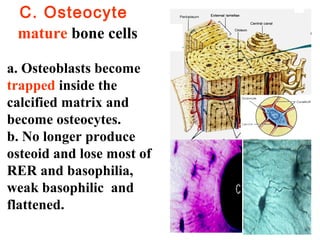
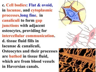
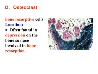
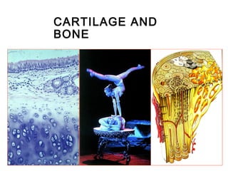
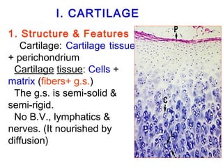
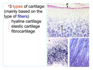
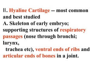
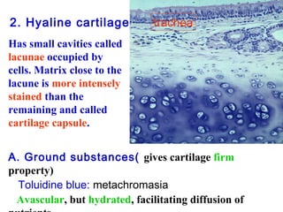
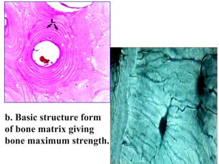
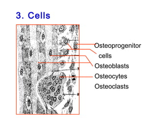
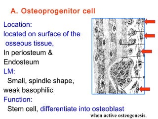
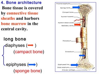
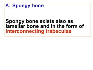
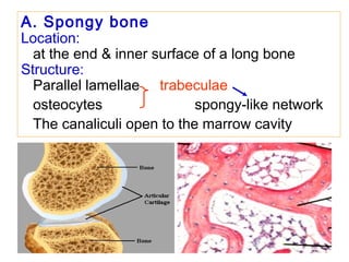
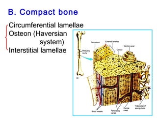
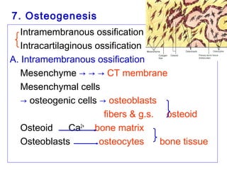
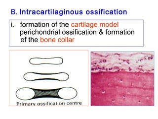
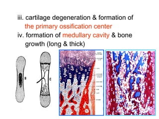
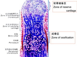
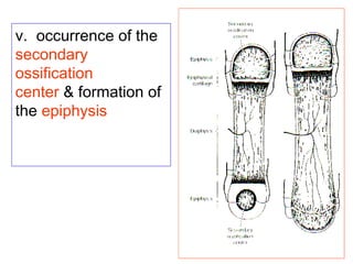
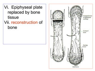
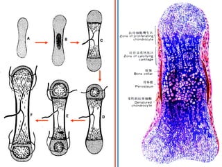
Cartilage and bone are connective tissues that provide structure and support. Cartilage is composed of chondrocytes within a firm matrix, and there are three types: hyaline, elastic, and fibrocartilage. Bone tissue contains osteoprogenitor cells, osteoblasts, osteocytes, and osteoclasts embedded within an organic and inorganic matrix. Compact bone contains concentric osteons and interstitial lamellae that maximize strength. Bone develops through intramembranous or endochondral ossification involving cartilage models and growth plates.
CONTENTS
FORMATION OF BONE
CLASSIFICATION OF BONES
STRUCTURE OF BONE
BLOOD SUPPLY
COMPOSITION OF BONE
FRACTURE HEALING
CARTILAGE
TYPES OF CARTILAGE
BONE (syn – Os; Osteon)
Osseous tissue, a specialised form of dense connective
tissue consisting of bone cells (osteocytes)
Embedded in a matrix of calcified intercelluar
substance
Bone matrix contains collagen fibres and the minerals
calcium phosphate and calcium carbonate
Cartilage is a resilient and smooth elastic connective tissue, a rubber-like padding that covers and protects the ends of long bones at the joints, and is a structural component of the rib cage, the ear, the nose, the bronchial tubes, the intervertebral discs, and many other body components.
Histology
Junqueira’s Basic Histology Text and Atlas, 15th Ed
Bone tissue also called (osseous tissue) is a type of specialized dense connective tissue.
Histology
Junqueira’s Basic Histology Text and Atlas, 15th Ed
Epithelium cellstissues histology
1. Chapter 4 Tissues and Histology • Tissues - collections of similar cells and the substances surrounding them • Tissue classification based on structure of cells, composition of noncellular extracellular matrix, and cell function • Major types of adult tissues – Epithelial – Connective – Muscle – Nervous • Histology: Microscopic Study of Tissues – Biopsy: removal of tissues for diagnostic purposes – Autopsy: examination of organs of a dead body to determine cause of death
The Indian Dental Academy is the Leader in continuing dental education , training dentists in all aspects of dentistry and
offering a wide range of dental certified courses in different formats.for more details please visit
www.indiandentalacademy.com
The Indian Dental Academy is the Leader in continuing dental education , training dentists in all aspects of dentistry and
offering a wide range of dental certified courses in different formats.for more details please visit
www.indiandentalacademy.com
It is the preparation of tissues for microscopic examination.
It is an effective diagnostic tool in clinical pathology.
Histological preparations reveal normal tissue structure, tissue abnormalities and cancerous conditions.
Bone physiologynew /certified fixed orthodontic courses by Indian dental acad...Indian dental academy
The Indian Dental Academy is the Leader in continuing dental education , training dentists in all aspects of dentistry and offering a wide range of dental certified courses in different formats.
Indian dental academy provides dental crown & Bridge,rotary endodontics,fixed orthodontics,
Dental implants courses.for details pls visit www.indiandentalacademy.com ,or call
0091-9248678078
in this pdf you will know about one of the most important structure in the human body its bone we will know about its origin , types , function and other things
The bone of the skeleton is a mineralized vascular type of connective tissue with a solid matrix. The alveolar process is the bony extension of the mandible and maxilla that provides the necessary support for the teeth and serves as a site of attachment for the periodontal ligament fibers. By its resorption and deposition, it also compensates for tooth movement.
Cartilage is a connective tissue structure that is composed of a collagen and proteoglycan-rich matrix and a single cell type: the chondrocyte. Cartilage is unique among connective tissues in that it lacks blood vessels and nerves and receives its nutrition solely by diffusion
Description :
The Indian Dental Academy is the Leader in continuing dental education , training dentists in all aspects of dentistry and
offering a wide range of dental certified courses in different formats.for more details please visit
www.indiandentalacademy.com
3. I . C A R CTaILrtAilaGgEe
Ⅰ. Introduction
A. A rigid tissue, capable of
providing support, connection
and sliding surface for joints.
B. Composed of firm
intercellular material (cartilage
matrix) and cells
(chondrocytes).
4. I. CARTILAGE
Cartilage
1. Structure & Features
Cartilage: Cartilage tissue
+ perichondrium
Cartilage tissue: Cells +
matrix (fibers+ g.s.)
The g.s. is semi-solid &
semi-rigid.
No B.V., lymphatics &
nerves. (It nourished by
diffusion)
5. 3 types of cartilage:
(mainly based on the
type of fibers)
hyaline cartilage
elastic cartilage
fibrocartilage
6. Ⅱ. Hyaline Cartilage -- most common
and best studied
A. Skeleton of early embryo;
supporting structures of respiratory
passages (nose through bronchi;
larynx,
trachea etc), ventral ends of ribs and
articular ends of bones in a joint.
7. 2. Hyaline cartilage trachea
Has small cavities called
lacunae occupied by
cells. Matrix close to the
lacune is more intensely
stained than the
remaining and called
cartilage capsule.
A. Ground substances( gives cartilage firm
property)
Toluidine blue: metachromasia
Avascular, but hydrated, facilitating diffusion of
nutrients.
8. Chemical composition is similar to
that of L.C.T. ( more chondroitin so
basophilic in H & E sections. ) &
chondronectin(ChN)
Cartilage lacunae:
matrix cavity
Cartilage capsule:
on the surface of
the lacunae;
strong basophilic
cap
9. B. Fibers
Collagenous fibril (D=10-
20nm) made up of type II collagen,
invisible in LM (delicate
& same refractive index as
the g.s.)
10. C. Chondrocytes
1. Young chondrocytes
are flat and singly fill
lacunae at the
periphery.
2.Toward the center,
cells round up and
mature, and may
appear in groups
designated isogenous
groups, derived from a
single cell.
Located in
cartilage lacunae
Basophilic
11. LM:
Peripheral chondrocytes:
less mature; small &
flat; single;
Central chondrocytes:
more mature; round;
isogenous group.
EM: shows abundant RER
and well-developed Golgi
complex,responsible for
synthesis and secretion
of matrix.
12. D. Perichondrium: Dense connective tissue sheath
around the cartilage.
Outer layer: more collagenous fibers; is protective
Inner layer: more b.v. & cells, containing
osteoprogenitor cells
(→ chondrocytes
→ new cartilage
tissue)
Function:
protection, nutrition,
repairing
13. E. Cartilage growth & regeneration
Appositional growth:
Osteoprogenitor cells in perichodrium
produce new layer of cartilage tissue
from outside
Interstitial growth:
Chondrocytes: increase the number of
cells (mitosis) & the amount of matrix
from within
15. 4. Fibrocartilage
Location:
intervertebral disk;
symphysis pubis;
end of tendons etc.
Structure:
collagenous fiber
(A large number, in
parallel bands),
less g.s. (acidophilic)
& chondrocytes
16. Bone II. Bone
Ⅰ. Bone Tissue (Osseous Tissue)
Specialized connective tissue with
calcified matrix, thus firmer(hard &
rigid ) than cartilage, and provides
support, movement, protection and a
storage site for calcium.
17. II. Bone
Bone
1. Characteristics:
• bone tissue + medullary
cavity & bone marrow +
periosteum & endosteum
• bone tissue= cells +
bone matrix (fibers +
calcified g.s.)
18. 2. Bone matrix
Organic (35%); inorganic (65%)
• Organic part:
collagenous fibers (90%)
amorphous g.s.:
proteins & GAG ( chondroitin s., keratin s.) is
much less than in cartilage.
to unit the fibers together.
Glycoproteins:
osteocalcin
osteonectin
osteopontin
19. Function: be responsible to
toughness & resilience, Imparts
resilience to bone.
• Inorganic part (bone mineral):
Calcium phosphate in the form of
hydroxyapatite crystal
be responsible to hardness & rigidity
Imparts hardness to bone.
20. • bone lamella : the form that bone
matrix arranged in
a. plywood-like layers (lamellae) with the fibers
parallel in one layer and rectangular
between adjacent layers. apatite lie
alongside the collagen fibrils. The bone so
called lamellar bone
b. * lacunae & canaliculi
in the lamellae
23. A. Osteoprogenitor cell
Location:
located on surface of the
osseous tissue,
In periosteum &
Endosteum
LM:
Small, spindle shape,
weak basophilic
Function:
Stem cell, differentiate into osteoblast
when active osteogenesis.
25. B. Osteoblast
Location: at surface of
bone tissue in a layer;
LM: cuboidal cells, strong
basophilic, large pale
nuclear w/ obvious
nucleolus
EM:
short processes &
gap junctions;
rich in rER & Gl.
indicate active protein
synthesis.
26. ※Newly-formed, uncalcified
organic matrix is called osteoid.
※Osteoid is soon calcified with
calcium salt deposition initiated
by matrix vesicles probably
derived from osteoblasts.
28. C. Osteocyte
mature bone cells
a. Osteoblasts become
trapped inside the
calcified matrix and
become osteocytes.
b. No longer produce
osteoid and lose most of
RER and basophilia,
weak basophilic and
flattened.
29. c. Cell bodies: Flat & ovoid,
in lacunae, and cytoplasmic
processes,long fine, in
canaliculi to form gap
junctions with adjacent
osteocytes, providing for
intercellular communication.
d. tissue fluid fills in
lacunae & canaliculi,
Osteocytes and their processes
are bathed in tissue fluid,
which are from blood vessels
in Haversian canals.
30. Function:
Maintain & renew the bone
matrix;
Exchange the Ca2+ b/w tissue fluid
and blood, stable of blood Ca2+
31. D. Osteoclast
bone resorptive cells
Location:
a. Often found in
depression on the
bone surface
involved in bone
resorption.
32. b. Giant multinucleate(2-
50), irregular in shape
and acidophilic, formed
by fusion of monocytes.
c. Ruffled border
(irregular microvilli) on
the surface adjacent to
bone and lysosomes in
the cytoplasm are
evidence of bone
resorption.
33. Lysosomal enzymes and
organic acids are released
into extracellular space
and break down organic
and mineral components
of bone.
EM:
Mv.(ruffled border);
Lysosomes;
Phagocytosis vacuoles
Others: Mit. rER.
34. Function:
Clear zone: attached the bone surface to
form a closed microenvironment
Osteoclasts release lysosomal enzymes
(proteinases, carbonic anhydrase), & acids
(citric acid, lactic acid) to dissolve the bone
matrix
Mv.: increase the absorptive area & absorb
the dissolved products
* belong to mononuclear phagocyte system
35.
36. 4. Bone architecture
Bone tissue is covered
by connective tissue
sheaths and harbors
bone marrow in the
central cavity.
long bone
diaphyses ( )
(campact bone)
epiphyses ( )
(sponge bone)
37. A. Spongy bone
Spongy bone exists also as
lamellar bone and in the form of
interconnecting trabeculae
38. A. Spongy bone
Location:
at the end & inner surface of a long bone
Structure:
Parallel lamellae trabeculae
osteocytes spongy-like network
The canaliculi open to the marrow cavity
39. B. Compact bone
Circumferential lamellae
Osteon (Haversian
system)
Interstitial lamellae
40. Circumferential lamellae :
follow the outer and inner
circumferences of the diaphysis.
arranged in concentric circles.
outer~: 10-40 layers
inner~: fewer layers
41. Osteons or
Haversian systems
the structural unit of
compact bone. It
contains several
concentrically
arranged lamellae
and a central canal
(Haversian canal)
with CT, blood
vessels, nerves, and
cells.
42. 4. Volkmanns canals
crossly perforate the
circumferential lamellae
and bring blood vessels
into the compact bone.
5. Between or within
bone lamellae are living
cells (osteocytes) in
lacunae connected to
one another by
canaliculi, the
innermost of which
connected to the central
canals.
43. b. Osteon (Haversian system)
• Cylindrical units;
• 8-20 layers of Osteon
lamellae (Haversian~ )
Central canal (b.v. / n.f. /
tissue fluid in it),
the canaliculi open to the
canals;
• cement line
wraps around the unit;
• Volkmann’s canals
(perforating canals)
communicate w/ the
central canals.
44. c. Interstitial lamellae:
Irregular lamellae
and fill spaces b/w the
osteons;
It is the remanets of
osteons during
reconstruction.
I.L
os
45. 5. Periosteum a layer of dense connective tissue
covering the external surface of bone
outer layer:
dense c.t., thicker;
the perforating f.
(Sharpey’s fibers) fix
the periosteum to
bone;
inner layer:
containing
osteoprogenitor
cells,small b.v. & n.
46. 6. Endosteum a thin layer of bone lining
cells on inner surface of bone.
thin c.t. & a layer of bone lining cells (special
osteoprogenitor cells) w/ processes.
Differentiate into osteoblasts for ossification
Attract osteoclasts
Lining the surface of
the marrow cavities
to maintain the stability
of Ca2+ in lacune
47. 5. Periosteum
Osteoprogenitor cells present in both
throughout life -- can give rise to
osteoblasts in case of fracture.
48. Qestions
What is the cartilage and the cartilage
tissue?
How many cartilage types in the human
body?
Describe the Hyaline cartilage.
What is the bone and the bone tissue?
Describe the structure and function of the
cells in bone tissue.
Describe the architecture of the compact
bone.