SlideShare a Scribd company logo
1 of 130
1
Thoracic Aorta
THE THORACIC
AORTA, SVC,
IVC, AZV, HAZV,
THORACIC PART
OF
SYMPATHETIC
TRUNK
2
Thoracic Aorta
Agenda
‫‏‬
Anatomy
‫‏‬
Definitions
‫‏‬
Epidemiology
‫‏‬
Etiology
‫‏‬
Pathophysiology
‫‏‬
Presentation
‫‏‬
Diagnosis
‫‏‬
Natural History
‫‏‬
Treatment
‫‏‬
Prognosis
‫‏‬
Underwriting Considerations
3
Thoracic Aorta
Anatomy
4
Thoracic Aorta
Definitions
‫‏‬
Normal Dimensions
– Mid-descending 26-28 mm
‫‏‬
Dilation (Ballooning, Bulging, Ectasia)
‫‏‬
Aneurysm
– Types
• Saccular
• Fusiform
– Definition
• When the diameter exceeds 4 cm or diameter exceeds 1.5 times normal
‫‏‬
Dissection
– Tear in vessel wall results in false lumen
– Types
• Type A – involves ascending aorta
• Type B – involves descending aorta
5
Thoracic Aorta
Normal Aortic Dimensions
Hager A. et al.; J Thorac Cardiovasc Surg 2002;123:1060-1066
Company Confidential
©2010 Genworth Financial, Inc. All rights reserved.
‫‏‬ THE AORTA AND ITS MAJOR BRANCHES
‫‏‬ The aorta commences at the aortic valve, above the vestibule of the
left ventricle and terminates in the abdomen at the level of the fourth
lumbar vertebra (L4), where it bifurcates to form the right and left
common iliac arteries. It is an elastic artery and it is divisible into
four parts, viz:-
 The Ascending thoracic aorta.
 The Arch of the aorta.
 The Descending thoracic aorta and
 The Abdominal aorta.
Company Confidential
©2010 Genworth Financial, Inc. All rights reserved.
‫‏‬ ASCENDING THORACIC AORTA:
‫‏‬ This is located in the middle mediastinum and measures 5cm long. It
has the following branches through which it supplies the heart:
Right coronary artery.
Left coronary artery
Company Confidential
©2010 Genworth Financial, Inc. All rights reserved.
‫‏‬ THE ARCH OF THE AORTA:
‫‏‬ This is located in the superior mediastinum.
‫‏‬ It is the continuation of the ascending aorta.
‫‏‬ Its branches are:
• The Brachiocephalic trunk. This gives rise to:
Right Common Carotid artery.
Right Subclavian artery.
• Left Common Carotid artery.
• Left Subclavian artery.
Company Confidential
©2010 Genworth Financial, Inc. All rights reserved.
‫‏‬
‫‏‬ Each common carotid artery:
‫‏‬ Bifurcates to give rise to an:
 internal carotid artery and
 external carotid artery.
‫‏‬ The internal carotid continues into the cranial cavity to supply cranial
contents while the external carotid supplies the head and neck
region.
Company Confidential
©2010 Genworth Financial, Inc. All rights reserved.
‫‏‬ The Subclavian artery:
‫‏‬ The subclavian artery supplies structures in the head and neck and
the thoracic regions through the following branches:
Vertebral artery.
The thyrocervical artery.
The internal thoracic (Mammary) art.
The costocervical artery
The dorsal scapular artery
‫‏‬ Each Subclavian artery continues as the axillary artery at the outer
margin (Lateral margin) of the first rib.
‫‏‬ The axillary artery is the main arterial supply to the upper limb.
Company Confidential
©2010 Genworth Financial, Inc. All rights reserved.
‫‏‬ THE DESCENDING THORACIC AORTA
‫‏‬ This artery is the direct continuation of the arch of the aorta.
‫‏‬ It passes through the posterior mediastinum and terminates
posterior to the median arcuate ligament of the diaphragm at the
level of the 12th thoracic vertebra (T12).
‫‏‬ At this point it continues into the abdominal cavity as the Abdominal
aorta.
Company Confidential
©2010 Genworth Financial, Inc. All rights reserved.
‫‏‬ The branches of the Thoracic Aorta
include:
• 9 pairs of Posterior intercostal
arteries.
• A pair of subcostal arteries.
• 2 left bronchial art.
• 2 esophageal art.
• Pericardial branches (Unknown
number).
• Mediastinal branches (Unknown
number).
• A pair of Superior Phrenic art. (Right &
Left).
Company Confidential
©2010 Genworth Financial, Inc. All rights reserved.
Company Confidential
©2010 Genworth Financial, Inc. All rights reserved.
‫‏‬ THE ABDOMINAL AORTA
‫‏‬ This is the continuation of the
descending thoracic aorta from the
level of T12.
‫‏‬ It is located on the posterior abdominal
wall and terminates at the level of L4.
‫‏‬ Here it bifurcates to give rise to the
right and left Common iliac arteries.
Company Confidential
©2010 Genworth Financial, Inc. All rights reserved.
‫‏‬ The branches of the Abdominal Aorta include:
• Coeliac trunk. ( Foregut)
• Superior mesenteric art. (Midgut)
• Inferior mesenteric art. (Hindgut)
• Median sacral art. (Posterior pelvic wall)
• 2 Renal art. (The Kidneys)
• 1 Middle suprarenal art.
• 2 Testicular/Ovarian art.
• 2 Inferior phrenic art.
• 4 pairs of Lumbar art.
Company Confidential
©2010 Genworth Financial, Inc. All rights reserved.
‫‏‬ Each of the Common iliac arteries will bifurcate at the level of the
sacroiliac joint to give rise to Internal and External iliac arteries.
‫‏‬ The internal iliac artery will supply the pelvic, perineal(genital) and
gluteal regions.
‫‏‬ The external iliac continues as the Femoral artery beyond the inguinal
ligament to supply the entire lower limb.
Company Confidential
©2010 Genworth Financial, Inc. All rights reserved.
20
Thoracic Aorta
Aortic Aneurysm
21
Thoracic Aorta
Aortic Aneurysm
(A) Tomodensitometric and (B) echocardiographic views of an aortic root
aneurysm.
Nataf P , Lansac E Heart 2006;92:1345-1352
22
Thoracic Aorta
Aortic Aneurysm
http://www.medscape.com/viewarticle/406630_15
Figure 23. Atherosclerotic vascular dis-ease in an
aortic aneurysm. Axial postcontrast image
(window = 440, level = 40) reveals a large
contrast collection projecting from the
undersurface of the aortic arch, consistent with
aneurysm (arrow). the low attention material
within the aneurysm represents thrombus
23
Thoracic Aorta
Aortic Aneurysm
http://www.medscape.com/viewarticle/406630_15
Figure 24. Aortic aneurysm
rupture. Axial postcontrast image
(window = 440, level = 40)
through the aortic arch reveals
an aortic aneurysm with contrast
penetrating the thrombus within
the aneurysm (open arrow).
24
Thoracic Aorta
Aortic Dissection
25
Thoracic Aorta
Aortic Dissection
http://www.medscape.com/viewarticle/406630_15
Figure 12. Stanford type B (Debakey Type III)
aortic dissection: descending thoracic aorta.
(A) Axial postcontrast image (window = 440,
level = 40) reveals intimal flap (arrow), t = true
lumen, f = false lumen. (B) Oblique sagittal
reconstruction reveals complex nature of the
intimal flap (arrows).
26
Thoracic Aorta
Epidemiology
‫‏‬
Thoracic aneurysms
– Prevalence greater than 3-4% of those over 65
– 6 cases per 100,000 person-years
– Incidence increasing
– In the top 15 causes of death
– Thoracic aortic aneurysm – rupture 3.5/100,000 persons
‫‏‬
Thoracic aortic dissection
– 2000 new cases/year
– Acute - 3.5/100,000 persons
– Male:Female ratio 2:1
27
Thoracic Aorta
Etiologies
‫‏‬
Underlying Etiologies
– Atherosclerosis
– Marfan’s
– Type IV Ehlers-Danlos
– Infection (syphillis)
– Arteritis (giant cell, Takayasu, Behcet’s)
– Trauma
‫‏‬
Risk Factors
– Smoking
– COPD
– HTN
– Male gender
– Older age
– High BMI
– Abnormal aortic valve (e.g., bicuspid valve)
– Family history
28
Thoracic Aorta
Presentation
‫‏‬
Aneurysm
– Most asymptomatic
– Superior vena cava syndrome
– Hoarseness
– Bronchial obstruction
– Dysphagia
– Hemoptysis
– Paralysis/paraplegia
– Lower extremity embolism
‫‏‬
Dissection
– Chest/back/neck pain
– Neurologic signs
– Horner syndrome
– Hoarseness
– Acute aortic regurgitation
29
Thoracic Aorta
Diagnosis
‫‏‬
Chest x-ray
– Widened mediastinum
‫‏‬
Echocardiogram
– Transthoracic – aortic root
– Transesophageal – ascending and descending
‫‏‬
Aortography
– Delineates the lumen
‫‏‬
CT scan
– Most widely used diagnostic tool
‫‏‬
MRI
– Avoids contrast dye
30
Thoracic Aorta
Echocardiography
Nataf P , Lansac E Heart 2006;92:1345-1352
31
Thoracic Aorta
Natural History
0
5
10
15
20
25
<2.75
cm/m2
2.75-4.25
cm/m2
>4.25
cm/m2
Aortic Size Index (ASI)
Annual
Risk
of
Rupture
http://emedicine.medscape.com/article/242904=print
ASI = aortic dia (cm)/body surface area (m2)
32
Thoracic Aorta
Natural History
‫‏‬
Yearly Rupture or Dissection Rates for Thoracic Aortic Aneurysms: Simple
Prediction Based on Size
– 304 patients; 58.9% male; median age 65.8
– Aneurysm size – 43.7% were 4.0-4.9 cm
– Location – 72% ascending
– Follow up – average 43.1 months
– End points
Davies RR, et al. Ann Thorac Surg 2002;73:17
44
Death alone
15
Dissection alone
5
Rupture alone
4
Rupture and death (no dissection)
5
Dissection, death (no rupture)
2
Dissection, rupture (no death)
2
Dissection, rupture and death
No. Patients
Events
33
Thoracic Aorta
Natural History
Davies RR, et al. Ann Thorac Surg 2002;73:17
Cumulative incidence of acute dissection or rupture as a function of initial aneurysm size.
34
Thoracic Aorta
Natural History
Davies RR, et al. Ann Thorac Surg 2002;73:17
Kaplan-Meier cumulative hazard function of rupture or dissection.
35
Thoracic Aorta
Natural History
Davies RR, et al. Ann Thorac Surg 2002;73:17
Average yearly rates of negative outcomes during the first 5 years after presentation
36
Thoracic Aorta
Natural History
Davies RR, et al. Ann Thorac Surg 2002;73:17
Kaplan-Meier cumulative hazard function of rupture.
37
Thoracic Aorta
Natural History
Kaplan-Meier cumulative survival before operative repair.
Davies RR, et al. Ann Thorac Surg 2002;73:17
38
Thoracic Aorta
Natural History
Davies RR, et al. Ann Thorac Surg 2002;73:17
Kaplan-Meier cumulative survival
39
Thoracic Aorta
Natural History
Davies RR, et al. Ann Thorac Surg 2002;73:17
Kaplan-Meier cumulative survival
40
Thoracic Aorta
Natural History
Davies RR, et al. Ann Thorac Surg 2002;73:17
Kaplan-Meier cumulative survival
41
Thoracic Aorta
Treatment - Aneurysm
‫‏‬
Medical
– BP control
– Smoking cessation
– No heavy lifting
‫‏‬
Surgical
– Dacron tube graft
– Ascending – may need to replace valve
– Arch – graft
– Descending – graft, stent grafts
42
Thoracic Aorta
Treatment - Dissection
‫‏‬
Type A
– Surgical
‫‏‬
Type B
– Medical
– Surgical
• Acute with rupture, leak or distal ischemia.
43
Thoracic Aorta
Treatment – Indications for Intervention
‫‏‬
Aortic size
– Ascending diameter >5.5 cm
– Descending diameter >6.5 cm
– Growth rate >1 cm/yr (avg ascending 0.07 cm/yr; descending 0.19 cm/yr)
‫‏‬
Symptomatic aneurysm
‫‏‬
Traumatic rupture
‫‏‬
Pseudoaneurysm
‫‏‬
Large saccular aneurysm
‫‏‬
Mycotic aneurysm
‫‏‬
Aortic coarctation
‫‏‬
Bronchial compression
‫‏‬
Aortobronchial or aortoesophageal fistuala
44
Thoracic Aorta
Treatment - Surgical
Composite valve and graft replacement.
Nataf P , Lansac E Heart 2006;92:1345-1352
45
Thoracic Aorta
Complications
‫‏‬
Bleeding
‫‏‬
CVA – 2-5%
‫‏‬
CHF
‫‏‬
Respiratory failure
‫‏‬
Graft leaks
‫‏‬
Fistula formation
‫‏‬
Spinal cord damage
‫‏‬
Renal failure
46
Thoracic Aorta
Prognosis
‫‏‬
Aneurysm
– Early post-op mortality 4-10%; lower for descending aneurysm repair; much
higher for aortic arch repair
– Stroke occurs 2-5%
– Renal failure requiring dialysis – 7%
– Spinal cord injury – 3%
‫‏‬
Dissection
– Treated 10-yr survival rate 60%
– Type A
• 30% mortality surgical
• 60% mortality medical
– Type B
• 10% mortality medical
• 30% mortality surgical
47
Thoracic Aorta
Underwriting Approach and Considerations
‫‏‬
Obtain cardiology and/or vascular medical records
‫‏‬
Review serial echos/scans as available
‫‏‬
Review blood pressure control
‫‏‬
Higher Risk
– Aneurysm >5 cm
– Poorly controlled blood pressure
– Increase in size >0.5 cm/yr
– Ongoing tobacco usage
– Associated cardiovascular disease (CAD, PVD, carotid disease)
– Non-atherosclerotic vascular disorders (Marfan’s, Ehlers-Danlos, etc)
‫‏‬
Lower Risk
– Aneurysm <5 cm/stable/ well followed
– Aneurysm repaired/stable
48
Thoracic Aorta
Summary
‫‏‬
Thoracic aortic dilation/aneurysm fairly common with age
‫‏‬
Risk factors are traditional cardiovascular risk factors
‫‏‬
Most are asymptomatic
‫‏‬
Thoracic aortic rupture rare
‫‏‬
Thoracic dissection rare
‫‏‬
Ascending aorta most common site of aneurysm formation
‫‏‬
Low risk for aneurysms less than 4 cm
In the end, it’s not what you call it………it’s size that matters!
REMEMBER
superior vena cava (SVC, also known as the
cava or cva)
‫‏‬ The superior vena cava (SVC, also known as the cava or cva) is a short,
but large diameter vein located in the anterior right
superior mediastinum. Its latin name is related to its large pipe
appearance in cadavers, 'cava' meaning 'hollow'.
49
Thoracic Aorta
‫‏‬ The superior vena cava is very important for the function of
the cardiovascular system, since it largely contributes to the input of
blood to the right atrium. Any hypertensive process in the right half of the
heart or in the pulmonary circulation retrogradely affects both superior
and inferior venae cavae. This is important since the veins are not
adjusted to high pressures, which can result with forming an aneurysm
or even rupture of the wall of the SVC.
‫‏‬ This article will discuss the anatomy and function of the superior vena
cava.
50
Thoracic Aorta
Key facts about the superior vena cavaTable quiz
Source Brachiocephalic vein, azygos vein
Draining area Upper half of the body (above the diaphragm)
Drains to Right atrium of heart
‫‏‬ Contents
‫‏‬ Anatomy
‫‏‬ Function
‫‏‬ Clinical notes
– Superior vena cava obstruction (SVCO)
– Superior vena cava syndrome (SVCS)
– Superior vena cava thrombosis
– Superior vena cava aneurysm
– Persistent left superior vena cava (PLSVC)
‫‏‬ Sources
51
Thoracic Aorta
‫‏‬ Anatomy
‫‏‬ Left brachiocephalic vein
‫‏‬ Vena brachiocephalica sinistra
‫‏‬ 1/3
‫‏‬ Synonyms: Left innominate vein, Vena anonyma sinistra
‫‏‬ Embryologically, the SVC is formed by the left and right brachiocephalic
veins (also known as the innominate veins) that also receive blood from
the upper limbs, certain parts of the head, one being the eyes, and neck.
‫‏‬ There is no valve that divides the SVC from the right atrium, which
conducts blood from right atrial and right ventricular contractions
upwards into the internal jugular vein (seen as the jugular venous
pressure) and sternocleidomastoid muscle.
52
Thoracic Aorta
‫‏‬ Positionally, the SVC begins behind the lower border of the 1st right
costal cartilage and descends vertically behind the 2nd and 3rd
intercostal spaces to drain into the right atrium at the level of the 3rd
costal cartilage. Its lower half is covered by a fibrous pericardium, which
is pierced by the SVC at the level of the 2nd costal cartilage.
53
Thoracic Aorta
Function
Superior vena cava coursing towards the right atrium of the
heart, returning deoxygenated blood from the body.
The SVC is one of the 2 large veins by which blood is returned from the body to the right side
of the heart. After circulating through the body systemically, deoxygenated blood returns to
the right atrium of the heart through either the SVC, which drains the upper body, or
the inferior vena cava (IVC) that drains everything below the diaphragm.
‫‏‬ Clinical notes
‫‏‬ Superior vena cava obstruction (SVCO)
‫‏‬ This usually refers to a partial or complete obstruction of the SVC,
often in the context of cancer (lung cancer, metastatic cancer, or
lymphoma). Clinically this obstruction can lead to enlarged veins in
the head and neck, and cause shortness of breath, cough, chest
pain, and difficulty swallowing).
‫‏‬ A clinical test known as Pemberton’s sign can be performed to
identify this condition. A positive Pemberton's sign is marked by
facial congestion and cyanosis (and/or respiratory distress) after 1
minute of having the patient elevate both arms until they touch the
sides of the face. This sign is indicative of superior vena
cava syndrome, commonly the result of a mass in the mediastinum.
54
Thoracic Aorta
‫‏‬ Superior vena cava syndrome (SVCS)
‫‏‬ This syndrome refers to a group of symptoms caused by the
obstruction of the SVC. More than 90% of the cases of SVC
obstruction are caused by cancer, most commonly bronchogenic
carcinoma, which includes small cell and non-small cell lung
carcinoma, Burkitt’s lymphoma, lymphoblastic lymphomas, pre-T-
cell lineage acute lymphoblastic leukemia, and other acute
leukemias.
‫‏‬ Characteristic symptoms are edema of the arms and face,
development of swollen collateral veins on the front of the chest
wall, shortness of breath, difficulty swallowing, stridor, cough, and
neurological symptoms (reduced alertness, etc. from edema in
the brain or airway compromise). Again, Pemberton’s sign can be
used to identify an SVCO.
55
Thoracic Aorta
‫‏‬ Superior vena cava thrombosis
‫‏‬ The thrombosis often occurs from a thrombus around a long-
term central venous catheter (CVC), especially in cancer patients with
permanent indwelling CVCs. CVC-related thrombosis is as high as
30% in adults. However, patients can be treated with thrombolytics
or anticoagulants, or by removal of the catheter.
‫‏‬ Superior vena cava aneurysm
‫‏‬ Venous aneurysms arising from the mediastinal systemic veins are
extremely rare, with the majority being fusiform (“spindle-shaped”)
aneurysms that arise from the SVC.
‫‏‬ Persistent left superior vena cava (PLSVC)
‫‏‬ A PLSVC is an embryologic remnant that is the most common
variation of the thoracic venous system, resulting from a failure to
involute during embryologic development.
56
Thoracic Aorta
The inferior vena cava (IVC) is the largest vein
of the human body. It is located at
the posterior abdominal wall on the right side of
the aorta. The IVC’s function is to carry
the venous blood from the lower
limbs and abdominopelvic region to the heart.
The inferior vena cava anatomy is essential
due to the vein’s great drainage area, which
also makes it a hot topic for anatomy exams.
For that reason, this page will cover the IVC
anatomy in a way that’s easy to read and
understand.
57
Thoracic Aorta
58
Thoracic Aorta
59
Thoracic Aorta
Key facts
Table quiz
Definition and functionThe vein that collects
deoxygenated blood from the abdomen,
pelvis and lower limbs and carries it to the
right atrium of the heartSourceCommon iliac
veins (L5)TributariesInferior Phrenic,
right Suprarenal, Renal, right Testicular
(gonadal), Lumbar, common Iliac
and Hepatic veins
Mnemonic: Portal System Returns To Liver
In Humans
Clinical relationsInferior vena cava
thrombosis 60
Thoracic Aorta
Anatomy
Inferior vena cava
Vena cava inferior
1/4
Synonyms: IVC
The inferior vena cava arises from the confluence
of the common iliac veins at the level of L5
vertebra, just inferior to the bifurcation of the
abdominal aorta. It then ascends the posterior
abdominal wall, to the right side of the aorta and
the bodies of the L3-L5 vertebrae. After passing
through its fossa on the posterior liver surface, the
IVC enters the thorax by traversing the inferior
vena caval foramen of the diaphragm.
61
Thoracic Aorta
62
Thoracic Aorta
‫‏‬ The tributaries of the IVC correspond to the branches of the abdominal
aorta. Note that some professors will want you to know at which
vertebral level the IVC gets its direct tributaries, so they are as follows:
‫‏‬ The direct tributaries are the inferior phrenic veins (T8), right suprarenal
(L1), renal (L1), right testicular (gonadal) (L2), lumbar (L1-L5),
common iliac (L5) and hepatic (T8). If you want an easy way to
remember them just memorise the mnemonic
' Portal System Returns To Liver In Humans'.
‫‏‬ Left gonadal and left suprarenal renal veins drain first into the left renal
vein
‫‏‬ The veins of the stomach, spleen, pancreas, small and large
intestines first empty into the hepatic portal vein. The hepatic portal vein
carries this blood to the liver to be processed and detoxified. Then, the
blood reaches the IVC through the hepatic veins.
‫‏‬ The inferior vena cava communicates with the superior vena
cava through the collateral vessels, which include the azygos
vein, lumbar veins, and vertebral venous plexuses.
63
Thoracic Aorta
64
Thoracic Aorta
‫‏‬ Function
‫‏‬ The IVC’s function is to convey the blood from the abdomen, pelvis,
and lower limbs to the right atrium of the heart. Additional IVC functions
are noticeable during some health disturbances, such as hepatic portal
vein obstruction or the obstruction of the IVC itself.
‫‏‬ Specialized vessels called
the portocaval (portosystemic) anastomoses open if the hepatic portal
vein is obstructed. The intestinal blood then bypasses the liver and
empties into the IVC directly. In cases where the IVC is occluded,
the collateral vessels to the superior vena cava open.
‫‏‬ Learn more about portocaval anastomoses with our article, then take this
specially designed quiz to consolidate everything you’ve learned about
the IVC and its tributaries.
65
Thoracic Aorta
‫‏‬ Inferior vena cava thrombosis
‫‏‬ Thrombosis of the inferior vena cava (IVCT) is a condition in which a
blood clot (thrombus) impedes the blood flow through the IVC. The
thrombus can be formed within the IVC itself, which is rare, or, more
commonly, travel from the deep veins of the legs in a condition called
deep venous thrombosis (DVT).
‫‏‬ IVC thrombosis may be caused by all the conditions that lead to venous
stasis. These include congenital abnormalities of the IVC,
immobilization, obesity, pregnancy, sedentary lifestyle, and tumors of
surrounding organs. IVCT presents with symptoms of venous
obstruction, such as pain and swelling of lower limbs and scrotum. IVCT
is diagnosed by ultrasound, CT,and MRI.
‫‏‬ Depending on the severity of the condition, the IVCT can be treated by
surgical removal of the thrombus, thrombolytic therapy, and
anticoagulant therapy. The treatment is necessary to prevent
disseminating of the thrombus into the pulmonary circulation (pulmonary
thromboembolism), which is an urgent medical condition.
‫‏‬ Sources
66
Thoracic Aorta
Azygos vein
and
Hemiazygos
vein
67
Thoracic Aorta
68
Veins of the thoracic wall
‫‏‬ The thoracic wall is drained anteriorly by the internal thoracic vein
and posteriorly by the azygos system of veins
69
‫‏‬ The azygos system of veins serves as an important anastomotic
pathway capable of returning venous blood from the lower part of
the body to the heart if the inferior vena cava is blocked.
70
‫‏‬ The major veins in the system are:
‫‏‬ Azygos vein, on the right
‫‏‬ Hemiazygos vein and the accessory hemiazygos vein, on the
left.
71
‫‏‬ Azygos vein
‫‏‬ The azygos vein arises opposite vertebra LI or LII
‫‏‬ It is formed by the union of the right ascending lumbar vein , the
right subcostal vein and lumber azygos veins .
72
73
‫‏‬ In addition to the posterior intercostal veins, the azygos vein
communicates with the vertebral venous plexuses.
74
75
‫‏‬ The azygos vein enters the thorax through the aortic hiatus of the
diaphragm, or it enters through or posterior to the right crus of the
diaphragm.
76
‫‏‬ It ascends through the posterior mediastinum, usually to the right
of the thoracic duct.
77
‫‏‬ At approximately vertebral level TIV, it arches anteriorly, over the
root of the right lung, to join the superior vena cava.
78
79
‫‏‬ Tributaries of the azygos vein
‫‏‬ Right superior intercostal vein
‫‏‬ 5th – 11th right posterior ICV
‫‏‬ Hemiazygos vein;
‫‏‬ Accessory hemiazygos vein
‫‏‬ Esophageal veins
‫‏‬ Mediastinal veins
‫‏‬ Pericardial veins
‫‏‬ Right bronchial veins
80
Hemiazygos vein
‫‏‬ The hemiazygos vein (inferior hemiazygos vein) usually arises at
the junction between the left ascending lumbar vein and the left
subcostal vein .
81
82
‫‏‬ The hemiazygos vein usually enters the thorax through the left crus
of the diaphragm, but may enter through the aortic hiatus.
83
‫‏‬ It ascends through the posterior mediastinum, on the left side, to
approximately vertebral level TIX.
‫‏‬ At this point, it crosses the vertebral column, posterior to the
thoracic aorta, esophagus, and thoracic duct, to enters the azygos
vein.
‫‏‬ The hemiazygos vein receives the inferior three intercostal veins, the
inferior oesophageal veins, and several small mediastinal branches.
84
85
‫‏‬ Accessory hemiazygos vein
‫‏‬ The accessory hemiazygos vein descends on the left side of upper thoracic vertebrae up to the
vertebral level TVIII . At this point, it crosses the vertebral column to join the azygos vein, or
ends in the hemiazygos vein, or has a connection to both veins. Usually, it also has a connection
superiorly to the left superior intercostal vein. Vessels that drain into the accessory hemiazygos
vein include: the fourth to eighth left posterior intercostal veins
86
87
Thoracic Part of
Sympathetic Chain
89
Thoracic Aorta
Sympathetic trunks
‫‏‬ The sympathetic trunks (sympathetic chain, gangliated cord) are a
paired bundle of nerve fibers that run from the base of the skull to
the coccyx.
Paravertebral sympathetic ganglia
Sympathetic trunks
‫‏‬ The sympathetic trunks are two parallel nerve cords extending on
either side of the vertebral column from the base of the skull to the
coccyx
‫‏‬ The ganglia and trunks are connected to adjacent spinal nerves by
gray rami communicantes throughout the length of the sympathetic
trunk and by white rami communicantes in the thoracic and upper
lumbar parts of the trunk (T1 to L2).
‫‏‬ The whiter rami are afferent fibers to the ganglion and found only
from T1 to L2 segments. These contain the preganglionic and
afferent sensory fibers.
‫‏‬ The gray rami communicans are present in all segments. They are
efferent fibers of the sympathetic ganglia. These contain the
postganglionic fibers.
‫‏‬ Parts of sympathetic chain
‫‏‬ Cervical part
‫‏‬ Thoracic part
‫‏‬ lumber Part
‫‏‬ Sacral part
Course & Relations
Thoracic part
of
Sympathetic
chain
Cervical part
of sympathetic
chain
Medial arcuate
lig. of
diaphragm
Abdominal part
of sympathetic
chain
Neck of 1st
rib
Company Confidential
©2010 Genworth Financial, Inc. All rights reserved.
Sympathetic
chain
Medial arcuate
ligament
Structure
Sympathetic
ganglion
Branches
- Rami communicants to
corresponding intercostal
nerves.
- To autonomic plexuses in
thorax.
- To aorta.
- To abdominal organs through
Greater
splanchnic
nerve
Applied Anatomy
1- Spinal anesthesia at high level
may lead to temporary drop of
blood pressure.
2- In case of essential
hypertension, cutting of
sympathetic chain between T2 &
T5 will lead to drop of blood
pressure.
Autonomic Plexes
1- Pulmonary
Plexuses.
2-Oesophageal
plexuses.
3- Cardiac plexuses
(superficial and deep).
1- Pulmonary Plexus
(Posterior & Anterior)
2- Oesophageal Plexus
(Anterior & Posterior)
3- Cardiac Plexus
(Superficial & Deep)
Lymphatic Drainage
Thoracic Lymph
Drainage
A- From Chest wall.
B- From thoracic contents.
A- From Chest Wall
1- Superficial lymph vessels:
- From front of chest to
pectoral lymph nodes,
parasternal or lower deep
cervical lymph nodes.
- From back of chest to
subscapular nodes.
2- Deep lymph vessels:
A- From anterior chest wall to
parasternal nodes.
B- Posterior part of chest wall to
intercostal lymph nodes (opposite
neck of ribs).
C- Four diaphragmatic nodes
(anterior, posterior and 2 lateral)
which drain diaphragm and liver.
B- from Thoracic
Contents
1- Anterior mediastinal
nodes.
2- Posterior mediastinal
nodes.
3- Nodes of lungs, bronchi
and trachea.
• Lymph vessels from:
-Anterior mediastinal nodes.
-Tracheal lymph nodes.
-Parasternal lymph nodes.
Will unit together to form broncho-
medistinal lymph vessels which joins
thoracic duct on left side and right
lymphatic duct in right side.
Deep lymph nodes of chest wall
Anterior
mediastinal
nodes
THANK YOU

More Related Content

Similar to AORTA SVC IVC AZV HAZV TTHRCSMPTHC 2022.ppt

Aortic dissection Nikhil
Aortic dissection NikhilAortic dissection Nikhil
Aortic dissection NikhilNikhil Vaishnav
 
Aortic dissection nikku ppt
Aortic dissection nikku pptAortic dissection nikku ppt
Aortic dissection nikku pptNikhil Vaishnav
 
Arterial supply of head and neck
Arterial supply of head and neckArterial supply of head and neck
Arterial supply of head and neckAvinash Rathore
 
imaging of aorta(branches of thorax) (1).pptx
imaging of aorta(branches of thorax) (1).pptximaging of aorta(branches of thorax) (1).pptx
imaging of aorta(branches of thorax) (1).pptxbalajiavanthika7559
 
Clinical Anatomy of the Heart, Pericardium and.pptx
Clinical Anatomy of the Heart, Pericardium and.pptxClinical Anatomy of the Heart, Pericardium and.pptx
Clinical Anatomy of the Heart, Pericardium and.pptxHafizMohd21
 
lecture 2- 1 Cardiovascular.pptx
lecture 2- 1 Cardiovascular.pptxlecture 2- 1 Cardiovascular.pptx
lecture 2- 1 Cardiovascular.pptxArabAlkhadam
 
Arteries of Head and Neck
Arteries of Head and NeckArteries of Head and Neck
Arteries of Head and NeckHimanshu Soni
 
Radiological approach to aortic aneurysm and acute diseases
Radiological approach to aortic aneurysm and acute diseasesRadiological approach to aortic aneurysm and acute diseases
Radiological approach to aortic aneurysm and acute diseasesMilan Silwal
 
Thoracic aortic aneurysm
Thoracic aortic aneurysmThoracic aortic aneurysm
Thoracic aortic aneurysmFredric Carson
 
Aortic dissection 01
Aortic dissection 01Aortic dissection 01
Aortic dissection 01Rakesh Sharma
 
An unusual origin of brachiocephalic and left common carotid arteries 2018
An unusual origin of brachiocephalic and left common carotid arteries 2018An unusual origin of brachiocephalic and left common carotid arteries 2018
An unusual origin of brachiocephalic and left common carotid arteries 2018Dr.Srikanth pawar
 
Presentation1.pptx, radiological vascular anatomy of the head and neck.
Presentation1.pptx, radiological vascular anatomy of the head and neck.Presentation1.pptx, radiological vascular anatomy of the head and neck.
Presentation1.pptx, radiological vascular anatomy of the head and neck.Abdellah Nazeer
 
Ascending aortic aneurysm and its clinical significance: A case report
Ascending aortic aneurysm and its clinical significance: A case reportAscending aortic aneurysm and its clinical significance: A case report
Ascending aortic aneurysm and its clinical significance: A case reportApollo Hospitals
 
Heart and its disease
Heart and its diseaseHeart and its disease
Heart and its diseasebhagya shree
 
CA anomalies on CT angiography
CA anomalies on CT angiographyCA anomalies on CT angiography
CA anomalies on CT angiographySahar Gamal
 
Aortic dissection dr.tapu
Aortic dissection dr.tapuAortic dissection dr.tapu
Aortic dissection dr.tapuNizam Uddin
 

Similar to AORTA SVC IVC AZV HAZV TTHRCSMPTHC 2022.ppt (20)

Aortic dissection Nikhil
Aortic dissection NikhilAortic dissection Nikhil
Aortic dissection Nikhil
 
Aortic dissection nikku ppt
Aortic dissection nikku pptAortic dissection nikku ppt
Aortic dissection nikku ppt
 
Arterial supply of head and neck
Arterial supply of head and neckArterial supply of head and neck
Arterial supply of head and neck
 
imaging of aorta(branches of thorax) (1).pptx
imaging of aorta(branches of thorax) (1).pptximaging of aorta(branches of thorax) (1).pptx
imaging of aorta(branches of thorax) (1).pptx
 
USG of Aorta and Coeliac axis
USG of Aorta and Coeliac axisUSG of Aorta and Coeliac axis
USG of Aorta and Coeliac axis
 
Clinical Anatomy of the Heart, Pericardium and.pptx
Clinical Anatomy of the Heart, Pericardium and.pptxClinical Anatomy of the Heart, Pericardium and.pptx
Clinical Anatomy of the Heart, Pericardium and.pptx
 
lecture 2- 1 Cardiovascular.pptx
lecture 2- 1 Cardiovascular.pptxlecture 2- 1 Cardiovascular.pptx
lecture 2- 1 Cardiovascular.pptx
 
Arteries of Head and Neck
Arteries of Head and NeckArteries of Head and Neck
Arteries of Head and Neck
 
Radiological approach to aortic aneurysm and acute diseases
Radiological approach to aortic aneurysm and acute diseasesRadiological approach to aortic aneurysm and acute diseases
Radiological approach to aortic aneurysm and acute diseases
 
Thoracic aortic aneurysm
Thoracic aortic aneurysmThoracic aortic aneurysm
Thoracic aortic aneurysm
 
Aortic dissection 01
Aortic dissection 01Aortic dissection 01
Aortic dissection 01
 
Cardiovascular system
Cardiovascular systemCardiovascular system
Cardiovascular system
 
An unusual origin of brachiocephalic and left common carotid arteries 2018
An unusual origin of brachiocephalic and left common carotid arteries 2018An unusual origin of brachiocephalic and left common carotid arteries 2018
An unusual origin of brachiocephalic and left common carotid arteries 2018
 
Presentation1.pptx, radiological vascular anatomy of the head and neck.
Presentation1.pptx, radiological vascular anatomy of the head and neck.Presentation1.pptx, radiological vascular anatomy of the head and neck.
Presentation1.pptx, radiological vascular anatomy of the head and neck.
 
Ascending aortic aneurysm and its clinical significance: A case report
Ascending aortic aneurysm and its clinical significance: A case reportAscending aortic aneurysm and its clinical significance: A case report
Ascending aortic aneurysm and its clinical significance: A case report
 
Heart and its disease
Heart and its diseaseHeart and its disease
Heart and its disease
 
CA anomalies on CT angiography
CA anomalies on CT angiographyCA anomalies on CT angiography
CA anomalies on CT angiography
 
Aortic dissection dr.tapu
Aortic dissection dr.tapuAortic dissection dr.tapu
Aortic dissection dr.tapu
 
Cardiovascular System Pathology 2014 edited by @jennings argwing
Cardiovascular System Pathology 2014 edited by @jennings argwingCardiovascular System Pathology 2014 edited by @jennings argwing
Cardiovascular System Pathology 2014 edited by @jennings argwing
 
Aortic dissection
Aortic dissectionAortic dissection
Aortic dissection
 

More from nidhi sharma

RADIOLOGY/Imaging of The Small Intestine
RADIOLOGY/Imaging of The Small IntestineRADIOLOGY/Imaging of The Small Intestine
RADIOLOGY/Imaging of The Small Intestinenidhi sharma
 
Male and Female Reproductive Systems 2024 jan. pptx human medical anatomy hum...
Male and Female Reproductive Systems 2024 jan. pptx human medical anatomy hum...Male and Female Reproductive Systems 2024 jan. pptx human medical anatomy hum...
Male and Female Reproductive Systems 2024 jan. pptx human medical anatomy hum...nidhi sharma
 
Prostate gland.pptx human anatomy human anatomy
Prostate gland.pptx  human anatomy  human anatomyProstate gland.pptx  human anatomy  human anatomy
Prostate gland.pptx human anatomy human anatomynidhi sharma
 
NEURON.pptx human medical anatomy medical science
NEURON.pptx human medical anatomy medical scienceNEURON.pptx human medical anatomy medical science
NEURON.pptx human medical anatomy medical sciencenidhi sharma
 
hepatobiliary system 2024.pptx hepatobiliary system
hepatobiliary system 2024.pptx hepatobiliary systemhepatobiliary system 2024.pptx hepatobiliary system
hepatobiliary system 2024.pptx hepatobiliary systemnidhi sharma
 
CELL COMPONENTS.pptx human body anatomy medical human anatomy
CELL COMPONENTS.pptx human body anatomy medical human anatomyCELL COMPONENTS.pptx human body anatomy medical human anatomy
CELL COMPONENTS.pptx human body anatomy medical human anatomynidhi sharma
 
BILIARY APPARATUS 2024.pptx human anatomy
BILIARY APPARATUS 2024.pptx human anatomyBILIARY APPARATUS 2024.pptx human anatomy
BILIARY APPARATUS 2024.pptx human anatomynidhi sharma
 
Human Male reproductive system human bdy
Human Male reproductive system human bdyHuman Male reproductive system human bdy
Human Male reproductive system human bdynidhi sharma
 
Bony anatomy of human body medical anatomy
Bony anatomy of human body medical anatomyBony anatomy of human body medical anatomy
Bony anatomy of human body medical anatomynidhi sharma
 
RADIO-ULNAR JOINT IS THE JOINT PRESENT IN HUMAN UPPER LIMB
RADIO-ULNAR JOINT IS THE JOINT PRESENT IN HUMAN UPPER LIMBRADIO-ULNAR JOINT IS THE JOINT PRESENT IN HUMAN UPPER LIMB
RADIO-ULNAR JOINT IS THE JOINT PRESENT IN HUMAN UPPER LIMBnidhi sharma
 
RADIO-ULNAR JOINT IS THE JOINT OF UPPER LIMB IN HUMAN BODY
RADIO-ULNAR JOINT IS THE JOINT OF UPPER LIMB IN HUMAN BODYRADIO-ULNAR JOINT IS THE JOINT OF UPPER LIMB IN HUMAN BODY
RADIO-ULNAR JOINT IS THE JOINT OF UPPER LIMB IN HUMAN BODYnidhi sharma
 
VENOUS DRAINAGE OF FACE.pptx
VENOUS DRAINAGE OF FACE.pptxVENOUS DRAINAGE OF FACE.pptx
VENOUS DRAINAGE OF FACE.pptxnidhi sharma
 
the_digestive_system
the_digestive_systemthe_digestive_system
the_digestive_systemnidhi sharma
 
triangles of neck.ppt
triangles of neck.ppttriangles of neck.ppt
triangles of neck.pptnidhi sharma
 

More from nidhi sharma (20)

RADIOLOGY/Imaging of The Small Intestine
RADIOLOGY/Imaging of The Small IntestineRADIOLOGY/Imaging of The Small Intestine
RADIOLOGY/Imaging of The Small Intestine
 
Male and Female Reproductive Systems 2024 jan. pptx human medical anatomy hum...
Male and Female Reproductive Systems 2024 jan. pptx human medical anatomy hum...Male and Female Reproductive Systems 2024 jan. pptx human medical anatomy hum...
Male and Female Reproductive Systems 2024 jan. pptx human medical anatomy hum...
 
Prostate gland.pptx human anatomy human anatomy
Prostate gland.pptx  human anatomy  human anatomyProstate gland.pptx  human anatomy  human anatomy
Prostate gland.pptx human anatomy human anatomy
 
NEURON.pptx human medical anatomy medical science
NEURON.pptx human medical anatomy medical scienceNEURON.pptx human medical anatomy medical science
NEURON.pptx human medical anatomy medical science
 
hepatobiliary system 2024.pptx hepatobiliary system
hepatobiliary system 2024.pptx hepatobiliary systemhepatobiliary system 2024.pptx hepatobiliary system
hepatobiliary system 2024.pptx hepatobiliary system
 
CELL COMPONENTS.pptx human body anatomy medical human anatomy
CELL COMPONENTS.pptx human body anatomy medical human anatomyCELL COMPONENTS.pptx human body anatomy medical human anatomy
CELL COMPONENTS.pptx human body anatomy medical human anatomy
 
BILIARY APPARATUS 2024.pptx human anatomy
BILIARY APPARATUS 2024.pptx human anatomyBILIARY APPARATUS 2024.pptx human anatomy
BILIARY APPARATUS 2024.pptx human anatomy
 
Human Male reproductive system human bdy
Human Male reproductive system human bdyHuman Male reproductive system human bdy
Human Male reproductive system human bdy
 
Bony anatomy of human body medical anatomy
Bony anatomy of human body medical anatomyBony anatomy of human body medical anatomy
Bony anatomy of human body medical anatomy
 
RADIO-ULNAR JOINT IS THE JOINT PRESENT IN HUMAN UPPER LIMB
RADIO-ULNAR JOINT IS THE JOINT PRESENT IN HUMAN UPPER LIMBRADIO-ULNAR JOINT IS THE JOINT PRESENT IN HUMAN UPPER LIMB
RADIO-ULNAR JOINT IS THE JOINT PRESENT IN HUMAN UPPER LIMB
 
RADIO-ULNAR JOINT IS THE JOINT OF UPPER LIMB IN HUMAN BODY
RADIO-ULNAR JOINT IS THE JOINT OF UPPER LIMB IN HUMAN BODYRADIO-ULNAR JOINT IS THE JOINT OF UPPER LIMB IN HUMAN BODY
RADIO-ULNAR JOINT IS THE JOINT OF UPPER LIMB IN HUMAN BODY
 
ULNAR NERVE.pptx
ULNAR NERVE.pptxULNAR NERVE.pptx
ULNAR NERVE.pptx
 
thorax.ppt
thorax.pptthorax.ppt
thorax.ppt
 
VENOUS DRAINAGE OF FACE.pptx
VENOUS DRAINAGE OF FACE.pptxVENOUS DRAINAGE OF FACE.pptx
VENOUS DRAINAGE OF FACE.pptx
 
the_digestive_system
the_digestive_systemthe_digestive_system
the_digestive_system
 
forearm.ppt
forearm.pptforearm.ppt
forearm.ppt
 
Rotator cuff.pptx
Rotator cuff.pptxRotator cuff.pptx
Rotator cuff.pptx
 
triangles of neck.ppt
triangles of neck.ppttriangles of neck.ppt
triangles of neck.ppt
 
Rectum.pptx
Rectum.pptxRectum.pptx
Rectum.pptx
 
Skin
SkinSkin
Skin
 

Recently uploaded

Biting mechanism of poisonous snakes.pdf
Biting mechanism of poisonous snakes.pdfBiting mechanism of poisonous snakes.pdf
Biting mechanism of poisonous snakes.pdfadityarao40181
 
Computed Fields and api Depends in the Odoo 17
Computed Fields and api Depends in the Odoo 17Computed Fields and api Depends in the Odoo 17
Computed Fields and api Depends in the Odoo 17Celine George
 
Final demo Grade 9 for demo Plan dessert.pptx
Final demo Grade 9 for demo Plan dessert.pptxFinal demo Grade 9 for demo Plan dessert.pptx
Final demo Grade 9 for demo Plan dessert.pptxAvyJaneVismanos
 
Organic Name Reactions for the students and aspirants of Chemistry12th.pptx
Organic Name Reactions  for the students and aspirants of Chemistry12th.pptxOrganic Name Reactions  for the students and aspirants of Chemistry12th.pptx
Organic Name Reactions for the students and aspirants of Chemistry12th.pptxVS Mahajan Coaching Centre
 
EPANDING THE CONTENT OF AN OUTLINE using notes.pptx
EPANDING THE CONTENT OF AN OUTLINE using notes.pptxEPANDING THE CONTENT OF AN OUTLINE using notes.pptx
EPANDING THE CONTENT OF AN OUTLINE using notes.pptxRaymartEstabillo3
 
Alper Gobel In Media Res Media Component
Alper Gobel In Media Res Media ComponentAlper Gobel In Media Res Media Component
Alper Gobel In Media Res Media ComponentInMediaRes1
 
KSHARA STURA .pptx---KSHARA KARMA THERAPY (CAUSTIC THERAPY)————IMP.OF KSHARA ...
KSHARA STURA .pptx---KSHARA KARMA THERAPY (CAUSTIC THERAPY)————IMP.OF KSHARA ...KSHARA STURA .pptx---KSHARA KARMA THERAPY (CAUSTIC THERAPY)————IMP.OF KSHARA ...
KSHARA STURA .pptx---KSHARA KARMA THERAPY (CAUSTIC THERAPY)————IMP.OF KSHARA ...M56BOOKSTORE PRODUCT/SERVICE
 
Crayon Activity Handout For the Crayon A
Crayon Activity Handout For the Crayon ACrayon Activity Handout For the Crayon A
Crayon Activity Handout For the Crayon AUnboundStockton
 
ECONOMIC CONTEXT - PAPER 1 Q3: NEWSPAPERS.pptx
ECONOMIC CONTEXT - PAPER 1 Q3: NEWSPAPERS.pptxECONOMIC CONTEXT - PAPER 1 Q3: NEWSPAPERS.pptx
ECONOMIC CONTEXT - PAPER 1 Q3: NEWSPAPERS.pptxiammrhaywood
 
Incoming and Outgoing Shipments in 1 STEP Using Odoo 17
Incoming and Outgoing Shipments in 1 STEP Using Odoo 17Incoming and Outgoing Shipments in 1 STEP Using Odoo 17
Incoming and Outgoing Shipments in 1 STEP Using Odoo 17Celine George
 
Capitol Tech U Doctoral Presentation - April 2024.pptx
Capitol Tech U Doctoral Presentation - April 2024.pptxCapitol Tech U Doctoral Presentation - April 2024.pptx
Capitol Tech U Doctoral Presentation - April 2024.pptxCapitolTechU
 
“Oh GOSH! Reflecting on Hackteria's Collaborative Practices in a Global Do-It...
“Oh GOSH! Reflecting on Hackteria's Collaborative Practices in a Global Do-It...“Oh GOSH! Reflecting on Hackteria's Collaborative Practices in a Global Do-It...
“Oh GOSH! Reflecting on Hackteria's Collaborative Practices in a Global Do-It...Marc Dusseiller Dusjagr
 
call girls in Kamla Market (DELHI) 🔝 >༒9953330565🔝 genuine Escort Service 🔝✔️✔️
call girls in Kamla Market (DELHI) 🔝 >༒9953330565🔝 genuine Escort Service 🔝✔️✔️call girls in Kamla Market (DELHI) 🔝 >༒9953330565🔝 genuine Escort Service 🔝✔️✔️
call girls in Kamla Market (DELHI) 🔝 >༒9953330565🔝 genuine Escort Service 🔝✔️✔️9953056974 Low Rate Call Girls In Saket, Delhi NCR
 
Interactive Powerpoint_How to Master effective communication
Interactive Powerpoint_How to Master effective communicationInteractive Powerpoint_How to Master effective communication
Interactive Powerpoint_How to Master effective communicationnomboosow
 
internship ppt on smartinternz platform as salesforce developer
internship ppt on smartinternz platform as salesforce developerinternship ppt on smartinternz platform as salesforce developer
internship ppt on smartinternz platform as salesforce developerunnathinaik
 
CARE OF CHILD IN INCUBATOR..........pptx
CARE OF CHILD IN INCUBATOR..........pptxCARE OF CHILD IN INCUBATOR..........pptx
CARE OF CHILD IN INCUBATOR..........pptxGaneshChakor2
 
DATA STRUCTURE AND ALGORITHM for beginners
DATA STRUCTURE AND ALGORITHM for beginnersDATA STRUCTURE AND ALGORITHM for beginners
DATA STRUCTURE AND ALGORITHM for beginnersSabitha Banu
 

Recently uploaded (20)

Biting mechanism of poisonous snakes.pdf
Biting mechanism of poisonous snakes.pdfBiting mechanism of poisonous snakes.pdf
Biting mechanism of poisonous snakes.pdf
 
Computed Fields and api Depends in the Odoo 17
Computed Fields and api Depends in the Odoo 17Computed Fields and api Depends in the Odoo 17
Computed Fields and api Depends in the Odoo 17
 
Final demo Grade 9 for demo Plan dessert.pptx
Final demo Grade 9 for demo Plan dessert.pptxFinal demo Grade 9 for demo Plan dessert.pptx
Final demo Grade 9 for demo Plan dessert.pptx
 
Model Call Girl in Tilak Nagar Delhi reach out to us at 🔝9953056974🔝
Model Call Girl in Tilak Nagar Delhi reach out to us at 🔝9953056974🔝Model Call Girl in Tilak Nagar Delhi reach out to us at 🔝9953056974🔝
Model Call Girl in Tilak Nagar Delhi reach out to us at 🔝9953056974🔝
 
Organic Name Reactions for the students and aspirants of Chemistry12th.pptx
Organic Name Reactions  for the students and aspirants of Chemistry12th.pptxOrganic Name Reactions  for the students and aspirants of Chemistry12th.pptx
Organic Name Reactions for the students and aspirants of Chemistry12th.pptx
 
EPANDING THE CONTENT OF AN OUTLINE using notes.pptx
EPANDING THE CONTENT OF AN OUTLINE using notes.pptxEPANDING THE CONTENT OF AN OUTLINE using notes.pptx
EPANDING THE CONTENT OF AN OUTLINE using notes.pptx
 
TataKelola dan KamSiber Kecerdasan Buatan v022.pdf
TataKelola dan KamSiber Kecerdasan Buatan v022.pdfTataKelola dan KamSiber Kecerdasan Buatan v022.pdf
TataKelola dan KamSiber Kecerdasan Buatan v022.pdf
 
Alper Gobel In Media Res Media Component
Alper Gobel In Media Res Media ComponentAlper Gobel In Media Res Media Component
Alper Gobel In Media Res Media Component
 
KSHARA STURA .pptx---KSHARA KARMA THERAPY (CAUSTIC THERAPY)————IMP.OF KSHARA ...
KSHARA STURA .pptx---KSHARA KARMA THERAPY (CAUSTIC THERAPY)————IMP.OF KSHARA ...KSHARA STURA .pptx---KSHARA KARMA THERAPY (CAUSTIC THERAPY)————IMP.OF KSHARA ...
KSHARA STURA .pptx---KSHARA KARMA THERAPY (CAUSTIC THERAPY)————IMP.OF KSHARA ...
 
Crayon Activity Handout For the Crayon A
Crayon Activity Handout For the Crayon ACrayon Activity Handout For the Crayon A
Crayon Activity Handout For the Crayon A
 
ECONOMIC CONTEXT - PAPER 1 Q3: NEWSPAPERS.pptx
ECONOMIC CONTEXT - PAPER 1 Q3: NEWSPAPERS.pptxECONOMIC CONTEXT - PAPER 1 Q3: NEWSPAPERS.pptx
ECONOMIC CONTEXT - PAPER 1 Q3: NEWSPAPERS.pptx
 
Incoming and Outgoing Shipments in 1 STEP Using Odoo 17
Incoming and Outgoing Shipments in 1 STEP Using Odoo 17Incoming and Outgoing Shipments in 1 STEP Using Odoo 17
Incoming and Outgoing Shipments in 1 STEP Using Odoo 17
 
OS-operating systems- ch04 (Threads) ...
OS-operating systems- ch04 (Threads) ...OS-operating systems- ch04 (Threads) ...
OS-operating systems- ch04 (Threads) ...
 
Capitol Tech U Doctoral Presentation - April 2024.pptx
Capitol Tech U Doctoral Presentation - April 2024.pptxCapitol Tech U Doctoral Presentation - April 2024.pptx
Capitol Tech U Doctoral Presentation - April 2024.pptx
 
“Oh GOSH! Reflecting on Hackteria's Collaborative Practices in a Global Do-It...
“Oh GOSH! Reflecting on Hackteria's Collaborative Practices in a Global Do-It...“Oh GOSH! Reflecting on Hackteria's Collaborative Practices in a Global Do-It...
“Oh GOSH! Reflecting on Hackteria's Collaborative Practices in a Global Do-It...
 
call girls in Kamla Market (DELHI) 🔝 >༒9953330565🔝 genuine Escort Service 🔝✔️✔️
call girls in Kamla Market (DELHI) 🔝 >༒9953330565🔝 genuine Escort Service 🔝✔️✔️call girls in Kamla Market (DELHI) 🔝 >༒9953330565🔝 genuine Escort Service 🔝✔️✔️
call girls in Kamla Market (DELHI) 🔝 >༒9953330565🔝 genuine Escort Service 🔝✔️✔️
 
Interactive Powerpoint_How to Master effective communication
Interactive Powerpoint_How to Master effective communicationInteractive Powerpoint_How to Master effective communication
Interactive Powerpoint_How to Master effective communication
 
internship ppt on smartinternz platform as salesforce developer
internship ppt on smartinternz platform as salesforce developerinternship ppt on smartinternz platform as salesforce developer
internship ppt on smartinternz platform as salesforce developer
 
CARE OF CHILD IN INCUBATOR..........pptx
CARE OF CHILD IN INCUBATOR..........pptxCARE OF CHILD IN INCUBATOR..........pptx
CARE OF CHILD IN INCUBATOR..........pptx
 
DATA STRUCTURE AND ALGORITHM for beginners
DATA STRUCTURE AND ALGORITHM for beginnersDATA STRUCTURE AND ALGORITHM for beginners
DATA STRUCTURE AND ALGORITHM for beginners
 

AORTA SVC IVC AZV HAZV TTHRCSMPTHC 2022.ppt

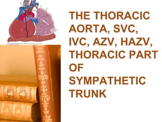
  • 1. 1 Thoracic Aorta THE THORACIC AORTA, SVC, IVC, AZV, HAZV, THORACIC PART OF SYMPATHETIC TRUNK
  • 4. 4 Thoracic Aorta Definitions ‫‏‬ Normal Dimensions – Mid-descending 26-28 mm ‫‏‬ Dilation (Ballooning, Bulging, Ectasia) ‫‏‬ Aneurysm – Types • Saccular • Fusiform – Definition • When the diameter exceeds 4 cm or diameter exceeds 1.5 times normal ‫‏‬ Dissection – Tear in vessel wall results in false lumen – Types • Type A – involves ascending aorta • Type B – involves descending aorta
  • 5. 5 Thoracic Aorta Normal Aortic Dimensions Hager A. et al.; J Thorac Cardiovasc Surg 2002;123:1060-1066
  • 6. Company Confidential ©2010 Genworth Financial, Inc. All rights reserved. ‫‏‬ THE AORTA AND ITS MAJOR BRANCHES ‫‏‬ The aorta commences at the aortic valve, above the vestibule of the left ventricle and terminates in the abdomen at the level of the fourth lumbar vertebra (L4), where it bifurcates to form the right and left common iliac arteries. It is an elastic artery and it is divisible into four parts, viz:-  The Ascending thoracic aorta.  The Arch of the aorta.  The Descending thoracic aorta and  The Abdominal aorta.
  • 7.
  • 8. Company Confidential ©2010 Genworth Financial, Inc. All rights reserved. ‫‏‬ ASCENDING THORACIC AORTA: ‫‏‬ This is located in the middle mediastinum and measures 5cm long. It has the following branches through which it supplies the heart: Right coronary artery. Left coronary artery
  • 9.
  • 10. Company Confidential ©2010 Genworth Financial, Inc. All rights reserved. ‫‏‬ THE ARCH OF THE AORTA: ‫‏‬ This is located in the superior mediastinum. ‫‏‬ It is the continuation of the ascending aorta. ‫‏‬ Its branches are: • The Brachiocephalic trunk. This gives rise to: Right Common Carotid artery. Right Subclavian artery. • Left Common Carotid artery. • Left Subclavian artery.
  • 11. Company Confidential ©2010 Genworth Financial, Inc. All rights reserved. ‫‏‬ ‫‏‬ Each common carotid artery: ‫‏‬ Bifurcates to give rise to an:  internal carotid artery and  external carotid artery. ‫‏‬ The internal carotid continues into the cranial cavity to supply cranial contents while the external carotid supplies the head and neck region.
  • 12. Company Confidential ©2010 Genworth Financial, Inc. All rights reserved. ‫‏‬ The Subclavian artery: ‫‏‬ The subclavian artery supplies structures in the head and neck and the thoracic regions through the following branches: Vertebral artery. The thyrocervical artery. The internal thoracic (Mammary) art. The costocervical artery The dorsal scapular artery ‫‏‬ Each Subclavian artery continues as the axillary artery at the outer margin (Lateral margin) of the first rib. ‫‏‬ The axillary artery is the main arterial supply to the upper limb.
  • 13. Company Confidential ©2010 Genworth Financial, Inc. All rights reserved. ‫‏‬ THE DESCENDING THORACIC AORTA ‫‏‬ This artery is the direct continuation of the arch of the aorta. ‫‏‬ It passes through the posterior mediastinum and terminates posterior to the median arcuate ligament of the diaphragm at the level of the 12th thoracic vertebra (T12). ‫‏‬ At this point it continues into the abdominal cavity as the Abdominal aorta.
  • 14. Company Confidential ©2010 Genworth Financial, Inc. All rights reserved. ‫‏‬ The branches of the Thoracic Aorta include: • 9 pairs of Posterior intercostal arteries. • A pair of subcostal arteries. • 2 left bronchial art. • 2 esophageal art. • Pericardial branches (Unknown number). • Mediastinal branches (Unknown number). • A pair of Superior Phrenic art. (Right & Left).
  • 15. Company Confidential ©2010 Genworth Financial, Inc. All rights reserved.
  • 16. Company Confidential ©2010 Genworth Financial, Inc. All rights reserved. ‫‏‬ THE ABDOMINAL AORTA ‫‏‬ This is the continuation of the descending thoracic aorta from the level of T12. ‫‏‬ It is located on the posterior abdominal wall and terminates at the level of L4. ‫‏‬ Here it bifurcates to give rise to the right and left Common iliac arteries.
  • 17. Company Confidential ©2010 Genworth Financial, Inc. All rights reserved. ‫‏‬ The branches of the Abdominal Aorta include: • Coeliac trunk. ( Foregut) • Superior mesenteric art. (Midgut) • Inferior mesenteric art. (Hindgut) • Median sacral art. (Posterior pelvic wall) • 2 Renal art. (The Kidneys) • 1 Middle suprarenal art. • 2 Testicular/Ovarian art. • 2 Inferior phrenic art. • 4 pairs of Lumbar art.
  • 18. Company Confidential ©2010 Genworth Financial, Inc. All rights reserved. ‫‏‬ Each of the Common iliac arteries will bifurcate at the level of the sacroiliac joint to give rise to Internal and External iliac arteries. ‫‏‬ The internal iliac artery will supply the pelvic, perineal(genital) and gluteal regions. ‫‏‬ The external iliac continues as the Femoral artery beyond the inguinal ligament to supply the entire lower limb.
  • 19. Company Confidential ©2010 Genworth Financial, Inc. All rights reserved.
  • 21. 21 Thoracic Aorta Aortic Aneurysm (A) Tomodensitometric and (B) echocardiographic views of an aortic root aneurysm. Nataf P , Lansac E Heart 2006;92:1345-1352
  • 22. 22 Thoracic Aorta Aortic Aneurysm http://www.medscape.com/viewarticle/406630_15 Figure 23. Atherosclerotic vascular dis-ease in an aortic aneurysm. Axial postcontrast image (window = 440, level = 40) reveals a large contrast collection projecting from the undersurface of the aortic arch, consistent with aneurysm (arrow). the low attention material within the aneurysm represents thrombus
  • 23. 23 Thoracic Aorta Aortic Aneurysm http://www.medscape.com/viewarticle/406630_15 Figure 24. Aortic aneurysm rupture. Axial postcontrast image (window = 440, level = 40) through the aortic arch reveals an aortic aneurysm with contrast penetrating the thrombus within the aneurysm (open arrow).
  • 25. 25 Thoracic Aorta Aortic Dissection http://www.medscape.com/viewarticle/406630_15 Figure 12. Stanford type B (Debakey Type III) aortic dissection: descending thoracic aorta. (A) Axial postcontrast image (window = 440, level = 40) reveals intimal flap (arrow), t = true lumen, f = false lumen. (B) Oblique sagittal reconstruction reveals complex nature of the intimal flap (arrows).
  • 26. 26 Thoracic Aorta Epidemiology ‫‏‬ Thoracic aneurysms – Prevalence greater than 3-4% of those over 65 – 6 cases per 100,000 person-years – Incidence increasing – In the top 15 causes of death – Thoracic aortic aneurysm – rupture 3.5/100,000 persons ‫‏‬ Thoracic aortic dissection – 2000 new cases/year – Acute - 3.5/100,000 persons – Male:Female ratio 2:1
  • 27. 27 Thoracic Aorta Etiologies ‫‏‬ Underlying Etiologies – Atherosclerosis – Marfan’s – Type IV Ehlers-Danlos – Infection (syphillis) – Arteritis (giant cell, Takayasu, Behcet’s) – Trauma ‫‏‬ Risk Factors – Smoking – COPD – HTN – Male gender – Older age – High BMI – Abnormal aortic valve (e.g., bicuspid valve) – Family history
  • 28. 28 Thoracic Aorta Presentation ‫‏‬ Aneurysm – Most asymptomatic – Superior vena cava syndrome – Hoarseness – Bronchial obstruction – Dysphagia – Hemoptysis – Paralysis/paraplegia – Lower extremity embolism ‫‏‬ Dissection – Chest/back/neck pain – Neurologic signs – Horner syndrome – Hoarseness – Acute aortic regurgitation
  • 29. 29 Thoracic Aorta Diagnosis ‫‏‬ Chest x-ray – Widened mediastinum ‫‏‬ Echocardiogram – Transthoracic – aortic root – Transesophageal – ascending and descending ‫‏‬ Aortography – Delineates the lumen ‫‏‬ CT scan – Most widely used diagnostic tool ‫‏‬ MRI – Avoids contrast dye
  • 30. 30 Thoracic Aorta Echocardiography Nataf P , Lansac E Heart 2006;92:1345-1352
  • 31. 31 Thoracic Aorta Natural History 0 5 10 15 20 25 <2.75 cm/m2 2.75-4.25 cm/m2 >4.25 cm/m2 Aortic Size Index (ASI) Annual Risk of Rupture http://emedicine.medscape.com/article/242904=print ASI = aortic dia (cm)/body surface area (m2)
  • 32. 32 Thoracic Aorta Natural History ‫‏‬ Yearly Rupture or Dissection Rates for Thoracic Aortic Aneurysms: Simple Prediction Based on Size – 304 patients; 58.9% male; median age 65.8 – Aneurysm size – 43.7% were 4.0-4.9 cm – Location – 72% ascending – Follow up – average 43.1 months – End points Davies RR, et al. Ann Thorac Surg 2002;73:17 44 Death alone 15 Dissection alone 5 Rupture alone 4 Rupture and death (no dissection) 5 Dissection, death (no rupture) 2 Dissection, rupture (no death) 2 Dissection, rupture and death No. Patients Events
  • 33. 33 Thoracic Aorta Natural History Davies RR, et al. Ann Thorac Surg 2002;73:17 Cumulative incidence of acute dissection or rupture as a function of initial aneurysm size.
  • 34. 34 Thoracic Aorta Natural History Davies RR, et al. Ann Thorac Surg 2002;73:17 Kaplan-Meier cumulative hazard function of rupture or dissection.
  • 35. 35 Thoracic Aorta Natural History Davies RR, et al. Ann Thorac Surg 2002;73:17 Average yearly rates of negative outcomes during the first 5 years after presentation
  • 36. 36 Thoracic Aorta Natural History Davies RR, et al. Ann Thorac Surg 2002;73:17 Kaplan-Meier cumulative hazard function of rupture.
  • 37. 37 Thoracic Aorta Natural History Kaplan-Meier cumulative survival before operative repair. Davies RR, et al. Ann Thorac Surg 2002;73:17
  • 38. 38 Thoracic Aorta Natural History Davies RR, et al. Ann Thorac Surg 2002;73:17 Kaplan-Meier cumulative survival
  • 39. 39 Thoracic Aorta Natural History Davies RR, et al. Ann Thorac Surg 2002;73:17 Kaplan-Meier cumulative survival
  • 40. 40 Thoracic Aorta Natural History Davies RR, et al. Ann Thorac Surg 2002;73:17 Kaplan-Meier cumulative survival
  • 41. 41 Thoracic Aorta Treatment - Aneurysm ‫‏‬ Medical – BP control – Smoking cessation – No heavy lifting ‫‏‬ Surgical – Dacron tube graft – Ascending – may need to replace valve – Arch – graft – Descending – graft, stent grafts
  • 42. 42 Thoracic Aorta Treatment - Dissection ‫‏‬ Type A – Surgical ‫‏‬ Type B – Medical – Surgical • Acute with rupture, leak or distal ischemia.
  • 43. 43 Thoracic Aorta Treatment – Indications for Intervention ‫‏‬ Aortic size – Ascending diameter >5.5 cm – Descending diameter >6.5 cm – Growth rate >1 cm/yr (avg ascending 0.07 cm/yr; descending 0.19 cm/yr) ‫‏‬ Symptomatic aneurysm ‫‏‬ Traumatic rupture ‫‏‬ Pseudoaneurysm ‫‏‬ Large saccular aneurysm ‫‏‬ Mycotic aneurysm ‫‏‬ Aortic coarctation ‫‏‬ Bronchial compression ‫‏‬ Aortobronchial or aortoesophageal fistuala
  • 44. 44 Thoracic Aorta Treatment - Surgical Composite valve and graft replacement. Nataf P , Lansac E Heart 2006;92:1345-1352
  • 45. 45 Thoracic Aorta Complications ‫‏‬ Bleeding ‫‏‬ CVA – 2-5% ‫‏‬ CHF ‫‏‬ Respiratory failure ‫‏‬ Graft leaks ‫‏‬ Fistula formation ‫‏‬ Spinal cord damage ‫‏‬ Renal failure
  • 46. 46 Thoracic Aorta Prognosis ‫‏‬ Aneurysm – Early post-op mortality 4-10%; lower for descending aneurysm repair; much higher for aortic arch repair – Stroke occurs 2-5% – Renal failure requiring dialysis – 7% – Spinal cord injury – 3% ‫‏‬ Dissection – Treated 10-yr survival rate 60% – Type A • 30% mortality surgical • 60% mortality medical – Type B • 10% mortality medical • 30% mortality surgical
  • 47. 47 Thoracic Aorta Underwriting Approach and Considerations ‫‏‬ Obtain cardiology and/or vascular medical records ‫‏‬ Review serial echos/scans as available ‫‏‬ Review blood pressure control ‫‏‬ Higher Risk – Aneurysm >5 cm – Poorly controlled blood pressure – Increase in size >0.5 cm/yr – Ongoing tobacco usage – Associated cardiovascular disease (CAD, PVD, carotid disease) – Non-atherosclerotic vascular disorders (Marfan’s, Ehlers-Danlos, etc) ‫‏‬ Lower Risk – Aneurysm <5 cm/stable/ well followed – Aneurysm repaired/stable
  • 48. 48 Thoracic Aorta Summary ‫‏‬ Thoracic aortic dilation/aneurysm fairly common with age ‫‏‬ Risk factors are traditional cardiovascular risk factors ‫‏‬ Most are asymptomatic ‫‏‬ Thoracic aortic rupture rare ‫‏‬ Thoracic dissection rare ‫‏‬ Ascending aorta most common site of aneurysm formation ‫‏‬ Low risk for aneurysms less than 4 cm In the end, it’s not what you call it………it’s size that matters! REMEMBER
  • 49. superior vena cava (SVC, also known as the cava or cva) ‫‏‬ The superior vena cava (SVC, also known as the cava or cva) is a short, but large diameter vein located in the anterior right superior mediastinum. Its latin name is related to its large pipe appearance in cadavers, 'cava' meaning 'hollow'. 49 Thoracic Aorta
  • 50. ‫‏‬ The superior vena cava is very important for the function of the cardiovascular system, since it largely contributes to the input of blood to the right atrium. Any hypertensive process in the right half of the heart or in the pulmonary circulation retrogradely affects both superior and inferior venae cavae. This is important since the veins are not adjusted to high pressures, which can result with forming an aneurysm or even rupture of the wall of the SVC. ‫‏‬ This article will discuss the anatomy and function of the superior vena cava. 50 Thoracic Aorta Key facts about the superior vena cavaTable quiz Source Brachiocephalic vein, azygos vein Draining area Upper half of the body (above the diaphragm) Drains to Right atrium of heart
  • 51. ‫‏‬ Contents ‫‏‬ Anatomy ‫‏‬ Function ‫‏‬ Clinical notes – Superior vena cava obstruction (SVCO) – Superior vena cava syndrome (SVCS) – Superior vena cava thrombosis – Superior vena cava aneurysm – Persistent left superior vena cava (PLSVC) ‫‏‬ Sources 51 Thoracic Aorta
  • 52. ‫‏‬ Anatomy ‫‏‬ Left brachiocephalic vein ‫‏‬ Vena brachiocephalica sinistra ‫‏‬ 1/3 ‫‏‬ Synonyms: Left innominate vein, Vena anonyma sinistra ‫‏‬ Embryologically, the SVC is formed by the left and right brachiocephalic veins (also known as the innominate veins) that also receive blood from the upper limbs, certain parts of the head, one being the eyes, and neck. ‫‏‬ There is no valve that divides the SVC from the right atrium, which conducts blood from right atrial and right ventricular contractions upwards into the internal jugular vein (seen as the jugular venous pressure) and sternocleidomastoid muscle. 52 Thoracic Aorta
  • 53. ‫‏‬ Positionally, the SVC begins behind the lower border of the 1st right costal cartilage and descends vertically behind the 2nd and 3rd intercostal spaces to drain into the right atrium at the level of the 3rd costal cartilage. Its lower half is covered by a fibrous pericardium, which is pierced by the SVC at the level of the 2nd costal cartilage. 53 Thoracic Aorta Function Superior vena cava coursing towards the right atrium of the heart, returning deoxygenated blood from the body. The SVC is one of the 2 large veins by which blood is returned from the body to the right side of the heart. After circulating through the body systemically, deoxygenated blood returns to the right atrium of the heart through either the SVC, which drains the upper body, or the inferior vena cava (IVC) that drains everything below the diaphragm.
  • 54. ‫‏‬ Clinical notes ‫‏‬ Superior vena cava obstruction (SVCO) ‫‏‬ This usually refers to a partial or complete obstruction of the SVC, often in the context of cancer (lung cancer, metastatic cancer, or lymphoma). Clinically this obstruction can lead to enlarged veins in the head and neck, and cause shortness of breath, cough, chest pain, and difficulty swallowing). ‫‏‬ A clinical test known as Pemberton’s sign can be performed to identify this condition. A positive Pemberton's sign is marked by facial congestion and cyanosis (and/or respiratory distress) after 1 minute of having the patient elevate both arms until they touch the sides of the face. This sign is indicative of superior vena cava syndrome, commonly the result of a mass in the mediastinum. 54 Thoracic Aorta
  • 55. ‫‏‬ Superior vena cava syndrome (SVCS) ‫‏‬ This syndrome refers to a group of symptoms caused by the obstruction of the SVC. More than 90% of the cases of SVC obstruction are caused by cancer, most commonly bronchogenic carcinoma, which includes small cell and non-small cell lung carcinoma, Burkitt’s lymphoma, lymphoblastic lymphomas, pre-T- cell lineage acute lymphoblastic leukemia, and other acute leukemias. ‫‏‬ Characteristic symptoms are edema of the arms and face, development of swollen collateral veins on the front of the chest wall, shortness of breath, difficulty swallowing, stridor, cough, and neurological symptoms (reduced alertness, etc. from edema in the brain or airway compromise). Again, Pemberton’s sign can be used to identify an SVCO. 55 Thoracic Aorta
  • 56. ‫‏‬ Superior vena cava thrombosis ‫‏‬ The thrombosis often occurs from a thrombus around a long- term central venous catheter (CVC), especially in cancer patients with permanent indwelling CVCs. CVC-related thrombosis is as high as 30% in adults. However, patients can be treated with thrombolytics or anticoagulants, or by removal of the catheter. ‫‏‬ Superior vena cava aneurysm ‫‏‬ Venous aneurysms arising from the mediastinal systemic veins are extremely rare, with the majority being fusiform (“spindle-shaped”) aneurysms that arise from the SVC. ‫‏‬ Persistent left superior vena cava (PLSVC) ‫‏‬ A PLSVC is an embryologic remnant that is the most common variation of the thoracic venous system, resulting from a failure to involute during embryologic development. 56 Thoracic Aorta
  • 57. The inferior vena cava (IVC) is the largest vein of the human body. It is located at the posterior abdominal wall on the right side of the aorta. The IVC’s function is to carry the venous blood from the lower limbs and abdominopelvic region to the heart. The inferior vena cava anatomy is essential due to the vein’s great drainage area, which also makes it a hot topic for anatomy exams. For that reason, this page will cover the IVC anatomy in a way that’s easy to read and understand. 57 Thoracic Aorta
  • 60. Key facts Table quiz Definition and functionThe vein that collects deoxygenated blood from the abdomen, pelvis and lower limbs and carries it to the right atrium of the heartSourceCommon iliac veins (L5)TributariesInferior Phrenic, right Suprarenal, Renal, right Testicular (gonadal), Lumbar, common Iliac and Hepatic veins Mnemonic: Portal System Returns To Liver In Humans Clinical relationsInferior vena cava thrombosis 60 Thoracic Aorta
  • 61. Anatomy Inferior vena cava Vena cava inferior 1/4 Synonyms: IVC The inferior vena cava arises from the confluence of the common iliac veins at the level of L5 vertebra, just inferior to the bifurcation of the abdominal aorta. It then ascends the posterior abdominal wall, to the right side of the aorta and the bodies of the L3-L5 vertebrae. After passing through its fossa on the posterior liver surface, the IVC enters the thorax by traversing the inferior vena caval foramen of the diaphragm. 61 Thoracic Aorta
  • 63. ‫‏‬ The tributaries of the IVC correspond to the branches of the abdominal aorta. Note that some professors will want you to know at which vertebral level the IVC gets its direct tributaries, so they are as follows: ‫‏‬ The direct tributaries are the inferior phrenic veins (T8), right suprarenal (L1), renal (L1), right testicular (gonadal) (L2), lumbar (L1-L5), common iliac (L5) and hepatic (T8). If you want an easy way to remember them just memorise the mnemonic ' Portal System Returns To Liver In Humans'. ‫‏‬ Left gonadal and left suprarenal renal veins drain first into the left renal vein ‫‏‬ The veins of the stomach, spleen, pancreas, small and large intestines first empty into the hepatic portal vein. The hepatic portal vein carries this blood to the liver to be processed and detoxified. Then, the blood reaches the IVC through the hepatic veins. ‫‏‬ The inferior vena cava communicates with the superior vena cava through the collateral vessels, which include the azygos vein, lumbar veins, and vertebral venous plexuses. 63 Thoracic Aorta
  • 65. ‫‏‬ Function ‫‏‬ The IVC’s function is to convey the blood from the abdomen, pelvis, and lower limbs to the right atrium of the heart. Additional IVC functions are noticeable during some health disturbances, such as hepatic portal vein obstruction or the obstruction of the IVC itself. ‫‏‬ Specialized vessels called the portocaval (portosystemic) anastomoses open if the hepatic portal vein is obstructed. The intestinal blood then bypasses the liver and empties into the IVC directly. In cases where the IVC is occluded, the collateral vessels to the superior vena cava open. ‫‏‬ Learn more about portocaval anastomoses with our article, then take this specially designed quiz to consolidate everything you’ve learned about the IVC and its tributaries. 65 Thoracic Aorta
  • 66. ‫‏‬ Inferior vena cava thrombosis ‫‏‬ Thrombosis of the inferior vena cava (IVCT) is a condition in which a blood clot (thrombus) impedes the blood flow through the IVC. The thrombus can be formed within the IVC itself, which is rare, or, more commonly, travel from the deep veins of the legs in a condition called deep venous thrombosis (DVT). ‫‏‬ IVC thrombosis may be caused by all the conditions that lead to venous stasis. These include congenital abnormalities of the IVC, immobilization, obesity, pregnancy, sedentary lifestyle, and tumors of surrounding organs. IVCT presents with symptoms of venous obstruction, such as pain and swelling of lower limbs and scrotum. IVCT is diagnosed by ultrasound, CT,and MRI. ‫‏‬ Depending on the severity of the condition, the IVCT can be treated by surgical removal of the thrombus, thrombolytic therapy, and anticoagulant therapy. The treatment is necessary to prevent disseminating of the thrombus into the pulmonary circulation (pulmonary thromboembolism), which is an urgent medical condition. ‫‏‬ Sources 66 Thoracic Aorta
  • 68. 68
  • 69. Veins of the thoracic wall ‫‏‬ The thoracic wall is drained anteriorly by the internal thoracic vein and posteriorly by the azygos system of veins 69
  • 70. ‫‏‬ The azygos system of veins serves as an important anastomotic pathway capable of returning venous blood from the lower part of the body to the heart if the inferior vena cava is blocked. 70
  • 71. ‫‏‬ The major veins in the system are: ‫‏‬ Azygos vein, on the right ‫‏‬ Hemiazygos vein and the accessory hemiazygos vein, on the left. 71
  • 72. ‫‏‬ Azygos vein ‫‏‬ The azygos vein arises opposite vertebra LI or LII ‫‏‬ It is formed by the union of the right ascending lumbar vein , the right subcostal vein and lumber azygos veins . 72
  • 73. 73
  • 74. ‫‏‬ In addition to the posterior intercostal veins, the azygos vein communicates with the vertebral venous plexuses. 74
  • 75. 75
  • 76. ‫‏‬ The azygos vein enters the thorax through the aortic hiatus of the diaphragm, or it enters through or posterior to the right crus of the diaphragm. 76
  • 77. ‫‏‬ It ascends through the posterior mediastinum, usually to the right of the thoracic duct. 77
  • 78. ‫‏‬ At approximately vertebral level TIV, it arches anteriorly, over the root of the right lung, to join the superior vena cava. 78
  • 79. 79
  • 80. ‫‏‬ Tributaries of the azygos vein ‫‏‬ Right superior intercostal vein ‫‏‬ 5th – 11th right posterior ICV ‫‏‬ Hemiazygos vein; ‫‏‬ Accessory hemiazygos vein ‫‏‬ Esophageal veins ‫‏‬ Mediastinal veins ‫‏‬ Pericardial veins ‫‏‬ Right bronchial veins 80
  • 81. Hemiazygos vein ‫‏‬ The hemiazygos vein (inferior hemiazygos vein) usually arises at the junction between the left ascending lumbar vein and the left subcostal vein . 81
  • 82. 82
  • 83. ‫‏‬ The hemiazygos vein usually enters the thorax through the left crus of the diaphragm, but may enter through the aortic hiatus. 83
  • 84. ‫‏‬ It ascends through the posterior mediastinum, on the left side, to approximately vertebral level TIX. ‫‏‬ At this point, it crosses the vertebral column, posterior to the thoracic aorta, esophagus, and thoracic duct, to enters the azygos vein. ‫‏‬ The hemiazygos vein receives the inferior three intercostal veins, the inferior oesophageal veins, and several small mediastinal branches. 84
  • 85. 85
  • 86. ‫‏‬ Accessory hemiazygos vein ‫‏‬ The accessory hemiazygos vein descends on the left side of upper thoracic vertebrae up to the vertebral level TVIII . At this point, it crosses the vertebral column to join the azygos vein, or ends in the hemiazygos vein, or has a connection to both veins. Usually, it also has a connection superiorly to the left superior intercostal vein. Vessels that drain into the accessory hemiazygos vein include: the fourth to eighth left posterior intercostal veins 86
  • 87. 87
  • 90. Sympathetic trunks ‫‏‬ The sympathetic trunks (sympathetic chain, gangliated cord) are a paired bundle of nerve fibers that run from the base of the skull to the coccyx.
  • 92.
  • 93. ‫‏‬ The sympathetic trunks are two parallel nerve cords extending on either side of the vertebral column from the base of the skull to the coccyx
  • 94. ‫‏‬ The ganglia and trunks are connected to adjacent spinal nerves by gray rami communicantes throughout the length of the sympathetic trunk and by white rami communicantes in the thoracic and upper lumbar parts of the trunk (T1 to L2).
  • 95. ‫‏‬ The whiter rami are afferent fibers to the ganglion and found only from T1 to L2 segments. These contain the preganglionic and afferent sensory fibers. ‫‏‬ The gray rami communicans are present in all segments. They are efferent fibers of the sympathetic ganglia. These contain the postganglionic fibers.
  • 96. ‫‏‬ Parts of sympathetic chain ‫‏‬ Cervical part ‫‏‬ Thoracic part ‫‏‬ lumber Part ‫‏‬ Sacral part
  • 98. Thoracic part of Sympathetic chain Cervical part of sympathetic chain Medial arcuate lig. of diaphragm Abdominal part of sympathetic chain Neck of 1st rib
  • 99. Company Confidential ©2010 Genworth Financial, Inc. All rights reserved. Sympathetic chain Medial arcuate ligament
  • 102. Branches - Rami communicants to corresponding intercostal nerves. - To autonomic plexuses in thorax. - To aorta. - To abdominal organs through
  • 103.
  • 104.
  • 105.
  • 106.
  • 108. Applied Anatomy 1- Spinal anesthesia at high level may lead to temporary drop of blood pressure. 2- In case of essential hypertension, cutting of sympathetic chain between T2 & T5 will lead to drop of blood pressure.
  • 109. Autonomic Plexes 1- Pulmonary Plexuses. 2-Oesophageal plexuses. 3- Cardiac plexuses (superficial and deep).
  • 111.
  • 112.
  • 114.
  • 116.
  • 117.
  • 119.
  • 120. Thoracic Lymph Drainage A- From Chest wall. B- From thoracic contents.
  • 121. A- From Chest Wall 1- Superficial lymph vessels: - From front of chest to pectoral lymph nodes, parasternal or lower deep cervical lymph nodes. - From back of chest to subscapular nodes.
  • 122. 2- Deep lymph vessels: A- From anterior chest wall to parasternal nodes. B- Posterior part of chest wall to intercostal lymph nodes (opposite neck of ribs). C- Four diaphragmatic nodes (anterior, posterior and 2 lateral) which drain diaphragm and liver.
  • 123. B- from Thoracic Contents 1- Anterior mediastinal nodes. 2- Posterior mediastinal nodes. 3- Nodes of lungs, bronchi and trachea.
  • 124. • Lymph vessels from: -Anterior mediastinal nodes. -Tracheal lymph nodes. -Parasternal lymph nodes. Will unit together to form broncho- medistinal lymph vessels which joins thoracic duct on left side and right lymphatic duct in right side.
  • 125. Deep lymph nodes of chest wall
  • 127.
  • 128.
  • 129.

Editor's Notes

  1. 1