Anatomy of the Rectum and Anal Canal
•Download as PPTX, PDF•
0 likes•46 views

The rectum is the lowest part of the gastrointestinal tract, extending from the sigmoid colon to the anal canal. It has two anteroposterior and three lateral curvatures and receives its blood supply from the inferior mesenteric artery. The anal canal extends from the anorectal junction to the anus. It contains two sphincter muscles - the internal anal sphincter, which is involuntary, and the external anal sphincter, which is voluntary.

Recommended
More Related Content
Similar to Anatomy of the Rectum and Anal Canal
Similar to Anatomy of the Rectum and Anal Canal (20)
More from nidhi sharma
More from nidhi sharma (20)
Recently uploaded
Recently uploaded (20)
Anatomy of the Rectum and Anal Canal
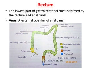
- 1. Rectum • The lowest part of gastrointestinal tract is formed by the rectum and anal canal • Anus external opening of anal canal (12 cm)
- 5. Course and direction • Downwards, backwards downwardsdownwards,forwards
- 6. Curvatures • Two anteroposterior • Three lateral Anteroposterior curves Lateral curves
- 8. Visceral relations • Anterior (male) Rectovesical pouch
- 10. • Posterior relation
- 11. Mucosal folds • Longitudinal folds transitory, lower part • Transverse folds/Houston’s valves/plicae transversales upper middle lower
- 12. Functional parts of rectum
- 13. Arterial supply Inferior mesenteric artery
- 14. • Superior rectal artery • Middle rectal arteries • Median sacral arteries supply the posterior wall of anorectal junction
- 15. Venous drainage
- 17. Lymphatic drainage • From upper half inferior mesenteric nodes • Lower half internal iliac nodes
- 18. Nerve supply • Sympathetic (L1, L2) Inhibitory to rectal musculature and motor to internal sphincter • Parasympathetic (S2, S3,S4)
- 19. Support of rectum • Pelvic floor • Fascia of Waldeyer • Lateral ligaments of rectum • Rectovesical fascia of Denonvilliers • Pelvic peritoneum • Perineal body
- 20. Clinical anatomy • Digital per rectum examination • Proctoscopy and sigmoidoscopy • Prolapse of rectum • Carcinoma
- 21. Anal canal • Terminal part of the large intestine • Situated below the level of pelvic diaphragm • 3.8 cm long • Extends from anorectal junction to the anus
- 22. Relations • Anterior • Posterior • Laterally ischiorectal fossae
- 23. • All round anal canal is surrounded by the sphincter muscles, the tone of which keeps the canal closed
- 24. Interior of anal canal (15mm) 8-10mm Of Morgagni
- 25. Interior of anal canal (15mm) 8-10mm Of Morgagni Upper part
- 26. Interior of anal canal (15mm) 8-10mm Of Morgagni • Middle part (pecten)
- 27. Interior of anal canal (15mm) 8-10mm Of Morgagni • lower part (cutaneous)
- 28. Anal sphincters • Internal anal sphincter involuntary • External anal sphincter voluntary
- 29. • Anorectal ring it is formed by the fusion of puborectalis , deep external sphincter and the internal sphincter • Rectal incontinence
- 30. Arterial supply • Above the pectinate line sup. Rectal art. • Below the pectinate line inf. Rectal art.
- 31. Venous drainage • Internal rectal venous plexus or haemorrhoidal plexus lies in the Submucosa above the pectinate line superior rectal vein • Veins present in the three anal columns at 3,7, and 11 o’clock position are large Primary internal piles
- 33. • External piles below the pectinate line • Painful • Do not bleed at stool
- 34. • External rectal venous plexus lies outside the muscular coat of the rectum and anal canal
- 35. Nerve supply • Above the pectinate line Symp. L1,L2 P.symp. S2,S3,S4 Below the pectinate line inferior rectal nerve (somatic) S2,S3,S4
- 36. • SPHINCTERS • Internal sphin. symp.( contraction) , p. symp. (relax) • External sphin. inferior rectal nerve, perineal branch of fourth sacral nerve