Bony anatomy of human body medical anatomy
•Download as PPTX, PDF•
0 likes•2 views

Bony anatomy.

Recommended
More Related Content
Similar to Bony anatomy of human body medical anatomy
Similar to Bony anatomy of human body medical anatomy (20)
More from nidhi sharma
More from nidhi sharma (20)
Recently uploaded
Recently uploaded (20)
Bony anatomy of human body medical anatomy
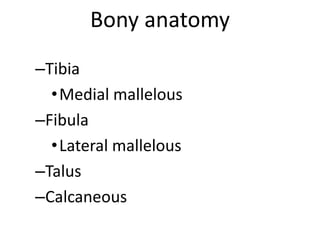
- 1. Bony anatomy –Tibia •Medial mallelous –Fibula •Lateral mallelous –Talus –Calcaneous
- 3. Soft Tissue Anatomy –Ligaments •Lateral aspect –Anterior tibiofibular –Posterior tibiofibular –Anterior talofibular –Posterior talofibular –Calcaneofibular
- 4. –Medial aspect • deltoid –Tendons • Achilles • Peroneus longus • Peroneus brevis • Anterior tibialis • Tibialis posterior • Flexor digitorium • Flexor hallicus longus
- 5. –Muscles •Gastrocnemius •Soleus •Tibialis anterior •Tibialis posterior •Flexor digitorium longus •Flexor hallicus longus •Peroneus longus •Peroneus brevis
- 7. Muscles That Move the Leg (Table 8.13, p.198) • These muscles connect the tibia or fibula to the femur or pelvic girdle. • The muscles either flex or extend the knee. • The muscles include the hamstring group and the quadriceps femoris group.
- 9. Gastrocnemius -is a very powerful superficial muscle .It runs from its two heads just above the knee to the heel, and is involved in standing, walking, running and jumping. Along with the soleus muscle it forms the calf muscle. Its function is plantar flexing the foot at the ankle joint and flexing the leg at the knee
- 10. . The Lateral Head originates from the Lateral Condyle of the femur, while the Medial Head originates from the Medial Condyle of the femur. Its other end forms a common tendon with the soleus muscle; this tendon is known as the calcaneal tendon or Achilles Tendon and inserts onto the posterior surface of the calcaneus, or mountain bone.
- 11. soleus • the soleus is a powerful muscle in the back part of the lower leg (the calf). It runs from just below the knee to the heel, and is involved in standing and walking. It is closely connected to the gastrocnemius muscle.
- 13. NERVE SUPPLY
- 15. Tibial nerve • The tibial nerve is a branch of the sciatic nerve. The tibial nerve passes through the popliteal fossa to pass below the arch of soleus. • In the popliteal fossa the nerve gives off branches to gastrocnemius, popliteus, soleus and plantaris muscles, an articular branch to the knee joint, and a cutaneous branch that will become the sural nerve. The sural nerve is joined by fibres from the common fibular nerve and runs down the calf to supply the lateral
- 16. • Lateral plantar nerve • The lateral plantar nerve supplies quadratus plantae, flexor digiti minimi, adductor hallucis, the interossei, three lumbricals. and abductor digiti minimi. Cutaneous innervation is to the lateral sole and lateral one and one half toes
- 17. BLOOD SUPPLY
- 18. posterior tibial artery • The posterior tibial artery –it is the large terminal branch of popliteal artery begins at the lower border of the Popliteus, opposite the interval between the tibia and fibula; it extends obliquely downward, and, as it descends, it approaches the tibial side of the leg, lying behind the tibia, • .
- 19. • and in the lower part of its course is situated midway between the medial malleolus and the medial process of the calcaneal tuberosity. Here it divides beneath the origin of the Adductor hallucis into the medial and lateral plantar arteries.
- 20. peroneal artery • In anatomy, the fibular artery (also known as the peroneal artery) supplies blood to the lateral compartment of the leg and is typically a branch of posterior tibial artery