SlideShare a Scribd company logo
1 of 25
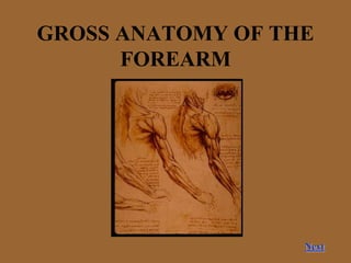
GROSS ANATOMY OF THE
FOREARM
Next
Main Menu
Bones of the forearm.
Superficial muscles of the anterior compartment.
Deep muscles of the anterior compartment.
Arteries and nerves of the anterior compartment.
Superficial muscles of the posterior compartment.
Deep muscles of the posterior compartment.
Arteries and nerves of the posterior compartment. Acknowledgements
Acknowledgements
• Supervision
Dr John Humpherson
• Anatomy Dissection Pictures
Gosling , Harris, Humpherson, Whitmore, Willan
Atlas of Human Anatomy, Gower Publishing, 1985
• Anatomy Drawings
Richard Neave & Medical Illustration
• Technical Help
Tony Bentley, Mike Mahon
Back
Bones of the forearm
The radius proximally articulates with the
humerus at the elbow joint. Distally it
articulates with the scaphoid and
lunate bones of the carpus, and with
the ulna at the distal radioulnar joint.
The ulna is the more medial of the two
bones. Its proximal end articulates with
the humerus at the elbow joint. Distally
it articulates with the radius. It is
excluded from the wrist joint by the
articular disc.
The interosseous membrane bind the
radius and the ulna together.
Head
Interosseous
membrane
Radius
Humerus
Tuberosity of
radius
Ulna
Head
Fig 1. Anterior view of bones of the forearm. Return to main menu
Superficial muscles of the anterior
compartment. Title page.
1
2
3
4
The superficial muscles of the anterior
compartment include pronator teres, flexor carpi
radialis, palmaris longus, and flexor carpi
ulnaris. Also included in this group is flexor
digitorum superficialis. The superficial group of
muscles all have the same origin, which is
attached to the medial epicondyle of the
humerus.
FOR FURTHER DETAILS CLICK ON EACH INDIVIDUAL
MUSCLE BELOW.
1. Pronator teres
2. Flexor carpi radialis
3. Flexor carpi ulnaris
4. Flexor digitorum superficialis
Palmaris longus (not included in the diagram).
Fig 2. Showing superficial muscles of the
posterior compartment.
Brachio-
radialis
Return to main menu
Pronator Teres
The pronator teres has two heads, the humeral head
and the ulnar head. The median nerve enters
the forearm between the two heads.
Origin :-
Humeral head- Medial epicondyle of the
humerus.
Ulnar head- Medial border of the coronoid
process of the ulna.
Insertion :-
Lateral aspect of the shaft of the radius.
Nerve supply :- Median nerve, C6 and C7.
Action :- Pronation of the forearm.
Flexion of the forearm.
Pronator teres
Radius
Fig 3. Pronator teres.
Return to title page
Flexor carpi radialis, palmaris longus, and flexor
carpi ulnaris
Flexor carpi radialis
Origin:- Medial epicondyle of the humerus.
Insertion:- Base of the second and third metacarpal
bones.
Nerve supply:- Median nerve, C6 and C7.
Action:- Flexes the hand at the wrist joint.
Abducts the hand at the wrist joint.
Palmaris longus
Origin:- Medial epicondyle of the humerus.
Insertion:- Flexor retinaculum and palmar aponeurosis.
Nerve supply:- Median nerve, C7 and C8
Action:- Flexes the hand at wrist joint.
Flexor carpi ulnaris.
Origin:-
Humeral head- Medial epicondyle of the humerus.
Ulnar head- Medial aspect of the olecranon process of the
ulna and the posterior border of the ulna.
Insertion:- Pisiform bone, hook of the hamate, and base
of the fifth metacarpal bone.
Nerve supply:- The ulnar nerve, C7, C8, and T1.
Action:- Flexes the hand at wrist joint.
Adducts the hand at wrist joint
Pronator
teres
Flexor
carpi
radialis
Palmaris
longus
Flexor
carpi
ulnaris
Fig 4. Flexor carpi radialis and ulnaris and palmaris longus
Return to title page
Flexor digitorum superficialis.
Origin:-
Humeroulnar head: Medial epicondyle of the humerus and
the medial margin of the coronoid process of the
ulna.
Radial head: Rising from the oblique line on the anterior
surface of the shaft of the radius.
Insertion:- The muscle belly gives rise to four tendons
distally. Each of the tendon attaches to the sides of
the middle phalanx of the four medial finger.
Nerve supply:- Median nerve, C8 and T1.
Action:- Flexes the middle phalanx of fingers.
Flexes the proximal phalanx of fingers (weak).
Flexes the wrist.
Flexor
digitorum
superficialis.
Fig 5. Flexor digitorum superficialis.
Return to title page
Deep Muscles of the anterior compartment.
Title page
The deep muscles of the forearm comprise
of the flexor pollicis longus, flexor
digitorum profundus and pronator
quadratus.
For further details click on each of the individual
muscles.
1. Flexor digitorum profundus.
2. Flexor pollicis longus.
Pronator quadratus not shown.
1
1
2
Brachio-
radialis
(cut)
Tendon of
carpi flexor
radialis (cut)
Fig 6. Deep muscles of the anterior
compartment.
Return to main
menu
Flexor digitorum profundus and flexor pollicis
longus
Flexor pollicis longus
Origin:- Middle of the anterior surface of the shaft of the
radius and from the adjoining part of the interosseous
membrane.
Insertion:- The tendon passes distally through the carpal
tunnel and attaches to the distal phalanx of the
thumb.
Nerve supply:- The anterior interosseous branch of the
median nerve.
Action:- Flexes the phalanges of the thumb.
Flexor digitorum profundus
Origin:- Upper three-quarters of the anteriomedial shaft of
the ulna.
Insertion:- The muscle divides into four tendons just prior
to traversing the carpal tunnel. They attach to the
distal phalanx of the four fingers.
Nerve supply:- The medial part is supplied by the ulnar
nerve, the lateral portion is supplied by the anterior
interosseous branch of the median nerve, C8 and T1.
Action:- Flexes the finger. Exclusive flexor of the distal
phalanx. Weak flexor of the wrist.
Flexor
pollicis
longus
Pronator
quadratus
Flexor
digitorum
profundus
Fig 7. Flexor digitorum profundus and flexor
pollicis longus.
Return to title page
Pronator Quadratus
Origin:- Lower quarter of the
anterior surface of the shaft of
the ulna.
Insertion:- Lower quarter of the
anterior surface of the shaft of
the radius.
Nerve supply:- The anterior
interosseous branch of the
median nerve.
Action:- Pronates the forearm.
Pronator
quadratus
Flexor
carpi
radialis
(cut)
Flexor
pollicis
longus Flexor
digitorum
profundus
Flexor
carpi
ulnaris
(cut)
Fig 8. Pronator quadratus.
Return to title
page
Arteries and Nerves of the anterior compartment.
Title page
Ulnar artery:- Larger than the radial artery. It
passes between the arch formed by the
radial and ulnar attachment of the flexor
digitorum superficialis and descends
through the anterior compartment. It enters
the palm of the hand in front of the flexor
retinaculum, and promptly divides in
superficial and deep palmer branches.
Branches-
Muscular branches- to the muscles of the anterior
compartment.
Recurrent branches- to the anastomosis around
the wrist joint
Branches to the anastomosis around the wrist joint
Common interosseous artery- arises in the upper
part of the ulnar artery and then divides in
the anterior and posterior interosseous
arteries.
Anterior interosseous artery:- arises from the
common interosseous artery and descends
in the anterior compartment to eventually
join the anastomosis around the wrist joint.
It supplies the deep flexor muscles, and
gives off nutrient branches to the the radius
and ulna.
Brachial
artery
Median
nerve
Ulnar
nerve
Radial
nerve
Ulnar
artery
Fig 9. Arteries and nerves of the
anterior compartment.
Next
Return to nerves
Arteries and Nerves of the anterior compartment.
Posterior interosseous artery- arises from the
common interosseous artery and and enters the
posterior compartment.
Radial artery:- It begins in the cubital fossa when the
brachial artery divides into the radial and ulna artery.
It passes distally, travels under the brachioradialis,
resting on the deep flexor muscles. The artery briefly
travels on the lateral side of the radius, before
travelling over the anterior surface of the radius. The
artery then winds around the lateral aspect of the
wrist, before entering the palm of the hand to form
the deep palmer arch.
Branches-
Muscular branches: to the neighbouring muscles.
Branches to the anastomosis around the wrist and elbow
joint.
Superficial palmer joint: arises just above the wrist,
frequently joins the ulnar artery to give rise to the
superficial palmer arch.
Radial
nerve
Superficial
radial nerve
Radial
artery
Brachial
artery
Median
nerve
Ulnar
nerve
Ulnar
artery
Median
nerve
Fig 10. Arteries and nerves of the
anterior compartment. Next
Return to nerves
Nerves of the anterior compartment
Median Nerve (fig 9, fig 10)
The median nerve leaves the cubital fossa in between the two heads of the pronator teres. It descends between the superficial and
deep flexor muscles. At the wrist it lies superficially, before entering the palm behind the flexor retinaculum.
Branches:-
Muscular branch:- all the superficial muscles of the anterior compartment except flexor carpi ulnaris.
Articular branches: to the elbow joint
Anterior interosseous nerve: arises from the median nerve as it emerges from the two heads of the pronator teres. (see below)
Palmer cutaneous Branch: Distributed to the skin over the lateral part of the palm.
Anterior interosseous nerve
It arises from the median nerve (see above) and then descends down the anterior surface of the interosseous membrane.
Branches-
Muscular branches: all the muscles of deep flexion in the anterior compartment except the medial part of digitorum profundus.
Articular branches: to the wrist joint, the distal radioulnar joint, and the joints of the carpus.
Ulnar Nerve(fig9, fig 10)
The ulnar nerve passes behind the medial epicondyle and enters the forearm between the two heads of flexor carpi ulnaris. It
descends between the flexor carpi ulnaris and the flexor digitorum profundus. The ulnar nerve becomes superficial at the wrist,
before entering the palm in front of the flexor retinaculum.
Branches-
Muscular branches: to the flexor carpi ulnaris and to the medial half of the flexor digitorum profundus.
Articular branches: to the elbow joint.
Palmar cutaneous branch: arises in the middle of the forearm, Supplies skin over the thenar eminence.
Dorsal cutaneous branch: arises in the distal third of the of the forearm, and passes medially to the dorsum of the hand.
Return to main menu
Superficial muscles of the posterior compartment.
Title page
The superficial muscles of the anterior compartment are mainly
concerned with the extension at wrist joint and of the digit. The
muscles in this group comprise of the Extensor carpi radialis brevis,
extensor digitorum, extensor digiti minimi, extensor carpi ulnaris,
and the two more lateral lying brachioradialis and extensor carpi
radialis longus.
NB- The anconeus also lies in the posterior compartment but is functionally
very different to the rest of the muscles in this group. Its action are
to aid the triceps in extension at the elbow joint.
Click below on each of the muscles for further detail
about each of the individual muscles.
1. Extensor carpi radialis longus
2. Extensor digitorum
3. Extensor carpi ulnaris
4. Extensor digiti minimi
Brachioradialis and extensor carpi brevis are not
included in the figure.
2
4
Anconeus
2
3
1
Fig 11. Superficial muscles of the posterior
compartment. Return to main menu
Extensor carpi radialis longus and brevis, and
extensor carpi ulnaris
Extensor carpi radialis longus
Origin:- From the lower third of the lateral supracondylar ridge of the
humerus.
Insertion:- Posterior surface of the base of the second metacarpal
bone.
Nerve supply:- Radial nerve, C6 and C7.
Action:- Extends and abducts the hand at the wrist joint.
Extensor carpi radialis brevis
Origin:- From the common tendon attached to the lateral epicondyle
of the humerus.
Insertion:- Posterior surface of the base of the third metacarpal bone.
Nerve supply:- Posterior interosseous nerve, C7 and C8.
Action:- Extends and abducts the hand at the wrist joint.
Extensor carpi ulnaris
Origin:- From the common tendon attached to the lateral epicondyle
of the humerus.
Insertion:- Posterior surface of the base of the fifth metacarpal bone.
Nerve supply:- Posterior interosseous nerve, C7 and C8
Action:- Extends and adducts the hand at wrist joint.
Extensor carpi
radialis longus
Extensor carpi
radialis brevis
Extensor
carpi ulnaris
Fig 12. Extensor carpi radialis brevis and
longus and extensor carpi ulnaris.
Return to title
page
Extensor digitorum and extensor digiti minimi
Extensor digitorum
Origin:- From the common tendon attached to the lateral
epicondyle of the humerus.
Insertion:- The muscle divides into four tendons which pass to
the fingers and form the dorsal expansion. On the dorsum
of the hand these are interconnected by fibrous tissue.
Near the proximal interphalangeal joint of each finger the
expansions divide into the central part, which inserts into
base of the middle phalanx, and the two lateral parts,
which insert into the base of the distal phalanx.
Nerve supply:- Posterior interosseous nerve, C7 and C8
Action:- Mainly it extends the metacarpophalangeal joint, but it
also assists in extending the proximal and distal
interphalangeal joint and the arm.
Extensor digiti minimi
Origin:- From the common tendon attached to the lateral
epicondyle of the humerus.
Insertion:- Via two tendons to the dorsal expansion for the
little finger.
Nerve supply:- Posterior interosseous nerve, C7 and C8
Action:- Assists in the extension of the little finger.
Extensor
digitorum
Extensor
digiti
minimi
Fig 13. Extensor digitorum and extensor
digiti minimi.
Return to title page
Brachioradialis
Origin:- From the upper twp thirds of
the lateral supracondylar ridge of
the humerus.
Insertion:- Base of the styloid process
of the radius.
Nerve supply:- Radial nerve, C5 and
C6.
Action:- It flexes the forearm (despite
being being served by an ‘extensor’
nerve), assists in rotating or
restoring the arm into midprone
position, depending on the initial
position.
Brachio-
radialis
Extensor carpi
radialis longus
Extensor carpi
radialis brevis
Flexor carpi
radialis
Fig 15. Brachioradialis (anterior
view).
Fig 14 Brachioradialis (lateral
view). Return to title page
Deep muscles in the posterior compartment. Title
page
The deep muscles of the fore arm consist
of three muscles that act on the thumb, the
extensor indicis and supinator. Except for
the supinator they all originate from only
the forearm bones.
For further details on each of the bones click on
the individual muscles.
1. Supinator.
2. Abductor pollicis longus.
3. Extensor pollicis longus.
4. Extensor indicis.
5. Extensor pollicis brevis.
1
2
4
3
5
Fig 16. Deep muscles of the posterior compartment.
Return to main menu
Extensor pollicis longus and brevis, and abductor
pollicis longus
Abductor pollicis longus
Origin:- Middle of the posterior surface of the shaft of the ulna and
radius.
Insertion:- Posterior surface of the base of the first metacarpal
bone.
Nerve supply:- The posterior interosseous nerve, C7 and C8.
Action:- Abducts and extends the thumb.
Extensor pollicis longus
Origin:- From the posterior surface of the ulna.
Insertion:- Posterior surface of the base of the distal phalanx of the
thumb.
Nerve supply:- The posterior interosseous nerve, C7 and C8.
Action:- Extends the thumb.
Extensor pollicis brevis
Origin:- Posterior surface of the radius.
Insertion:- Posterior surface of the proximal phalanx of the thumb
Nerve supply:- The posterior interosseous nerve, C7 and C8.
Action:- Extends the thumb.
Anatomical snuff box: medial boundary is the the tendon of the
extensor pollicis longus and the lateral boundary is the tendon
of the abductor pollicis longus and extensor pollicis brevis.
Abductor pollicis
longus.
Extensor
pollicis longus
Fig 17. Abductor pollicis longus, extensor
pollicis longus and brevis.
Extensor pollicis
brevis
Return to title page
Supinator
Supinator
The supinator consists of oblique
and a transverse head.
Origin:-
Oblique head: From the lateral
epicondyle of the humerus and
collateral ligament of the elbow
joint.
Transverse head: Supinator crest of
the ulna.
Insertion:- Both heads wind laterally
around the proximal part of the
radius, attaching to the anterior
aspect of the radius.
Nerve supply:- Posterior
interosseous nerve, C6 and C7.
Action:- Supinates the forearm
(mainly assists as the biceps
brachii is main supinator of the
forearm).
Transverse head
Oblique head
Fig 18. Supinator muscle
(anterior view). Return to title page
Extensor indicis
Origin:- Posterior surface of the
ulna.
Insertion:- Dorsal expansion of
the index finger
Nerve supply:- Posterior
interosseous nerve, C7 and
C8.
Action:- Extends the index
finger.
Return to title page
Extensor indicis
Fig 19. Extensor
pollicis
Arteries and nerves in the posterior
compartment. Title page
Arteries
Posterior interosseous artery:-
As discussed previously, in the cubital
fossa, the common interosseous artery
divides in the anterior and posterior
interosseous muscle. The posterior
interosseous artery lies between the
superficial and deep extensor muscles. It
terminates by anastomosing with the
anterior interosseous muscle and taking
part in the anastomosis around the wrist
joint.
Branches:
Muscular branch- to the neighbouring muscles.
Recurrent branch- takes part in the
anastomosis around the wrist joint.
Anterior interosseous artery (discussed
earlier):-
Enters the posterior compartment in the
distal third of the forearm. Supplies
branches to neighbouring muscles.
Radial artery (discussed earlier)
Has branches that supply the muscles in
the lateral aspect of compartment.
Next
Oblique head
of supinator (cut)
Posterior
interosseous
artery
Extensor
carpi ulnaris
Posterior
interosseous
nerve
Extensor carpi
radialis brevis
Fig 19. Posterior interosseous
artery and nerve.
Arteries and nerves in the posterior
compartment
Nerves
The posterior compartment of the arm is
supplied by branches of the radial nerve.
Posterior interosseous nerve (Deep branch):
It arises from the radial nerve just in front of the
lateral epicondyle of the humerus. It enters the
posterior compartment in between the two
heads of the supinator.
Branches:
Muscular branches: To most of the muscles of the
posterior compartment (see individual muscles
for detail).
Articular branches: To the elbow, wrist and carpal
joints.
Radial nerve:
Branches:
Muscular branches: supply the brachioradialis and
the extensor carpi radialis longus.
Articular branches: To the elbow joint.
Return to main menu
Posteror
interosseous
artery
Posteror
interosseous
nerve
Fig 20. Posteror interosseous
artery and nerve
THANK YOU

More Related Content

What's hot

Posterior compartment of leg
Posterior compartment of legPosterior compartment of leg
Posterior compartment of legAunum Iqbal
 
Front of forearm by vidya prashant
Front of forearm by vidya prashantFront of forearm by vidya prashant
Front of forearm by vidya prashantPrashant Bhavani
 
Anatomy of anterior compartment of forearm
Anatomy  of anterior compartment of forearmAnatomy  of anterior compartment of forearm
Anatomy of anterior compartment of forearmDr. Mohammad Mahmoud
 
venous & lymphatic drainage of upper limb
venous & lymphatic drainage of upper limbvenous & lymphatic drainage of upper limb
venous & lymphatic drainage of upper limbIallu Reddy
 
Anatomy of foot and ankle
Anatomy of foot and ankleAnatomy of foot and ankle
Anatomy of foot and ankleJunaid Ahmad
 
Hand anatomy 2017 new microsoft power point presentation
Hand anatomy 2017 new microsoft power point presentationHand anatomy 2017 new microsoft power point presentation
Hand anatomy 2017 new microsoft power point presentationessameahady
 
Atlas(C1) & Axis(C2) Vertebrae.pptx
Atlas(C1) & Axis(C2) Vertebrae.pptxAtlas(C1) & Axis(C2) Vertebrae.pptx
Atlas(C1) & Axis(C2) Vertebrae.pptxRahul Sharma
 
5 anterior forearm.ppt
5  anterior forearm.ppt5  anterior forearm.ppt
5 anterior forearm.pptmehrdad asgari
 
Anatomy of small joints of the foot
Anatomy of small joints of the footAnatomy of small joints of the foot
Anatomy of small joints of the footMohamed Ahmed Eladl
 
Arches of foot
Arches of foot  Arches of foot
Arches of foot drarunb4u
 
Anatomy of upper limb rehan
Anatomy of upper limb rehanAnatomy of upper limb rehan
Anatomy of upper limb rehanRehan Asad
 
Slideshow: Forearm Superficial Flexors
Slideshow: Forearm Superficial FlexorsSlideshow: Forearm Superficial Flexors
Slideshow: Forearm Superficial FlexorsThe Funky Professor
 

What's hot (20)

Posterior compartment of leg
Posterior compartment of legPosterior compartment of leg
Posterior compartment of leg
 
Front of forearm by vidya prashant
Front of forearm by vidya prashantFront of forearm by vidya prashant
Front of forearm by vidya prashant
 
Anatomy of anterior compartment of forearm
Anatomy  of anterior compartment of forearmAnatomy  of anterior compartment of forearm
Anatomy of anterior compartment of forearm
 
venous & lymphatic drainage of upper limb
venous & lymphatic drainage of upper limbvenous & lymphatic drainage of upper limb
venous & lymphatic drainage of upper limb
 
Anatomy of foot and ankle
Anatomy of foot and ankleAnatomy of foot and ankle
Anatomy of foot and ankle
 
Slideshow: Anterior Leg
Slideshow: Anterior LegSlideshow: Anterior Leg
Slideshow: Anterior Leg
 
Hand anatomy 2017 new microsoft power point presentation
Hand anatomy 2017 new microsoft power point presentationHand anatomy 2017 new microsoft power point presentation
Hand anatomy 2017 new microsoft power point presentation
 
Scapular region
Scapular region Scapular region
Scapular region
 
Hand anatomy
Hand anatomyHand anatomy
Hand anatomy
 
Atlas(C1) & Axis(C2) Vertebrae.pptx
Atlas(C1) & Axis(C2) Vertebrae.pptxAtlas(C1) & Axis(C2) Vertebrae.pptx
Atlas(C1) & Axis(C2) Vertebrae.pptx
 
5 anterior forearm.ppt
5  anterior forearm.ppt5  anterior forearm.ppt
5 anterior forearm.ppt
 
Anatomy of small joints of the foot
Anatomy of small joints of the footAnatomy of small joints of the foot
Anatomy of small joints of the foot
 
Wrist Joint
Wrist JointWrist Joint
Wrist Joint
 
Arches of foot
Arches of foot  Arches of foot
Arches of foot
 
The bones of the foot
The bones of the footThe bones of the foot
The bones of the foot
 
The wrist and hand
The wrist and handThe wrist and hand
The wrist and hand
 
Anatomy of upper limb rehan
Anatomy of upper limb rehanAnatomy of upper limb rehan
Anatomy of upper limb rehan
 
Slideshow: Forearm Superficial Flexors
Slideshow: Forearm Superficial FlexorsSlideshow: Forearm Superficial Flexors
Slideshow: Forearm Superficial Flexors
 
Slideshow: Forearm Deep Flexors
Slideshow: Forearm Deep FlexorsSlideshow: Forearm Deep Flexors
Slideshow: Forearm Deep Flexors
 
Vertebrae thoracic
Vertebrae thoracicVertebrae thoracic
Vertebrae thoracic
 

Similar to forearm.ppt

Anatomy of flexor compartment of forearm
Anatomy of  flexor compartment of forearm Anatomy of  flexor compartment of forearm
Anatomy of flexor compartment of forearm Shaifaly madan rustagi
 
Anatomy of Upper extremity
Anatomy of Upper extremityAnatomy of Upper extremity
Anatomy of Upper extremitySunil Pahari
 
Peripheral Nerves of Upper Limb
Peripheral Nerves of Upper LimbPeripheral Nerves of Upper Limb
Peripheral Nerves of Upper LimbSado Anatomist
 
RADIAL NERVE palsy.pptx
RADIAL NERVE palsy.pptxRADIAL NERVE palsy.pptx
RADIAL NERVE palsy.pptxhariramhalder
 
peripheral nerves of the upper limb - applied
peripheral nerves of the upper limb - appliedperipheral nerves of the upper limb - applied
peripheral nerves of the upper limb - appliedSumer Yadav
 
12peripheralnervesoftheupperlimb-120312033920-phpapp01-141228111052-conversio...
12peripheralnervesoftheupperlimb-120312033920-phpapp01-141228111052-conversio...12peripheralnervesoftheupperlimb-120312033920-phpapp01-141228111052-conversio...
12peripheralnervesoftheupperlimb-120312033920-phpapp01-141228111052-conversio...WdEaAlBoNy
 
4 arm region and cubital
4  arm region and cubital4  arm region and cubital
4 arm region and cubitalmehrdad asgari
 
UPPER LIMBS - WRIST AND HAND MAY 2022.pdf
UPPER LIMBS - WRIST AND HAND MAY 2022.pdfUPPER LIMBS - WRIST AND HAND MAY 2022.pdf
UPPER LIMBS - WRIST AND HAND MAY 2022.pdfManishaRao30
 
muscels of the upper Limb edited(1).pptx
muscels of the upper Limb edited(1).pptxmuscels of the upper Limb edited(1).pptx
muscels of the upper Limb edited(1).pptxPhebeLois1
 
Popliteal fossa & back of thigh
Popliteal fossa & back of thigh  Popliteal fossa & back of thigh
Popliteal fossa & back of thigh Prabhakar Yadav
 
The Hand Anatomy Part 1
The Hand Anatomy Part 1The Hand Anatomy Part 1
The Hand Anatomy Part 1nihattt
 

Similar to forearm.ppt (20)

Anatomy of flexor compartment of forearm
Anatomy of  flexor compartment of forearm Anatomy of  flexor compartment of forearm
Anatomy of flexor compartment of forearm
 
Anatomy of Upper extremity
Anatomy of Upper extremityAnatomy of Upper extremity
Anatomy of Upper extremity
 
Peripheral Nerves of Upper Limb
Peripheral Nerves of Upper LimbPeripheral Nerves of Upper Limb
Peripheral Nerves of Upper Limb
 
RADIAL NERVE palsy.pptx
RADIAL NERVE palsy.pptxRADIAL NERVE palsy.pptx
RADIAL NERVE palsy.pptx
 
peripheral nerve injuries
peripheral nerve injuriesperipheral nerve injuries
peripheral nerve injuries
 
13 ms Forearm.ppt
13 ms Forearm.ppt13 ms Forearm.ppt
13 ms Forearm.ppt
 
740_Hand.ppt
740_Hand.ppt740_Hand.ppt
740_Hand.ppt
 
peripheral nerves of the upper limb - applied
peripheral nerves of the upper limb - appliedperipheral nerves of the upper limb - applied
peripheral nerves of the upper limb - applied
 
Median nerve
Median nerveMedian nerve
Median nerve
 
12peripheralnervesoftheupperlimb-120312033920-phpapp01-141228111052-conversio...
12peripheralnervesoftheupperlimb-120312033920-phpapp01-141228111052-conversio...12peripheralnervesoftheupperlimb-120312033920-phpapp01-141228111052-conversio...
12peripheralnervesoftheupperlimb-120312033920-phpapp01-141228111052-conversio...
 
4 arm region and cubital
4  arm region and cubital4  arm region and cubital
4 arm region and cubital
 
Nil
NilNil
Nil
 
UPPER LIMBS - WRIST AND HAND MAY 2022.pdf
UPPER LIMBS - WRIST AND HAND MAY 2022.pdfUPPER LIMBS - WRIST AND HAND MAY 2022.pdf
UPPER LIMBS - WRIST AND HAND MAY 2022.pdf
 
muscels of the upper Limb edited(1).pptx
muscels of the upper Limb edited(1).pptxmuscels of the upper Limb edited(1).pptx
muscels of the upper Limb edited(1).pptx
 
Forearm muscles
Forearm musclesForearm muscles
Forearm muscles
 
Muscles of human body
Muscles of human bodyMuscles of human body
Muscles of human body
 
Popliteal fossa & back of thigh
Popliteal fossa & back of thigh  Popliteal fossa & back of thigh
Popliteal fossa & back of thigh
 
The Hand Anatomy Part 1
The Hand Anatomy Part 1The Hand Anatomy Part 1
The Hand Anatomy Part 1
 
Radialnerve.pptx
Radialnerve.pptxRadialnerve.pptx
Radialnerve.pptx
 
elbow joint .ppt
elbow joint .pptelbow joint .ppt
elbow joint .ppt
 

More from nidhi sharma

RADIOLOGY/Imaging of The Small Intestine
RADIOLOGY/Imaging of The Small IntestineRADIOLOGY/Imaging of The Small Intestine
RADIOLOGY/Imaging of The Small Intestinenidhi sharma
 
Male and Female Reproductive Systems 2024 jan. pptx human medical anatomy hum...
Male and Female Reproductive Systems 2024 jan. pptx human medical anatomy hum...Male and Female Reproductive Systems 2024 jan. pptx human medical anatomy hum...
Male and Female Reproductive Systems 2024 jan. pptx human medical anatomy hum...nidhi sharma
 
Prostate gland.pptx human anatomy human anatomy
Prostate gland.pptx  human anatomy  human anatomyProstate gland.pptx  human anatomy  human anatomy
Prostate gland.pptx human anatomy human anatomynidhi sharma
 
NEURON.pptx human medical anatomy medical science
NEURON.pptx human medical anatomy medical scienceNEURON.pptx human medical anatomy medical science
NEURON.pptx human medical anatomy medical sciencenidhi sharma
 
hepatobiliary system 2024.pptx hepatobiliary system
hepatobiliary system 2024.pptx hepatobiliary systemhepatobiliary system 2024.pptx hepatobiliary system
hepatobiliary system 2024.pptx hepatobiliary systemnidhi sharma
 
CELL COMPONENTS.pptx human body anatomy medical human anatomy
CELL COMPONENTS.pptx human body anatomy medical human anatomyCELL COMPONENTS.pptx human body anatomy medical human anatomy
CELL COMPONENTS.pptx human body anatomy medical human anatomynidhi sharma
 
BILIARY APPARATUS 2024.pptx human anatomy
BILIARY APPARATUS 2024.pptx human anatomyBILIARY APPARATUS 2024.pptx human anatomy
BILIARY APPARATUS 2024.pptx human anatomynidhi sharma
 
Human Male reproductive system human bdy
Human Male reproductive system human bdyHuman Male reproductive system human bdy
Human Male reproductive system human bdynidhi sharma
 
Bony anatomy of human body medical anatomy
Bony anatomy of human body medical anatomyBony anatomy of human body medical anatomy
Bony anatomy of human body medical anatomynidhi sharma
 
RADIO-ULNAR JOINT IS THE JOINT PRESENT IN HUMAN UPPER LIMB
RADIO-ULNAR JOINT IS THE JOINT PRESENT IN HUMAN UPPER LIMBRADIO-ULNAR JOINT IS THE JOINT PRESENT IN HUMAN UPPER LIMB
RADIO-ULNAR JOINT IS THE JOINT PRESENT IN HUMAN UPPER LIMBnidhi sharma
 
RADIO-ULNAR JOINT IS THE JOINT OF UPPER LIMB IN HUMAN BODY
RADIO-ULNAR JOINT IS THE JOINT OF UPPER LIMB IN HUMAN BODYRADIO-ULNAR JOINT IS THE JOINT OF UPPER LIMB IN HUMAN BODY
RADIO-ULNAR JOINT IS THE JOINT OF UPPER LIMB IN HUMAN BODYnidhi sharma
 
VENOUS DRAINAGE OF FACE.pptx
VENOUS DRAINAGE OF FACE.pptxVENOUS DRAINAGE OF FACE.pptx
VENOUS DRAINAGE OF FACE.pptxnidhi sharma
 
AORTA SVC IVC AZV HAZV TTHRCSMPTHC 2022.ppt
AORTA SVC IVC AZV HAZV TTHRCSMPTHC 2022.pptAORTA SVC IVC AZV HAZV TTHRCSMPTHC 2022.ppt
AORTA SVC IVC AZV HAZV TTHRCSMPTHC 2022.pptnidhi sharma
 
the_digestive_system
the_digestive_systemthe_digestive_system
the_digestive_systemnidhi sharma
 
triangles of neck.ppt
triangles of neck.ppttriangles of neck.ppt
triangles of neck.pptnidhi sharma
 

More from nidhi sharma (20)

RADIOLOGY/Imaging of The Small Intestine
RADIOLOGY/Imaging of The Small IntestineRADIOLOGY/Imaging of The Small Intestine
RADIOLOGY/Imaging of The Small Intestine
 
Male and Female Reproductive Systems 2024 jan. pptx human medical anatomy hum...
Male and Female Reproductive Systems 2024 jan. pptx human medical anatomy hum...Male and Female Reproductive Systems 2024 jan. pptx human medical anatomy hum...
Male and Female Reproductive Systems 2024 jan. pptx human medical anatomy hum...
 
Prostate gland.pptx human anatomy human anatomy
Prostate gland.pptx  human anatomy  human anatomyProstate gland.pptx  human anatomy  human anatomy
Prostate gland.pptx human anatomy human anatomy
 
NEURON.pptx human medical anatomy medical science
NEURON.pptx human medical anatomy medical scienceNEURON.pptx human medical anatomy medical science
NEURON.pptx human medical anatomy medical science
 
hepatobiliary system 2024.pptx hepatobiliary system
hepatobiliary system 2024.pptx hepatobiliary systemhepatobiliary system 2024.pptx hepatobiliary system
hepatobiliary system 2024.pptx hepatobiliary system
 
CELL COMPONENTS.pptx human body anatomy medical human anatomy
CELL COMPONENTS.pptx human body anatomy medical human anatomyCELL COMPONENTS.pptx human body anatomy medical human anatomy
CELL COMPONENTS.pptx human body anatomy medical human anatomy
 
BILIARY APPARATUS 2024.pptx human anatomy
BILIARY APPARATUS 2024.pptx human anatomyBILIARY APPARATUS 2024.pptx human anatomy
BILIARY APPARATUS 2024.pptx human anatomy
 
Human Male reproductive system human bdy
Human Male reproductive system human bdyHuman Male reproductive system human bdy
Human Male reproductive system human bdy
 
Bony anatomy of human body medical anatomy
Bony anatomy of human body medical anatomyBony anatomy of human body medical anatomy
Bony anatomy of human body medical anatomy
 
RADIO-ULNAR JOINT IS THE JOINT PRESENT IN HUMAN UPPER LIMB
RADIO-ULNAR JOINT IS THE JOINT PRESENT IN HUMAN UPPER LIMBRADIO-ULNAR JOINT IS THE JOINT PRESENT IN HUMAN UPPER LIMB
RADIO-ULNAR JOINT IS THE JOINT PRESENT IN HUMAN UPPER LIMB
 
RADIO-ULNAR JOINT IS THE JOINT OF UPPER LIMB IN HUMAN BODY
RADIO-ULNAR JOINT IS THE JOINT OF UPPER LIMB IN HUMAN BODYRADIO-ULNAR JOINT IS THE JOINT OF UPPER LIMB IN HUMAN BODY
RADIO-ULNAR JOINT IS THE JOINT OF UPPER LIMB IN HUMAN BODY
 
ULNAR NERVE.pptx
ULNAR NERVE.pptxULNAR NERVE.pptx
ULNAR NERVE.pptx
 
thorax.ppt
thorax.pptthorax.ppt
thorax.ppt
 
VENOUS DRAINAGE OF FACE.pptx
VENOUS DRAINAGE OF FACE.pptxVENOUS DRAINAGE OF FACE.pptx
VENOUS DRAINAGE OF FACE.pptx
 
AORTA SVC IVC AZV HAZV TTHRCSMPTHC 2022.ppt
AORTA SVC IVC AZV HAZV TTHRCSMPTHC 2022.pptAORTA SVC IVC AZV HAZV TTHRCSMPTHC 2022.ppt
AORTA SVC IVC AZV HAZV TTHRCSMPTHC 2022.ppt
 
the_digestive_system
the_digestive_systemthe_digestive_system
the_digestive_system
 
Rotator cuff.pptx
Rotator cuff.pptxRotator cuff.pptx
Rotator cuff.pptx
 
triangles of neck.ppt
triangles of neck.ppttriangles of neck.ppt
triangles of neck.ppt
 
Rectum.pptx
Rectum.pptxRectum.pptx
Rectum.pptx
 
Skin
SkinSkin
Skin
 

Recently uploaded

Capitol Tech U Doctoral Presentation - April 2024.pptx
Capitol Tech U Doctoral Presentation - April 2024.pptxCapitol Tech U Doctoral Presentation - April 2024.pptx
Capitol Tech U Doctoral Presentation - April 2024.pptxCapitolTechU
 
Difference Between Search & Browse Methods in Odoo 17
Difference Between Search & Browse Methods in Odoo 17Difference Between Search & Browse Methods in Odoo 17
Difference Between Search & Browse Methods in Odoo 17Celine George
 
Painted Grey Ware.pptx, PGW Culture of India
Painted Grey Ware.pptx, PGW Culture of IndiaPainted Grey Ware.pptx, PGW Culture of India
Painted Grey Ware.pptx, PGW Culture of IndiaVirag Sontakke
 
ENGLISH 7_Q4_LESSON 2_ Employing a Variety of Strategies for Effective Interp...
ENGLISH 7_Q4_LESSON 2_ Employing a Variety of Strategies for Effective Interp...ENGLISH 7_Q4_LESSON 2_ Employing a Variety of Strategies for Effective Interp...
ENGLISH 7_Q4_LESSON 2_ Employing a Variety of Strategies for Effective Interp...JhezDiaz1
 
AmericanHighSchoolsprezentacijaoskolama.
AmericanHighSchoolsprezentacijaoskolama.AmericanHighSchoolsprezentacijaoskolama.
AmericanHighSchoolsprezentacijaoskolama.arsicmarija21
 
Introduction to AI in Higher Education_draft.pptx
Introduction to AI in Higher Education_draft.pptxIntroduction to AI in Higher Education_draft.pptx
Introduction to AI in Higher Education_draft.pptxpboyjonauth
 
Like-prefer-love -hate+verb+ing & silent letters & citizenship text.pdf
Like-prefer-love -hate+verb+ing & silent letters & citizenship text.pdfLike-prefer-love -hate+verb+ing & silent letters & citizenship text.pdf
Like-prefer-love -hate+verb+ing & silent letters & citizenship text.pdfMr Bounab Samir
 
Enzyme, Pharmaceutical Aids, Miscellaneous Last Part of Chapter no 5th.pdf
Enzyme, Pharmaceutical Aids, Miscellaneous Last Part of Chapter no 5th.pdfEnzyme, Pharmaceutical Aids, Miscellaneous Last Part of Chapter no 5th.pdf
Enzyme, Pharmaceutical Aids, Miscellaneous Last Part of Chapter no 5th.pdfSumit Tiwari
 
call girls in Kamla Market (DELHI) 🔝 >༒9953330565🔝 genuine Escort Service 🔝✔️✔️
call girls in Kamla Market (DELHI) 🔝 >༒9953330565🔝 genuine Escort Service 🔝✔️✔️call girls in Kamla Market (DELHI) 🔝 >༒9953330565🔝 genuine Escort Service 🔝✔️✔️
call girls in Kamla Market (DELHI) 🔝 >༒9953330565🔝 genuine Escort Service 🔝✔️✔️9953056974 Low Rate Call Girls In Saket, Delhi NCR
 
Full Stack Web Development Course for Beginners
Full Stack Web Development Course  for BeginnersFull Stack Web Development Course  for Beginners
Full Stack Web Development Course for BeginnersSabitha Banu
 
Historical philosophical, theoretical, and legal foundations of special and i...
Historical philosophical, theoretical, and legal foundations of special and i...Historical philosophical, theoretical, and legal foundations of special and i...
Historical philosophical, theoretical, and legal foundations of special and i...jaredbarbolino94
 
POINT- BIOCHEMISTRY SEM 2 ENZYMES UNIT 5.pptx
POINT- BIOCHEMISTRY SEM 2 ENZYMES UNIT 5.pptxPOINT- BIOCHEMISTRY SEM 2 ENZYMES UNIT 5.pptx
POINT- BIOCHEMISTRY SEM 2 ENZYMES UNIT 5.pptxSayali Powar
 
Meghan Sutherland In Media Res Media Component
Meghan Sutherland In Media Res Media ComponentMeghan Sutherland In Media Res Media Component
Meghan Sutherland In Media Res Media ComponentInMediaRes1
 
Employee wellbeing at the workplace.pptx
Employee wellbeing at the workplace.pptxEmployee wellbeing at the workplace.pptx
Employee wellbeing at the workplace.pptxNirmalaLoungPoorunde1
 
DATA STRUCTURE AND ALGORITHM for beginners
DATA STRUCTURE AND ALGORITHM for beginnersDATA STRUCTURE AND ALGORITHM for beginners
DATA STRUCTURE AND ALGORITHM for beginnersSabitha Banu
 

Recently uploaded (20)

Capitol Tech U Doctoral Presentation - April 2024.pptx
Capitol Tech U Doctoral Presentation - April 2024.pptxCapitol Tech U Doctoral Presentation - April 2024.pptx
Capitol Tech U Doctoral Presentation - April 2024.pptx
 
Difference Between Search & Browse Methods in Odoo 17
Difference Between Search & Browse Methods in Odoo 17Difference Between Search & Browse Methods in Odoo 17
Difference Between Search & Browse Methods in Odoo 17
 
9953330565 Low Rate Call Girls In Rohini Delhi NCR
9953330565 Low Rate Call Girls In Rohini  Delhi NCR9953330565 Low Rate Call Girls In Rohini  Delhi NCR
9953330565 Low Rate Call Girls In Rohini Delhi NCR
 
Painted Grey Ware.pptx, PGW Culture of India
Painted Grey Ware.pptx, PGW Culture of IndiaPainted Grey Ware.pptx, PGW Culture of India
Painted Grey Ware.pptx, PGW Culture of India
 
ESSENTIAL of (CS/IT/IS) class 06 (database)
ESSENTIAL of (CS/IT/IS) class 06 (database)ESSENTIAL of (CS/IT/IS) class 06 (database)
ESSENTIAL of (CS/IT/IS) class 06 (database)
 
Model Call Girl in Tilak Nagar Delhi reach out to us at 🔝9953056974🔝
Model Call Girl in Tilak Nagar Delhi reach out to us at 🔝9953056974🔝Model Call Girl in Tilak Nagar Delhi reach out to us at 🔝9953056974🔝
Model Call Girl in Tilak Nagar Delhi reach out to us at 🔝9953056974🔝
 
ENGLISH 7_Q4_LESSON 2_ Employing a Variety of Strategies for Effective Interp...
ENGLISH 7_Q4_LESSON 2_ Employing a Variety of Strategies for Effective Interp...ENGLISH 7_Q4_LESSON 2_ Employing a Variety of Strategies for Effective Interp...
ENGLISH 7_Q4_LESSON 2_ Employing a Variety of Strategies for Effective Interp...
 
AmericanHighSchoolsprezentacijaoskolama.
AmericanHighSchoolsprezentacijaoskolama.AmericanHighSchoolsprezentacijaoskolama.
AmericanHighSchoolsprezentacijaoskolama.
 
Introduction to AI in Higher Education_draft.pptx
Introduction to AI in Higher Education_draft.pptxIntroduction to AI in Higher Education_draft.pptx
Introduction to AI in Higher Education_draft.pptx
 
Model Call Girl in Bikash Puri Delhi reach out to us at 🔝9953056974🔝
Model Call Girl in Bikash Puri  Delhi reach out to us at 🔝9953056974🔝Model Call Girl in Bikash Puri  Delhi reach out to us at 🔝9953056974🔝
Model Call Girl in Bikash Puri Delhi reach out to us at 🔝9953056974🔝
 
Like-prefer-love -hate+verb+ing & silent letters & citizenship text.pdf
Like-prefer-love -hate+verb+ing & silent letters & citizenship text.pdfLike-prefer-love -hate+verb+ing & silent letters & citizenship text.pdf
Like-prefer-love -hate+verb+ing & silent letters & citizenship text.pdf
 
Enzyme, Pharmaceutical Aids, Miscellaneous Last Part of Chapter no 5th.pdf
Enzyme, Pharmaceutical Aids, Miscellaneous Last Part of Chapter no 5th.pdfEnzyme, Pharmaceutical Aids, Miscellaneous Last Part of Chapter no 5th.pdf
Enzyme, Pharmaceutical Aids, Miscellaneous Last Part of Chapter no 5th.pdf
 
call girls in Kamla Market (DELHI) 🔝 >༒9953330565🔝 genuine Escort Service 🔝✔️✔️
call girls in Kamla Market (DELHI) 🔝 >༒9953330565🔝 genuine Escort Service 🔝✔️✔️call girls in Kamla Market (DELHI) 🔝 >༒9953330565🔝 genuine Escort Service 🔝✔️✔️
call girls in Kamla Market (DELHI) 🔝 >༒9953330565🔝 genuine Escort Service 🔝✔️✔️
 
Full Stack Web Development Course for Beginners
Full Stack Web Development Course  for BeginnersFull Stack Web Development Course  for Beginners
Full Stack Web Development Course for Beginners
 
Historical philosophical, theoretical, and legal foundations of special and i...
Historical philosophical, theoretical, and legal foundations of special and i...Historical philosophical, theoretical, and legal foundations of special and i...
Historical philosophical, theoretical, and legal foundations of special and i...
 
POINT- BIOCHEMISTRY SEM 2 ENZYMES UNIT 5.pptx
POINT- BIOCHEMISTRY SEM 2 ENZYMES UNIT 5.pptxPOINT- BIOCHEMISTRY SEM 2 ENZYMES UNIT 5.pptx
POINT- BIOCHEMISTRY SEM 2 ENZYMES UNIT 5.pptx
 
Meghan Sutherland In Media Res Media Component
Meghan Sutherland In Media Res Media ComponentMeghan Sutherland In Media Res Media Component
Meghan Sutherland In Media Res Media Component
 
OS-operating systems- ch04 (Threads) ...
OS-operating systems- ch04 (Threads) ...OS-operating systems- ch04 (Threads) ...
OS-operating systems- ch04 (Threads) ...
 
Employee wellbeing at the workplace.pptx
Employee wellbeing at the workplace.pptxEmployee wellbeing at the workplace.pptx
Employee wellbeing at the workplace.pptx
 
DATA STRUCTURE AND ALGORITHM for beginners
DATA STRUCTURE AND ALGORITHM for beginnersDATA STRUCTURE AND ALGORITHM for beginners
DATA STRUCTURE AND ALGORITHM for beginners
 

forearm.ppt

  • 1. GROSS ANATOMY OF THE FOREARM Next
  • 2. Main Menu Bones of the forearm. Superficial muscles of the anterior compartment. Deep muscles of the anterior compartment. Arteries and nerves of the anterior compartment. Superficial muscles of the posterior compartment. Deep muscles of the posterior compartment. Arteries and nerves of the posterior compartment. Acknowledgements
  • 3. Acknowledgements • Supervision Dr John Humpherson • Anatomy Dissection Pictures Gosling , Harris, Humpherson, Whitmore, Willan Atlas of Human Anatomy, Gower Publishing, 1985 • Anatomy Drawings Richard Neave & Medical Illustration • Technical Help Tony Bentley, Mike Mahon Back
  • 4. Bones of the forearm The radius proximally articulates with the humerus at the elbow joint. Distally it articulates with the scaphoid and lunate bones of the carpus, and with the ulna at the distal radioulnar joint. The ulna is the more medial of the two bones. Its proximal end articulates with the humerus at the elbow joint. Distally it articulates with the radius. It is excluded from the wrist joint by the articular disc. The interosseous membrane bind the radius and the ulna together. Head Interosseous membrane Radius Humerus Tuberosity of radius Ulna Head Fig 1. Anterior view of bones of the forearm. Return to main menu
  • 5. Superficial muscles of the anterior compartment. Title page. 1 2 3 4 The superficial muscles of the anterior compartment include pronator teres, flexor carpi radialis, palmaris longus, and flexor carpi ulnaris. Also included in this group is flexor digitorum superficialis. The superficial group of muscles all have the same origin, which is attached to the medial epicondyle of the humerus. FOR FURTHER DETAILS CLICK ON EACH INDIVIDUAL MUSCLE BELOW. 1. Pronator teres 2. Flexor carpi radialis 3. Flexor carpi ulnaris 4. Flexor digitorum superficialis Palmaris longus (not included in the diagram). Fig 2. Showing superficial muscles of the posterior compartment. Brachio- radialis Return to main menu
  • 6. Pronator Teres The pronator teres has two heads, the humeral head and the ulnar head. The median nerve enters the forearm between the two heads. Origin :- Humeral head- Medial epicondyle of the humerus. Ulnar head- Medial border of the coronoid process of the ulna. Insertion :- Lateral aspect of the shaft of the radius. Nerve supply :- Median nerve, C6 and C7. Action :- Pronation of the forearm. Flexion of the forearm. Pronator teres Radius Fig 3. Pronator teres. Return to title page
  • 7. Flexor carpi radialis, palmaris longus, and flexor carpi ulnaris Flexor carpi radialis Origin:- Medial epicondyle of the humerus. Insertion:- Base of the second and third metacarpal bones. Nerve supply:- Median nerve, C6 and C7. Action:- Flexes the hand at the wrist joint. Abducts the hand at the wrist joint. Palmaris longus Origin:- Medial epicondyle of the humerus. Insertion:- Flexor retinaculum and palmar aponeurosis. Nerve supply:- Median nerve, C7 and C8 Action:- Flexes the hand at wrist joint. Flexor carpi ulnaris. Origin:- Humeral head- Medial epicondyle of the humerus. Ulnar head- Medial aspect of the olecranon process of the ulna and the posterior border of the ulna. Insertion:- Pisiform bone, hook of the hamate, and base of the fifth metacarpal bone. Nerve supply:- The ulnar nerve, C7, C8, and T1. Action:- Flexes the hand at wrist joint. Adducts the hand at wrist joint Pronator teres Flexor carpi radialis Palmaris longus Flexor carpi ulnaris Fig 4. Flexor carpi radialis and ulnaris and palmaris longus Return to title page
  • 8. Flexor digitorum superficialis. Origin:- Humeroulnar head: Medial epicondyle of the humerus and the medial margin of the coronoid process of the ulna. Radial head: Rising from the oblique line on the anterior surface of the shaft of the radius. Insertion:- The muscle belly gives rise to four tendons distally. Each of the tendon attaches to the sides of the middle phalanx of the four medial finger. Nerve supply:- Median nerve, C8 and T1. Action:- Flexes the middle phalanx of fingers. Flexes the proximal phalanx of fingers (weak). Flexes the wrist. Flexor digitorum superficialis. Fig 5. Flexor digitorum superficialis. Return to title page
  • 9. Deep Muscles of the anterior compartment. Title page The deep muscles of the forearm comprise of the flexor pollicis longus, flexor digitorum profundus and pronator quadratus. For further details click on each of the individual muscles. 1. Flexor digitorum profundus. 2. Flexor pollicis longus. Pronator quadratus not shown. 1 1 2 Brachio- radialis (cut) Tendon of carpi flexor radialis (cut) Fig 6. Deep muscles of the anterior compartment. Return to main menu
  • 10. Flexor digitorum profundus and flexor pollicis longus Flexor pollicis longus Origin:- Middle of the anterior surface of the shaft of the radius and from the adjoining part of the interosseous membrane. Insertion:- The tendon passes distally through the carpal tunnel and attaches to the distal phalanx of the thumb. Nerve supply:- The anterior interosseous branch of the median nerve. Action:- Flexes the phalanges of the thumb. Flexor digitorum profundus Origin:- Upper three-quarters of the anteriomedial shaft of the ulna. Insertion:- The muscle divides into four tendons just prior to traversing the carpal tunnel. They attach to the distal phalanx of the four fingers. Nerve supply:- The medial part is supplied by the ulnar nerve, the lateral portion is supplied by the anterior interosseous branch of the median nerve, C8 and T1. Action:- Flexes the finger. Exclusive flexor of the distal phalanx. Weak flexor of the wrist. Flexor pollicis longus Pronator quadratus Flexor digitorum profundus Fig 7. Flexor digitorum profundus and flexor pollicis longus. Return to title page
  • 11. Pronator Quadratus Origin:- Lower quarter of the anterior surface of the shaft of the ulna. Insertion:- Lower quarter of the anterior surface of the shaft of the radius. Nerve supply:- The anterior interosseous branch of the median nerve. Action:- Pronates the forearm. Pronator quadratus Flexor carpi radialis (cut) Flexor pollicis longus Flexor digitorum profundus Flexor carpi ulnaris (cut) Fig 8. Pronator quadratus. Return to title page
  • 12. Arteries and Nerves of the anterior compartment. Title page Ulnar artery:- Larger than the radial artery. It passes between the arch formed by the radial and ulnar attachment of the flexor digitorum superficialis and descends through the anterior compartment. It enters the palm of the hand in front of the flexor retinaculum, and promptly divides in superficial and deep palmer branches. Branches- Muscular branches- to the muscles of the anterior compartment. Recurrent branches- to the anastomosis around the wrist joint Branches to the anastomosis around the wrist joint Common interosseous artery- arises in the upper part of the ulnar artery and then divides in the anterior and posterior interosseous arteries. Anterior interosseous artery:- arises from the common interosseous artery and descends in the anterior compartment to eventually join the anastomosis around the wrist joint. It supplies the deep flexor muscles, and gives off nutrient branches to the the radius and ulna. Brachial artery Median nerve Ulnar nerve Radial nerve Ulnar artery Fig 9. Arteries and nerves of the anterior compartment. Next Return to nerves
  • 13. Arteries and Nerves of the anterior compartment. Posterior interosseous artery- arises from the common interosseous artery and and enters the posterior compartment. Radial artery:- It begins in the cubital fossa when the brachial artery divides into the radial and ulna artery. It passes distally, travels under the brachioradialis, resting on the deep flexor muscles. The artery briefly travels on the lateral side of the radius, before travelling over the anterior surface of the radius. The artery then winds around the lateral aspect of the wrist, before entering the palm of the hand to form the deep palmer arch. Branches- Muscular branches: to the neighbouring muscles. Branches to the anastomosis around the wrist and elbow joint. Superficial palmer joint: arises just above the wrist, frequently joins the ulnar artery to give rise to the superficial palmer arch. Radial nerve Superficial radial nerve Radial artery Brachial artery Median nerve Ulnar nerve Ulnar artery Median nerve Fig 10. Arteries and nerves of the anterior compartment. Next Return to nerves
  • 14. Nerves of the anterior compartment Median Nerve (fig 9, fig 10) The median nerve leaves the cubital fossa in between the two heads of the pronator teres. It descends between the superficial and deep flexor muscles. At the wrist it lies superficially, before entering the palm behind the flexor retinaculum. Branches:- Muscular branch:- all the superficial muscles of the anterior compartment except flexor carpi ulnaris. Articular branches: to the elbow joint Anterior interosseous nerve: arises from the median nerve as it emerges from the two heads of the pronator teres. (see below) Palmer cutaneous Branch: Distributed to the skin over the lateral part of the palm. Anterior interosseous nerve It arises from the median nerve (see above) and then descends down the anterior surface of the interosseous membrane. Branches- Muscular branches: all the muscles of deep flexion in the anterior compartment except the medial part of digitorum profundus. Articular branches: to the wrist joint, the distal radioulnar joint, and the joints of the carpus. Ulnar Nerve(fig9, fig 10) The ulnar nerve passes behind the medial epicondyle and enters the forearm between the two heads of flexor carpi ulnaris. It descends between the flexor carpi ulnaris and the flexor digitorum profundus. The ulnar nerve becomes superficial at the wrist, before entering the palm in front of the flexor retinaculum. Branches- Muscular branches: to the flexor carpi ulnaris and to the medial half of the flexor digitorum profundus. Articular branches: to the elbow joint. Palmar cutaneous branch: arises in the middle of the forearm, Supplies skin over the thenar eminence. Dorsal cutaneous branch: arises in the distal third of the of the forearm, and passes medially to the dorsum of the hand. Return to main menu
  • 15. Superficial muscles of the posterior compartment. Title page The superficial muscles of the anterior compartment are mainly concerned with the extension at wrist joint and of the digit. The muscles in this group comprise of the Extensor carpi radialis brevis, extensor digitorum, extensor digiti minimi, extensor carpi ulnaris, and the two more lateral lying brachioradialis and extensor carpi radialis longus. NB- The anconeus also lies in the posterior compartment but is functionally very different to the rest of the muscles in this group. Its action are to aid the triceps in extension at the elbow joint. Click below on each of the muscles for further detail about each of the individual muscles. 1. Extensor carpi radialis longus 2. Extensor digitorum 3. Extensor carpi ulnaris 4. Extensor digiti minimi Brachioradialis and extensor carpi brevis are not included in the figure. 2 4 Anconeus 2 3 1 Fig 11. Superficial muscles of the posterior compartment. Return to main menu
  • 16. Extensor carpi radialis longus and brevis, and extensor carpi ulnaris Extensor carpi radialis longus Origin:- From the lower third of the lateral supracondylar ridge of the humerus. Insertion:- Posterior surface of the base of the second metacarpal bone. Nerve supply:- Radial nerve, C6 and C7. Action:- Extends and abducts the hand at the wrist joint. Extensor carpi radialis brevis Origin:- From the common tendon attached to the lateral epicondyle of the humerus. Insertion:- Posterior surface of the base of the third metacarpal bone. Nerve supply:- Posterior interosseous nerve, C7 and C8. Action:- Extends and abducts the hand at the wrist joint. Extensor carpi ulnaris Origin:- From the common tendon attached to the lateral epicondyle of the humerus. Insertion:- Posterior surface of the base of the fifth metacarpal bone. Nerve supply:- Posterior interosseous nerve, C7 and C8 Action:- Extends and adducts the hand at wrist joint. Extensor carpi radialis longus Extensor carpi radialis brevis Extensor carpi ulnaris Fig 12. Extensor carpi radialis brevis and longus and extensor carpi ulnaris. Return to title page
  • 17. Extensor digitorum and extensor digiti minimi Extensor digitorum Origin:- From the common tendon attached to the lateral epicondyle of the humerus. Insertion:- The muscle divides into four tendons which pass to the fingers and form the dorsal expansion. On the dorsum of the hand these are interconnected by fibrous tissue. Near the proximal interphalangeal joint of each finger the expansions divide into the central part, which inserts into base of the middle phalanx, and the two lateral parts, which insert into the base of the distal phalanx. Nerve supply:- Posterior interosseous nerve, C7 and C8 Action:- Mainly it extends the metacarpophalangeal joint, but it also assists in extending the proximal and distal interphalangeal joint and the arm. Extensor digiti minimi Origin:- From the common tendon attached to the lateral epicondyle of the humerus. Insertion:- Via two tendons to the dorsal expansion for the little finger. Nerve supply:- Posterior interosseous nerve, C7 and C8 Action:- Assists in the extension of the little finger. Extensor digitorum Extensor digiti minimi Fig 13. Extensor digitorum and extensor digiti minimi. Return to title page
  • 18. Brachioradialis Origin:- From the upper twp thirds of the lateral supracondylar ridge of the humerus. Insertion:- Base of the styloid process of the radius. Nerve supply:- Radial nerve, C5 and C6. Action:- It flexes the forearm (despite being being served by an ‘extensor’ nerve), assists in rotating or restoring the arm into midprone position, depending on the initial position. Brachio- radialis Extensor carpi radialis longus Extensor carpi radialis brevis Flexor carpi radialis Fig 15. Brachioradialis (anterior view). Fig 14 Brachioradialis (lateral view). Return to title page
  • 19. Deep muscles in the posterior compartment. Title page The deep muscles of the fore arm consist of three muscles that act on the thumb, the extensor indicis and supinator. Except for the supinator they all originate from only the forearm bones. For further details on each of the bones click on the individual muscles. 1. Supinator. 2. Abductor pollicis longus. 3. Extensor pollicis longus. 4. Extensor indicis. 5. Extensor pollicis brevis. 1 2 4 3 5 Fig 16. Deep muscles of the posterior compartment. Return to main menu
  • 20. Extensor pollicis longus and brevis, and abductor pollicis longus Abductor pollicis longus Origin:- Middle of the posterior surface of the shaft of the ulna and radius. Insertion:- Posterior surface of the base of the first metacarpal bone. Nerve supply:- The posterior interosseous nerve, C7 and C8. Action:- Abducts and extends the thumb. Extensor pollicis longus Origin:- From the posterior surface of the ulna. Insertion:- Posterior surface of the base of the distal phalanx of the thumb. Nerve supply:- The posterior interosseous nerve, C7 and C8. Action:- Extends the thumb. Extensor pollicis brevis Origin:- Posterior surface of the radius. Insertion:- Posterior surface of the proximal phalanx of the thumb Nerve supply:- The posterior interosseous nerve, C7 and C8. Action:- Extends the thumb. Anatomical snuff box: medial boundary is the the tendon of the extensor pollicis longus and the lateral boundary is the tendon of the abductor pollicis longus and extensor pollicis brevis. Abductor pollicis longus. Extensor pollicis longus Fig 17. Abductor pollicis longus, extensor pollicis longus and brevis. Extensor pollicis brevis Return to title page
  • 21. Supinator Supinator The supinator consists of oblique and a transverse head. Origin:- Oblique head: From the lateral epicondyle of the humerus and collateral ligament of the elbow joint. Transverse head: Supinator crest of the ulna. Insertion:- Both heads wind laterally around the proximal part of the radius, attaching to the anterior aspect of the radius. Nerve supply:- Posterior interosseous nerve, C6 and C7. Action:- Supinates the forearm (mainly assists as the biceps brachii is main supinator of the forearm). Transverse head Oblique head Fig 18. Supinator muscle (anterior view). Return to title page
  • 22. Extensor indicis Origin:- Posterior surface of the ulna. Insertion:- Dorsal expansion of the index finger Nerve supply:- Posterior interosseous nerve, C7 and C8. Action:- Extends the index finger. Return to title page Extensor indicis Fig 19. Extensor pollicis
  • 23. Arteries and nerves in the posterior compartment. Title page Arteries Posterior interosseous artery:- As discussed previously, in the cubital fossa, the common interosseous artery divides in the anterior and posterior interosseous muscle. The posterior interosseous artery lies between the superficial and deep extensor muscles. It terminates by anastomosing with the anterior interosseous muscle and taking part in the anastomosis around the wrist joint. Branches: Muscular branch- to the neighbouring muscles. Recurrent branch- takes part in the anastomosis around the wrist joint. Anterior interosseous artery (discussed earlier):- Enters the posterior compartment in the distal third of the forearm. Supplies branches to neighbouring muscles. Radial artery (discussed earlier) Has branches that supply the muscles in the lateral aspect of compartment. Next Oblique head of supinator (cut) Posterior interosseous artery Extensor carpi ulnaris Posterior interosseous nerve Extensor carpi radialis brevis Fig 19. Posterior interosseous artery and nerve.
  • 24. Arteries and nerves in the posterior compartment Nerves The posterior compartment of the arm is supplied by branches of the radial nerve. Posterior interosseous nerve (Deep branch): It arises from the radial nerve just in front of the lateral epicondyle of the humerus. It enters the posterior compartment in between the two heads of the supinator. Branches: Muscular branches: To most of the muscles of the posterior compartment (see individual muscles for detail). Articular branches: To the elbow, wrist and carpal joints. Radial nerve: Branches: Muscular branches: supply the brachioradialis and the extensor carpi radialis longus. Articular branches: To the elbow joint. Return to main menu Posteror interosseous artery Posteror interosseous nerve Fig 20. Posteror interosseous artery and nerve