Download to read offline


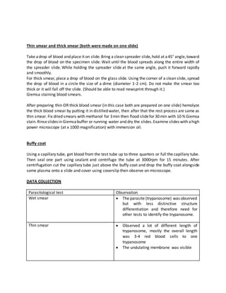


Trypanosomes are microscopic unicellular parasites that infect various hosts. The document discusses diagnosing trypanosomes from a blood sample using different tests. Thin blood smears stained with Giemsa revealed trypanosomes around 3-4 red blood cells in length with visible undulating membranes. Observation of moving trypanosomes in the buffy coat confirmed the presence of the parasites. The tests identified the parasite as a trypanosome based on its morphology seen under the microscope.


![Trypanosoma [1]](https://cdn.slidesharecdn.com/ss_thumbnails/trypanosomaseminar1-170312074241-thumbnail.jpg?width=640&height=640&fit=bounds)


