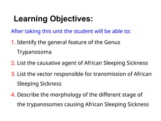
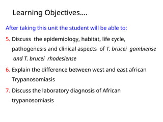
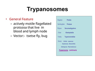
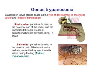
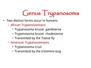
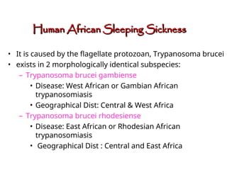
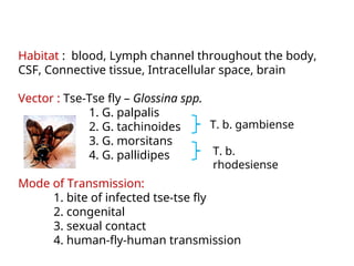
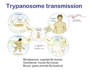
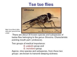
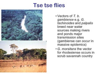
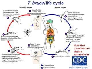
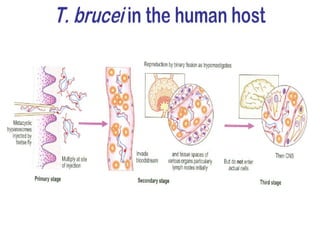
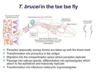
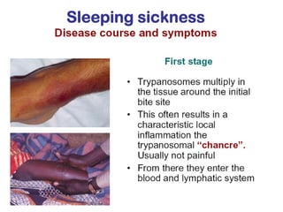
Learning Objectives:
After taking this unit the student will be able to:
1. Identify the general feature of the Genus
Trypanosoma
2. List the causative agent of African Sleeping Sickness
3. List the vector responsible for transmission of African
Sleeping Sickness
4. Describe the morphology of the different stage of
the trypanosomes causing African Sleeping Sickness
Learning Objectives….
After taking this unit the student will be able to:
5. Discuss the epidemiology, habitat, life cycle,
pathogenesis and clinical aspects of T. brucei gambiense
and T. brucei rhodesiense
6. Explain the difference between west and east african
Trypanosomiasis
7. Discuss the laboratory diagnosis of African
trypanosomiasis
Outline
• Common feature of the Genus trypanosoma
• Classification of the Genus trypanosoma
• Trypanosoma brucei gambiense and Trypanosoma
brucei rhodesiense
– Epidemiology
– Morphology
– Transmission and life cycle
– Pathology and clinical manifestation
• Laboratory diagnosis
– Microscopy
– Immunological assay
Trypanosomes
• General Feature
– actively motile flagellated
protozoa that live in
blood and lymph node
– Vector:- tsetse fly, bug
Classified in to two groups based on the type of development in the insect
vector and mode of transmission
• Two distinct forms occur in humans
– African Trypanosomiasis
• Trypanosoma brucei gambiense
• Trypanosoma brucei rhodesiense
• Transmited by the Tsetse fly
– American Trypanosomiasis
• Trypanosoma cruzi
• Transmited by the tritomine bug
Genus Trypanosoma
African Trypanosomiasis
• It is caused by the flagellate protozoan, Trypanosoma brucei
• exists in 2 morphologically identical subspecies:
– Trypanosoma brucei gambiense
• Disease: West African or Gambian African
trypanosomiasis
• Geographical Dist: Central & West Africa
– Trypanosoma brucei rhodesiense
• Disease: East African or Rhodesian African
trypanosomiasis
• Geographical Dist : Central and East Africa
Human African Sleeping Sickness
confined to tropical Africa between 15°N and 20°S,
Morphology
– spindle – shaped elongated body
(polymorphic -from long slender to short and blunt)
– Measuring 14-33 u X 1.5-3.5 u
– nucleus centrally located with karyosome
– kinetoplast found at the posterior end of the body
– undulating membrane originating from the
blepharoplast
– Anterior flagellum runs along the edge of undulating
membrane
– volutin granules scattered in the cytoplasm
2 stages of development of trypanosoma brucei infection in stained
preparation
1. Trypomastigote
- seen among vertebrate host
(human, others like dog, goat and
cattle)
- can be seen in the blood stream, CSF
and lymphatics
2. Epimastigote
- seen among invertebrate host
(tse-tse fly)
Habitat : blood, Lymph channel throughout the body,
CSF, Connective tissue, Intracellular space, brain
Vector : Tse-Tse fly – Glossina spp.
1. G. palpalis
2. G. tachinoides
3. G. morsitans
4. G. pallidipes
Mode of Transmission:
1. bite of infected tse-tse fly
2. congenital
3. sexual contact
4. human-fly-human transmission
T. b. gambiense
T. b.
rhodesiense
Laboratory diagnosis
• A definitive diagnosis requires detection of trypanosomes
– In blood,
– Lymph nodes,
– CSF,
– Skin chancre aspirates, or
– Bone marrow.
• Empiric treatment with subsequent symptomatic
improvement is the usual diagnosis in areas where
diagnostic studies are not readily available.
• Lymph node aspirate
– Is commonly used as a rapid test for trypanosomes at a high dry
magnification (X 400).
– It requires immediate search for parasites because they are
mobile for only 15-20 minutes.
• Blood smear
– A wet smear of unstained blood for mobile trypanosomes, again
for 15-20 minutes
– Giemsa-stained thick smear (more sensitive)
– wright and leishman stains are inadequate.
– Better assays
– Hematocrit centrifugation technique for buffy coat examination,
– Miniature anion-exchange centrifugation technique, which filters
out the red cells but not the trypanosomes.
• Chancre aspirate
– Can be used as a wet preparation, especially in the east african
form of the disease, but a blood smear is more sensitive.
• CSF assay
– Lumbar puncture should always be performed in
patients with parasitemia or lymphadenopathy.
– The double centrifugation technique is the most
sensitive method to detect the trypanosomes.
– Other CSF findings include elevated WBC count,
elevated IgM, and elevated total protein levels.
A: Trypansoma brucei sp. in thick blood smears stained with Giemsa.
B: Trypanosoma brucei sp. in thin blood smears stained with Giemsa
A B
C: Trypanosoma brucei sp. in thin blood smears stained with Wright-Giemsa.
D: Trypanosoma brucei sp. in a thin blood smear stained with Giemsa
• Other Tests:
Serologic antibody detection- Field diagnosis
– The standard serologic assay to diagnose West
African trypanosomiasis is the card agglutination test
for trypanosomiasis (CATT).
– The CATT
• can be conducted in the field without electricity
• results are available in only 10 minutes.
• It is highly sensitive (96%) but less specific because of cross-
reactivity with animal trypanosomes.
– Commercial antibody tests for Eastern African
trypanosomiasis are not available.
• Other tests developed but not frequently used clinically
include:
• antibody detection in the CSF and enzyme-linked
immunoassay (ELISA), polymerase chain reaction
(PCR).
• General
– In African trypanosomiasis, the most significant
abnormalities are anemia, hypergammaglobulinemia,
elevated erythrocyte sedimentation rate (ESR),
thrombocytopenia, and hypoalbuminemia, but not
eosinophilia or abnormal liver function.
– In the West African form of the disease, the total
immunoglobulin M (IgM) level is notably higher in
blood and CSF (along with high CSF protein).
Human African Trypanosomiasis, Summury
West African East African
Agent: T. brucei gambiense T. brucei rhodesiense
Vector: Riverine tsetse fly savanna tsetse fly
Distribution: west/central africa east/south africa
Reservoir: human antelope/cattle
Disease: chronic (years) rapid progression: 1-4
weeks
Mortality: 70% 100%
At risk: rural persons rural, visitors to game
reserves

Trypanosomes presented by DavidManyiel.ppt

  • 3.
    Learning Objectives: After takingthis unit the student will be able to: 1. Identify the general feature of the Genus Trypanosoma 2. List the causative agent of African Sleeping Sickness 3. List the vector responsible for transmission of African Sleeping Sickness 4. Describe the morphology of the different stage of the trypanosomes causing African Sleeping Sickness
  • 4.
    Learning Objectives…. After takingthis unit the student will be able to: 5. Discuss the epidemiology, habitat, life cycle, pathogenesis and clinical aspects of T. brucei gambiense and T. brucei rhodesiense 6. Explain the difference between west and east african Trypanosomiasis 7. Discuss the laboratory diagnosis of African trypanosomiasis
  • 5.
    Outline • Common featureof the Genus trypanosoma • Classification of the Genus trypanosoma • Trypanosoma brucei gambiense and Trypanosoma brucei rhodesiense – Epidemiology – Morphology – Transmission and life cycle – Pathology and clinical manifestation • Laboratory diagnosis – Microscopy – Immunological assay
  • 6.
    Trypanosomes • General Feature –actively motile flagellated protozoa that live in blood and lymph node – Vector:- tsetse fly, bug
  • 7.
    Classified in totwo groups based on the type of development in the insect vector and mode of transmission
  • 8.
    • Two distinctforms occur in humans – African Trypanosomiasis • Trypanosoma brucei gambiense • Trypanosoma brucei rhodesiense • Transmited by the Tsetse fly – American Trypanosomiasis • Trypanosoma cruzi • Transmited by the tritomine bug Genus Trypanosoma
  • 9.
  • 12.
    • It iscaused by the flagellate protozoan, Trypanosoma brucei • exists in 2 morphologically identical subspecies: – Trypanosoma brucei gambiense • Disease: West African or Gambian African trypanosomiasis • Geographical Dist: Central & West Africa – Trypanosoma brucei rhodesiense • Disease: East African or Rhodesian African trypanosomiasis • Geographical Dist : Central and East Africa Human African Sleeping Sickness Human African Sleeping Sickness
  • 13.
    confined to tropicalAfrica between 15°N and 20°S,
  • 14.
    Morphology – spindle –shaped elongated body (polymorphic -from long slender to short and blunt) – Measuring 14-33 u X 1.5-3.5 u – nucleus centrally located with karyosome – kinetoplast found at the posterior end of the body – undulating membrane originating from the blepharoplast – Anterior flagellum runs along the edge of undulating membrane – volutin granules scattered in the cytoplasm
  • 16.
    2 stages ofdevelopment of trypanosoma brucei infection in stained preparation 1. Trypomastigote - seen among vertebrate host (human, others like dog, goat and cattle) - can be seen in the blood stream, CSF and lymphatics 2. Epimastigote - seen among invertebrate host (tse-tse fly)
  • 17.
    Habitat : blood,Lymph channel throughout the body, CSF, Connective tissue, Intracellular space, brain Vector : Tse-Tse fly – Glossina spp. 1. G. palpalis 2. G. tachinoides 3. G. morsitans 4. G. pallidipes Mode of Transmission: 1. bite of infected tse-tse fly 2. congenital 3. sexual contact 4. human-fly-human transmission T. b. gambiense T. b. rhodesiense
  • 33.
    Laboratory diagnosis • Adefinitive diagnosis requires detection of trypanosomes – In blood, – Lymph nodes, – CSF, – Skin chancre aspirates, or – Bone marrow. • Empiric treatment with subsequent symptomatic improvement is the usual diagnosis in areas where diagnostic studies are not readily available.
  • 34.
    • Lymph nodeaspirate – Is commonly used as a rapid test for trypanosomes at a high dry magnification (X 400). – It requires immediate search for parasites because they are mobile for only 15-20 minutes. • Blood smear – A wet smear of unstained blood for mobile trypanosomes, again for 15-20 minutes – Giemsa-stained thick smear (more sensitive) – wright and leishman stains are inadequate. – Better assays – Hematocrit centrifugation technique for buffy coat examination, – Miniature anion-exchange centrifugation technique, which filters out the red cells but not the trypanosomes. • Chancre aspirate – Can be used as a wet preparation, especially in the east african form of the disease, but a blood smear is more sensitive.
  • 35.
    • CSF assay –Lumbar puncture should always be performed in patients with parasitemia or lymphadenopathy. – The double centrifugation technique is the most sensitive method to detect the trypanosomes. – Other CSF findings include elevated WBC count, elevated IgM, and elevated total protein levels.
  • 37.
    A: Trypansoma bruceisp. in thick blood smears stained with Giemsa. B: Trypanosoma brucei sp. in thin blood smears stained with Giemsa A B
  • 38.
    C: Trypanosoma bruceisp. in thin blood smears stained with Wright-Giemsa. D: Trypanosoma brucei sp. in a thin blood smear stained with Giemsa
  • 39.
    • Other Tests: Serologicantibody detection- Field diagnosis – The standard serologic assay to diagnose West African trypanosomiasis is the card agglutination test for trypanosomiasis (CATT). – The CATT • can be conducted in the field without electricity • results are available in only 10 minutes. • It is highly sensitive (96%) but less specific because of cross- reactivity with animal trypanosomes. – Commercial antibody tests for Eastern African trypanosomiasis are not available. • Other tests developed but not frequently used clinically include: • antibody detection in the CSF and enzyme-linked immunoassay (ELISA), polymerase chain reaction (PCR).
  • 41.
    • General – InAfrican trypanosomiasis, the most significant abnormalities are anemia, hypergammaglobulinemia, elevated erythrocyte sedimentation rate (ESR), thrombocytopenia, and hypoalbuminemia, but not eosinophilia or abnormal liver function. – In the West African form of the disease, the total immunoglobulin M (IgM) level is notably higher in blood and CSF (along with high CSF protein).
  • 44.
    Human African Trypanosomiasis,Summury West African East African Agent: T. brucei gambiense T. brucei rhodesiense Vector: Riverine tsetse fly savanna tsetse fly Distribution: west/central africa east/south africa Reservoir: human antelope/cattle Disease: chronic (years) rapid progression: 1-4 weeks Mortality: 70% 100% At risk: rural persons rural, visitors to game reserves