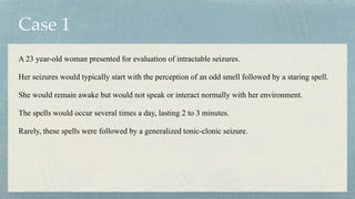
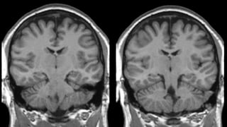
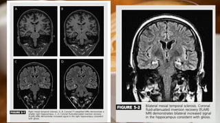
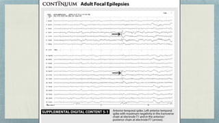
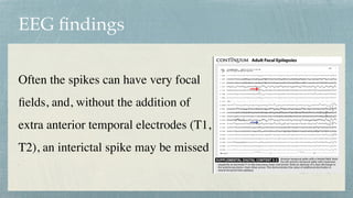
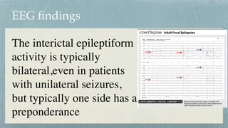
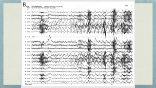
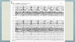
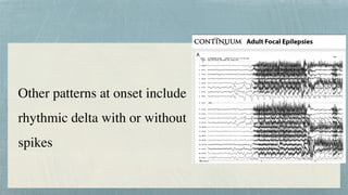
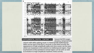
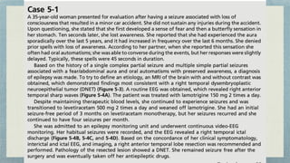
2. Case 1
A 23 year-old woman presented for evaluation of intractable seizures.
Her seizures would typically start with the perception of an odd smell followed by a staring spell.
She would remain awake but would not speak or interact normally with her environment.
The spells would occur several times a day, lasting 2 to 3 minutes.
Rarely, these spells were followed by a generalized tonic-clonic seizure.
3.
4. Temporal lobe epilepsy
Temporal-lobe epilepsy is the most common type of epilepsy in adults.
It can be divided into 2 main categories :Lateral(neocortical) and medial onset
5. Mesial Temporal lobe epilepsy
Often begins during late adolescence or early adulthood
Prolonged febrile seizures believed to be risk factor for development of hippocampal sclerosis(a
common cause of mesial temporal lobe epilepsy)
Hippocompal sclerosis is characterised by atrophy and gliosis of the hippocampus (unilateral or
bilateral)
Other aetiologies of mesial temporal lobe epilepsy ie cavernous
hemangiomas,gliosis,encephalomalacia secondary to prior injury and tumour ie dysembroplasric
neuroepithelial tumours
6.
7. Clinical features
Many patients with mesial temporal lobe epilepsy experience auras, which can
include psychic sensations (déjà vu and jamais vu), a gastric rising sensation, a
sensation of “butter
fl
ies” in their stomach, fear, or olfactory symptoms (usually
an unpleasant chemical or burning smell). The aura is typically followed by
loss of consciousness/ awareness with prominent oral and manual automatisms
in patients with dominant temporal lobe seizures, while nondominant temporal
lobe seizures may have preserved awareness with oral and manual automatisms
8. Clinical features
Dystonic limb posturing during the ictus has been described and is very predictive of seizure onset starting in the mesial
temporal lobe contralateral to the dystonic limb.
It is common for the patient to have manual automatisms in the hand ipsilateral to the seizure focus.
Symptoms associated with nondominant temporal lobe seizures include ictal spitting, vomiting, and urinary urge.
The seizures typically last 60 to 90 seconds and are often followed by a period of confusion/disorientation, more prominent in
patients with dominant temporal lobe seizures.
After dominant temporal lobe seizures, prominent postictal symptoms may be attributed to involvement of the language centers
in the ictal discharge.
Seizures from the mesial temporal region rarely secondarily generalize, but when they do, it is typically very late into the
seizure in contrast to neo- ortical temporal lobe or extratemporal lobe epilepsy, which frequently secondarily generalizes earlier
in the seizure.
9. Clinical features
Several clinical features help to lateralize the seizure focus to the ipsilateral temporal
lobe regardless of dominance.
These include postictal nose wiping, ictal unilateral eye blinking and ictal piloerection.
The hand that wipes the nose
fi
rst after a seizure is typically ipsilateral to the seizure
focus.
10.
11. EEG
fi
ndings
In mesial temporal lobe epilepsy, the interictal EEG is
associated with anterior temporal spikes/sharp waves
that are maximum in electrodes T1/F7 or T2/F8 (FT9/
FT10 in the 10/10 system)
12. EEG
fi
ndings
Often the spikes can have very focal
fi
elds, and, without the addition of
extra anterior temporal electrodes (T1,
T2), an interictal spike may be missed
14. Sleep deprivation can often activate focal epileptiform
discharges; therefore, an EEG with sleep recording is
recommended during the diagnostic workup.
The presence of mid or posterior temporal spikes may
suggest a more widespread epileptogenic network
even in the presence of clear mesial temporal
pathology
15. The ictal EEG is typically characterized by rhythmic
alpha or theta activity that evolves into higher-
amplitude rhythmic delta or theta activity that may be
sharply contoured or contain discrete spikes
21. Often, the initial ictal discharge may be preceded
by an initial sharp wave or suppression of the
normal EEG patterns or epileptiform activity
that was occurring immediately before the ictus
22. The seizure discharge may remain limited to the mesial
temporal structures, but often spreads to the remainder of the
ipsilateral hemisphere and contralateral temporal lobe.
During the postictal state, unilateral temporal lobe slowing that
is typically ipsilateral to the seizure onset may be seen.
23. Other patterns at onset include
rhythmic delta with or without
spikes
24.
25. Imaging
An MRI of the brain without contrast is recommended in the evaluation
of a patient with suspected mesial temporal lobe epilepsy
.
An enhanced MRI may be used dependent on local imaging protocols and
suspected etiologies.
It is preferable to include coronal T1- and T2-weighted images with thin
cuts (less than 3 mm) through the temporal lobes to properly evaluate the
hippocampus.
26. Imaging
Key imaging characteristics to evaluate for hippocampal
sclerosis are asymmetric size (smaller on the affected side) and
increased T2 signal in the hippocampus consistent with gliosis.
Inclusion of gradient echo imaging may assist in the
identi
fi
cation of vascular etiologies
27. Imaging
Fluorodeoxyglucose positron emission tomography (FDG-PET) scan
imaging has been extensively studied in patients undergoing presurgical
evaluation, and unilateral temporal lobe hypometabolism ipsilateral to the
seizure focus is a positive predictor of a good outcome after epilepsy surgery.
FDG-PET scanning is reserved only for patients who are being considered
for epilepsy surgery since the presence or absence of temporal lobe
hypometabolism does not impact medical treatment.
28. Genetics?
Familial forms of mesial temporal lobe epilepsy have been
described. The majority of reports describe families with
psychic auras and simple or complex partial seizures. The
imaging studies are normal, and the disease course is benign
for most patients. The exact genetic mutation is not known