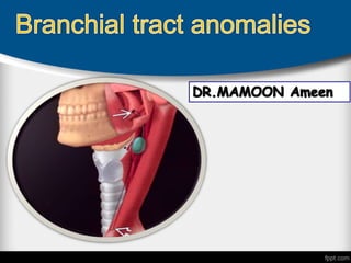
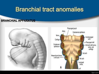
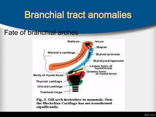
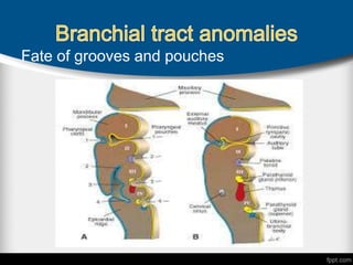
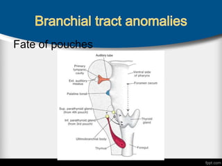
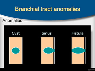
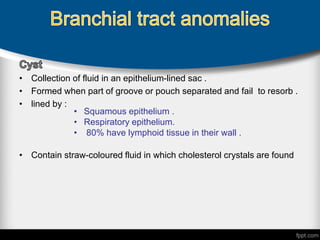
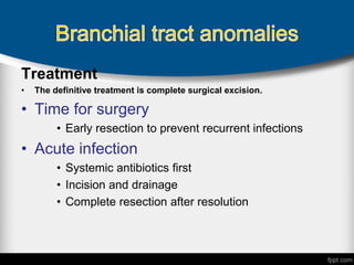
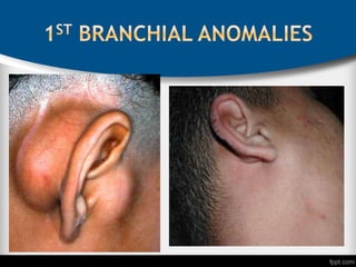
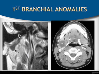
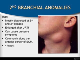
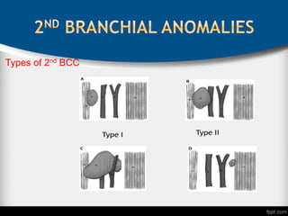
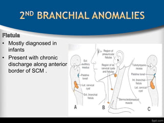
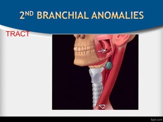
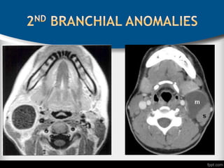
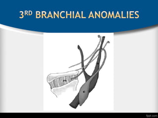
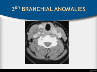
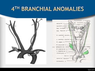
Branchial anomalies result from improper
development of the branchial apparatus
Fate of branchial arches
Fate of grooves and pouches
Fate of pouches
Anomalies
• Collection of fluid in an epithelium-lined sac .
• Formed when part of groove or pouch separated and fail to resorb .
• lined by :
• Contain straw-coloured fluid in which cholesterol crystals are found
• Squamous epithelium .
• Respiratory epithelium.
• 80% have lymphoid tissue in their wall .
:
 Represent persistence of both the cleft and the
corresponding pouch forming a communication that
is epithelial lined.
 The fistula lies caudal to the structures derived from
that particular arch and connects the skin to the
foregut.
 Lined by stratified squamous ,columnar , or ciliated
epithelium .
 Blind-ended track leading from an epithelial
surface into deeper tissues (partial fistula)
 Occur when groove or pouch fails to resorb
Diagnosis
• Upper airway endoscopy
• Pharyngeal opening
• Tonsillar fossa
• Pyriform sinus
• FNAC
• To clarify the diagnosis
• To rule out metastatic CA
• Ultrasound
• Round mass with uniform low echogenicity and lack of internal
septations
• Diagnosis
CT scan
• is first choice investigation
• Homogeneous lesion with low attenuation centrally and a smooth
enhancing rim
MRI
• Hypointense on T1 and hyperintense on T2
Fluroscopic or CT fistulography
• Inject radioopac dye into the fistula or sinus to delineate course
Barium swallow Esophageography
• for 3rd and 4th anomalies
Treatment
• The definitive treatment is complete surgical excision.
• Time for surgery
• Early resection to prevent recurrent infections
• Acute infection
• Systemic antibiotics first
• Incision and drainage
• Complete resection after resolution
 Can present as cysts, sinuses or fistulae located
between the EAC and the submandibular area.
 Represent 1% of all branchial anomalies
 Female > male
 Involve EAC or occasionally, the middle ear
 Course Close to parotid gland ,superficial lobe.
Symptoms:
 Otorrhea
 Parotid swelling
 Mandible pit discharge
 Unilateral facial palsy
Two types :
• Ectodermally derived
• Duplication of the external
auditory canal (EAC).
• immediately anterior ,inferior or
posterior to the pinna
• course lateral to the facial nerve,
.
• Ectodermal and mesodermal
derived tissues
• Terminate in EAC
• Behind or below the mandible
• Always suprahyaoid
• pass medial to the facial nerve
• More common than type I
Treatment
• Standard cervico-mastiod-facial parotidectomy incision
with facial nerve dissection and superficial
parotidectomy.
• Lacrimal probes can help locate tract
• Most common and represent 90-95% of branchial
anomalies.
• Cyst >fistula
• Cysts manifest as smooth , soft masses in the lateral
neck located anterior and deep to SCM.
• Fistulae tend to manifest as recurrent neck infections
following URTI
• Mostly diagnosed at 2nd
and 3rd decade
• Enlarged after URTI
• Can cause pressure
symptoms
• Commonly along the
anterior border of SCM.
• 4 types :
Types of 2nd BCC
• Mostly diagnosed in
infants
• Present with chronic
discharge along anterior
border of SCM .
TRACT
Treatment
• Transverse incision over skin fold
• Transvers elliptical incision made around the external opening and the
tract identified
Treatment
• surgeon must dissect around the cyst bed to exclude
associated fistula or tract
• Exploration of associated tract with complete excision
• Monofilament or probe to cannaulate the fistula tract
• Finger assisted to identify internal opening in tonsillar
fossa
Treatment
• The tract must be carefully ligated and divided at its entry
into the fossa
• The spinal accessory, hypoglossal, and vagus nerves must
identified to be protected from injury during the dissection.
• Cysts lying medial to carotid sheath are more easily
approached trans-orally
• Very Rare
• Mid or lower anterior border of SCM and at the level of
superior pole of thyroid
• Internal Opening to pyriform fossa
• This anomaly is also closely related to the thyroid gland,
which
when inflamed, may cause thyroiditis.
• Enlarged rapidly after URTI
• Extremely rare
• A lateral cervical cyst with an internal Opening in
the pyriform sinus is a common occurrence .
• mostly in children
• In neonatal :present as lateral neck mass or abscess
with obstructive airway symptoms
• In children or adult: recurrent lateral neck abscess
and recurrent suppurative thyroiditis .
Treatment
 External approach
 Excision of the tract with endoscopic assissted cannaulation.
 Ligation and dividing the tract
 Ipsilateral hemithroidectomy with partial resection of thyroid
cartilage for 4th pouch anomaly
 Internal approach
 Endoscopic electric cauterization
 Endoscopic chemical cauterization with silver nitrate
 Permanent recurrent laryngeal nerve palsy.
 Post operative pharyngocutaneous fistula
 Hypoglossal nerve palsy.
Complications
Branchial anomalies
Branchial anomalies

Branchial anomalies

  • 2.
    Branchial anomalies resultfrom improper development of the branchial apparatus
  • 4.
  • 5.
    Fate of groovesand pouches
  • 6.
  • 7.
  • 8.
    • Collection offluid in an epithelium-lined sac . • Formed when part of groove or pouch separated and fail to resorb . • lined by : • Contain straw-coloured fluid in which cholesterol crystals are found • Squamous epithelium . • Respiratory epithelium. • 80% have lymphoid tissue in their wall .
  • 9.
    :  Represent persistenceof both the cleft and the corresponding pouch forming a communication that is epithelial lined.  The fistula lies caudal to the structures derived from that particular arch and connects the skin to the foregut.  Lined by stratified squamous ,columnar , or ciliated epithelium .
  • 10.
     Blind-ended trackleading from an epithelial surface into deeper tissues (partial fistula)  Occur when groove or pouch fails to resorb
  • 11.
    Diagnosis • Upper airwayendoscopy • Pharyngeal opening • Tonsillar fossa • Pyriform sinus • FNAC • To clarify the diagnosis • To rule out metastatic CA • Ultrasound • Round mass with uniform low echogenicity and lack of internal septations
  • 12.
    • Diagnosis CT scan •is first choice investigation • Homogeneous lesion with low attenuation centrally and a smooth enhancing rim MRI • Hypointense on T1 and hyperintense on T2 Fluroscopic or CT fistulography • Inject radioopac dye into the fistula or sinus to delineate course Barium swallow Esophageography • for 3rd and 4th anomalies
  • 13.
    Treatment • The definitivetreatment is complete surgical excision. • Time for surgery • Early resection to prevent recurrent infections • Acute infection • Systemic antibiotics first • Incision and drainage • Complete resection after resolution
  • 14.
     Can presentas cysts, sinuses or fistulae located between the EAC and the submandibular area.  Represent 1% of all branchial anomalies  Female > male  Involve EAC or occasionally, the middle ear  Course Close to parotid gland ,superficial lobe.
  • 15.
    Symptoms:  Otorrhea  Parotidswelling  Mandible pit discharge  Unilateral facial palsy Two types :
  • 16.
    • Ectodermally derived •Duplication of the external auditory canal (EAC). • immediately anterior ,inferior or posterior to the pinna • course lateral to the facial nerve, .
  • 17.
    • Ectodermal andmesodermal derived tissues • Terminate in EAC • Behind or below the mandible • Always suprahyaoid • pass medial to the facial nerve • More common than type I
  • 20.
    Treatment • Standard cervico-mastiod-facialparotidectomy incision with facial nerve dissection and superficial parotidectomy. • Lacrimal probes can help locate tract
  • 21.
    • Most commonand represent 90-95% of branchial anomalies. • Cyst >fistula • Cysts manifest as smooth , soft masses in the lateral neck located anterior and deep to SCM. • Fistulae tend to manifest as recurrent neck infections following URTI
  • 22.
    • Mostly diagnosedat 2nd and 3rd decade • Enlarged after URTI • Can cause pressure symptoms • Commonly along the anterior border of SCM. • 4 types :
  • 23.
  • 24.
    • Mostly diagnosedin infants • Present with chronic discharge along anterior border of SCM .
  • 25.
  • 27.
    Treatment • Transverse incisionover skin fold • Transvers elliptical incision made around the external opening and the tract identified
  • 28.
    Treatment • surgeon mustdissect around the cyst bed to exclude associated fistula or tract • Exploration of associated tract with complete excision • Monofilament or probe to cannaulate the fistula tract • Finger assisted to identify internal opening in tonsillar fossa
  • 29.
    Treatment • The tractmust be carefully ligated and divided at its entry into the fossa • The spinal accessory, hypoglossal, and vagus nerves must identified to be protected from injury during the dissection. • Cysts lying medial to carotid sheath are more easily approached trans-orally
  • 30.
    • Very Rare •Mid or lower anterior border of SCM and at the level of superior pole of thyroid • Internal Opening to pyriform fossa • This anomaly is also closely related to the thyroid gland, which when inflamed, may cause thyroiditis. • Enlarged rapidly after URTI
  • 33.
    • Extremely rare •A lateral cervical cyst with an internal Opening in the pyriform sinus is a common occurrence . • mostly in children • In neonatal :present as lateral neck mass or abscess with obstructive airway symptoms • In children or adult: recurrent lateral neck abscess and recurrent suppurative thyroiditis .
  • 36.
    Treatment  External approach Excision of the tract with endoscopic assissted cannaulation.  Ligation and dividing the tract  Ipsilateral hemithroidectomy with partial resection of thyroid cartilage for 4th pouch anomaly  Internal approach  Endoscopic electric cauterization  Endoscopic chemical cauterization with silver nitrate
  • 37.
     Permanent recurrentlaryngeal nerve palsy.  Post operative pharyngocutaneous fistula  Hypoglossal nerve palsy. Complications

Editor's Notes

  • #20 Coronal t1 mri for 1st bcc ..immediatly below and parallel to eac >>opening to eac
  • #26 Internal opening in tonsillar fossa >