Downloaded 1,458 times


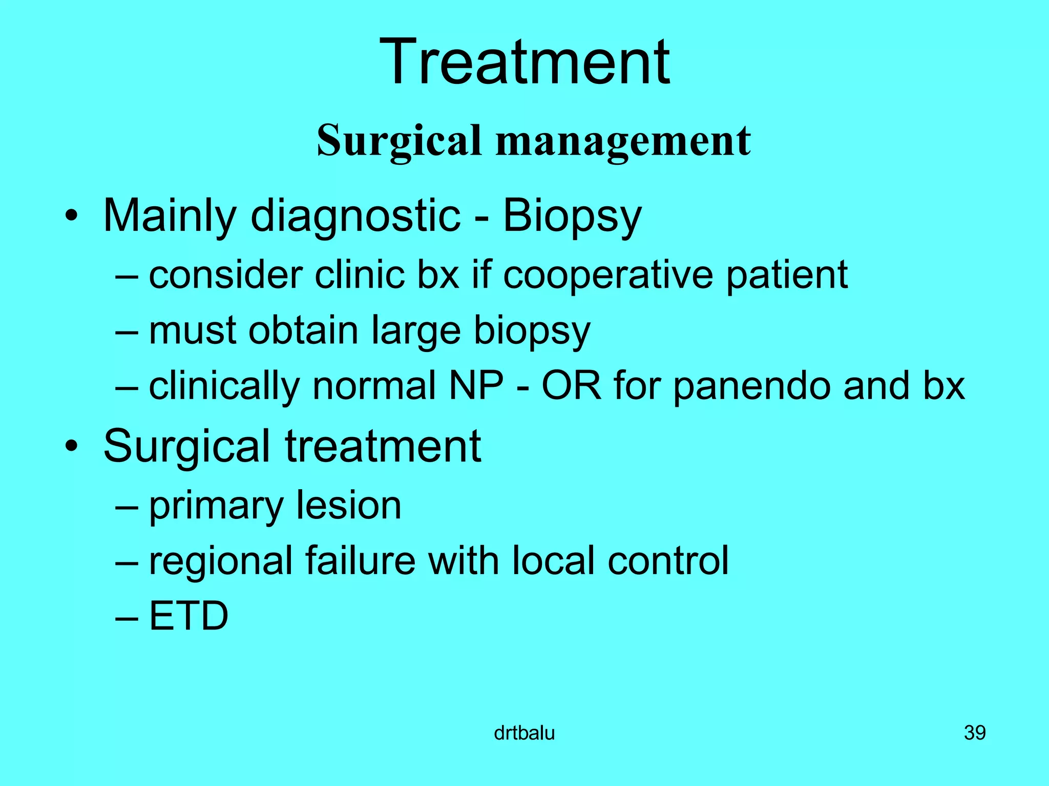


The document discusses the anatomy, histology, epidemiology, clinical features, diagnosis, staging, and treatment of nasopharyngeal carcinoma (NPC). Key points include: - NPC originates from the epithelial lining of the nasopharynx. - It has a strong association with Epstein-Barr virus. - Risk factors include genetic predisposition and environmental exposures like salted fish consumption. - Common symptoms are cervical lymphadenopathy, epistaxis, ear symptoms, and neurological deficits. - Diagnosis involves biopsy and serological testing for EBV markers. - Staging systems consider tumor size, node involvement, and serological factors. - Primary treatment is radiotherapy,


