Downloaded 325 times
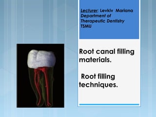

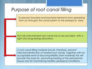
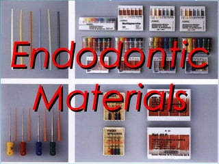
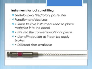
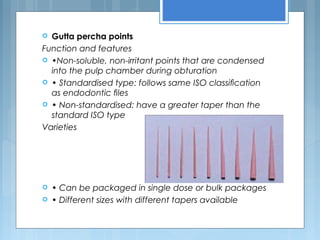
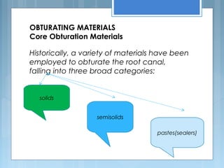
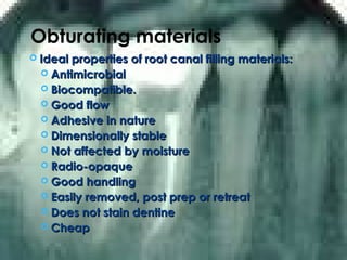
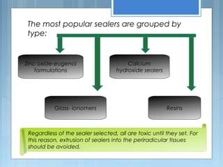
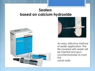
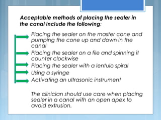
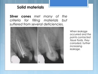
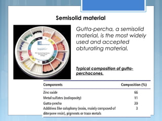
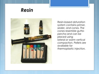
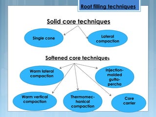

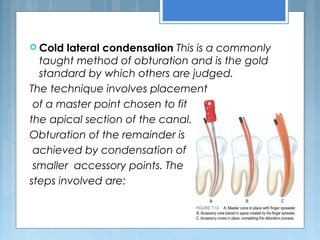
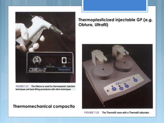
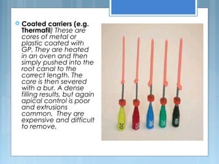
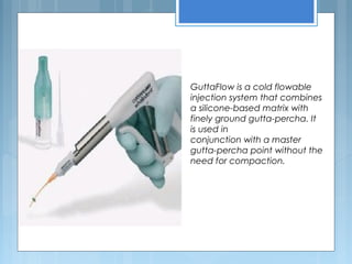
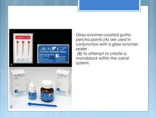
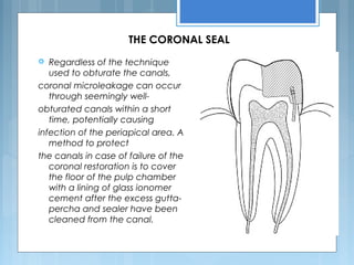


The document discusses root canal filling materials and techniques. It describes various instruments used for root canal filling like Lentulo spirals and spreaders. It discusses different obturation materials like gutta percha points, sealers based on zinc oxide-eugenol, calcium hydroxide, glass ionomers and resins. It also describes different root canal filling techniques like single cone, lateral condensation, warm lateral/vertical condensation and thermoplasticized techniques.


![CASE_PRESENTATION_ON_subdural_hematoma(SDH)[1 FINAL PPT]-1.pptx](https://cdn.slidesharecdn.com/ss_thumbnails/casepresentationonsubduralhematomasdh1finalppt-1-260129172522-d405d375-thumbnail.jpg?width=640&height=640&fit=bounds)


