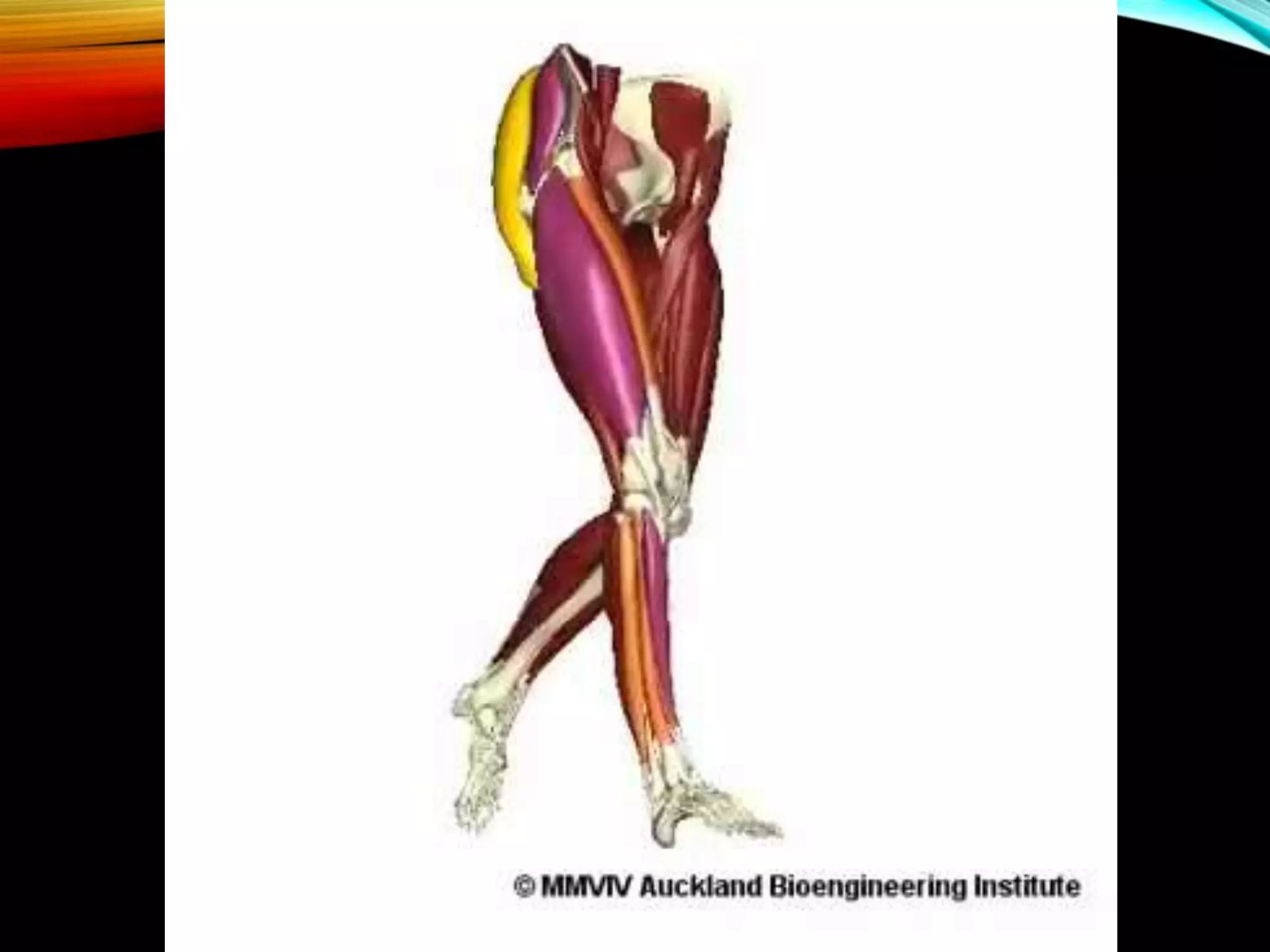
This document discusses foot drop, which is the inability to lift the front part of the foot. It can be caused by nerve injuries, neurological conditions, muscle weakness, or injuries. Symptoms include difficulty lifting the foot and dragging it when walking. Treatment depends on the underlying cause but may include bracing, physical therapy, nerve stimulation, or surgery to repair nerves or transfer tendons.
2. • Drop Foot
• The inability to lift the front part of the foot.
• Paralysis of anterior muscles of lower leg
• Inability to dorsiflex at the ankles and toes
• Causes the toes to drag along the ground while
walking.
• Can happen to one or both feet at the same time.
It can strike at any age.
• Temporary or permanent
3. CAUSES
• Injury to the peroneal nerve.
• sports injuries
• diabetes
• hip or knee replacement surgery
• spending long hours sitting cross-legged or squatting
• childbirth
• large amount of weight loss
• Injury to the nerve roots in the spine (L5)
4. • Neurological conditions that can contribute to foot
drop include:
• stroke
• multiple sclerosis (MS)
• cerebral palsy
• Charcot-Marie-Tooth disease
• Conditions that cause the muscles to progressively
weaken or deteriorate may cause foot drop:
• muscular dystrophy
• amyotrophic lateral sclerosis (Lou Gehrig’s disease)
• polio
5. • Rupture of Anterior Tibialis
• Fracture of fibula
• Compartment Syndrome
• Diabetes
• Alcohol Abuse
6. VULNERABILITY OF PERONEAL NERVE
• Funiculi of the peroneal nerve - larger and less
connective tissue
• Fewer autonomic fibers, so in any injury, motor and
sensory fibers bear the brunt of the trauma.
• More superficial course, especially at the fibular
neck
• Adheres closely to the periosteum of the proximal
fibula
8. SYMPTOMS
• Difficulty in lifting the foot.
• Dragging the foot on the floor as one walks.
• Slapping the foot down with each step.
• Raising thigh while walking (high stepping gait)
• Pain, weakness or numbness in the foot.
9. GAIT CYCLE
• Swing phase (SW): The period of time when the foot
is not in contact with the ground. In those cases
where the foot never leaves the ground (foot drag)
- phase when all portions of the foot are in forward
motion.
• Initial contact (IC): when the foot initially makes
contact with the ground; represents beginning of
the stance phase - foot strike.
• Terminal contact (TC): when the foot leaves the
ground - end of the stance phase or beginning of
the swing phase - foot off. .
10.
11.
12. FOOT DROP
• Drop foot SW: Greater flexion at the knee to
accommodate the inability to dorsiflex - stair
climbing movement.
• Drop foot IC: Instead of normal heel-toe foot strike,
foot may either slap the ground or the entire foot
may be planted on the ground all at once.
• Drop foot TC: Terminal contact is quite different -
inability to support their body weight – walker can
be used
13.
14.
15. IMAGING
• X-Ray
Post-Traumatic - tibia/fibula and ankle - any
bony injury.
Anatomic dysfunction (eg. Charcot joint)
• Ultrasonography
If bleeding is suspected in a patient with a hip
or knee prosthesis
• Magnetic Resonance Neurography
Tumor or a compressive mass lesion to the
peroneal nerve
16. ELECTROMYELOGRAM
• This study can confirm the type of neuropathy,
establish the site of the lesion, estimate extent of
injury, and provide a prognosis.
• Sequential studies are useful to monitor recovery
of acute lesions.
17. TREATMENT
• Depends on the underlying cause.
• If cause is successfully treated foot drop may
improve or even disappear.
• Medical treatment - Painful Paresthesia
• Sympathetic block
• Amitriptyline
• Nortriptyline
• Pregabalin
• Laproscopic Synovectomy
18. SPECIFIC TREATMENT
• Braces or splint
• Brace on the ankle and foot or splint that fits into the
shoe can help to hold the foot in the normal position
19. PHYSICAL THERAPY
• Exercises that strengthen the leg muscles
• Maintain the range of motion in knee and ankle
• Improve gait problems associated with foot drop.
21. SURGICAL REPAIR
• Foot drop due to direct trauma to the dorsiflexors
generally requires surgical repair.
• When nerve insult is the cause - restore the nerve
continuity - nerve grafting or repair.
• If there is no significant neuronal recovery at one
year - tendon transfer maybe considered.
• Bridal procedure
• Neurotendinous transpositon
22. BRIDALS PROCEDURE
• Tendon to bone attachment - posterior tibial tendon is
attatched to the second cuneiform bone.
• Tendon to tendon attachment
23. NEUROTENDINOUS TRANSPOSITION
• Lateral head of gastronemius is transposed to the
tendons of the anterior muscle group with simultaneous
transposition of the proximal end of deep peroneal
nerve.
• The nerve is sutured to the motor nerve of the
gartronemius
• Active voluntary dorsiflexion of foot
24. • AFTER TENDON TRANSFER
Cast and Non-Weight Bearing ambulation for 6
weeks
• PHYSIOTHERAPY
To correct gait abnormalities
• CHRONIC AND CONTRACTURE CASES
Achilles tendon lengthening
• In patients whom foot drop is due to neurologic and
anatomic factors (polio, charcot joint ) - Arthodesis
• Subtalar Stabilising procedure or Triple Arthodesis can be
done.
25. COMPLICATIONS
• Surgical procedure- wound infection may occur.
• Nerve graft failure
• In tendon transfer procedures- recurrent deformity
• In arthrodeses or fusion procedures- pseudoarthrosis,
delayed union, or nonunion.