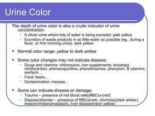
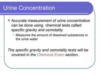
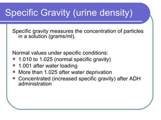
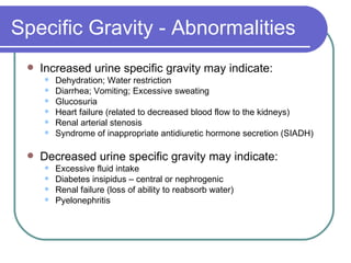
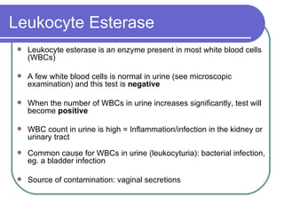
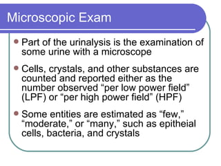
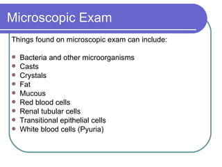
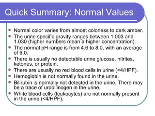
Urinalysis Jennifer Lyon, M.S., M.L.I.S. [email_address] Joanna Karpinski, M.L.I.S. [email_address]
Urinalysis A complete urinalysis consists of three distinct testing phases:  Physical examination  –  Evaluates the urine's color, clarity, and concentration  Chemical examination  –  Tests chemically for many substances that provide valuable information about health and disease only some of these are covered today Microscopic examination   Identifies and counts types of cells, casts, crystals, and other components (bacteria, mucous) that can be present in urine  Additional testing: urine culture (check to see if a microorganism is growing in the urine –  not covered today )
Why Is It Important? Urine composition provides information about:  The health and function of part of the body’s waste management system  State of kidney function Possible blood abnormalities Urine analysis can help a clinician: Identify infections and other diseases  Gauge generally the body’s hydration level
Sample Clinical Questions Is there a predictive value of acute kidney injury in ICU patients?  Is there a method for localizing the site of a urinary tract infection: upper (renal) vs. lower (bladder).  Which is the best procedure: rapid (quick-emptying) versus slow (periodically-clamped) voiding of urine through a catheter in patients with urinary retention?  What strategies to prevent urinary tract infection in the acutely ill adult population are supported by evidence  Is MRSA (methicillin-resistant staphylococcus aureus) a significant pathogen for urinary tract infections?
Physical Exam Color Concentration Clarity (Turbidity) (rarely) Odor
Urine Color The depth of urine color is also a crude indicator of urine concentration:  A dilute urine where lots of water is being excreted: pale yellow Excretion of waste products in as little water as possible (eg., during a fever, or first morning urine): dark yellow  Normal color range: yellow to dark amber Some color changes may not indicate disease: Drugs and vitamins: chloroquine, iron supplements, levodopa, nitrofurantoin, phenazopyridine, phenothiazines, phenytoin, B vitamins, warfarin… Food: beets… Contamination: menses… Some can indicate disease or damage: Trauma – presence of red blood cells(RBCs)-(red) Disease/disorder – presence of RBCs(red), chirrhosis(dark amber), melanin/melanoma(black), liver disease(neon yellow)
Urine Concentration Accurate measurement of urine concentration can be done using  chemical tests called specific gravity and osmolality Measures the amount of dissolved substances in the urine water  The specific gravity and osmolality tests will be covered in the  Chemical Exam  section.
Urine Clarity Urine clarity refers to how clear the urine is  Labs usually report clarity of the urine as clear slightly cloudy  cloudy turbid  “ Normal” urine can be clear or cloudy  Clinically unimportant substances that cause cloudiness: mucous, sperm and prostatic fluid, cells from the skin, normal urine crystals, and contaminants (eg. body lotions/powders)  Clinically important substances that cause cloudiness: Red blood cells, white blood cells, protein, and bacteria
Chemical Exam These tests are often done using a urine dipstick - a plastic stick that has patches of chemical indicators on it which is placed in the urine. The color changes of the patches provide the results. specific gravity/osmolality pH protein  hematuria (see also RBCs under microscopic exam) glucose ketones  leukocyte esterase nitrite bilirubin and urobilinogen
Specific Gravity (urine density) Specific gravity measures the concentration of particles in a solution (grams/ml).  Normal values under specific conditions: 1.010 to 1.025 (normal specific gravity)  1.001 after water loading  More than 1.025 after water deprivation  Concentrated (increased specific gravity) after ADH administration
Specific Gravity - Abnormalities Increased urine specific gravity may indicate: Dehydration; Water restriction  Diarrhea; Vomiting; Excessive sweating  Glucosuria Heart failure (related to decreased blood flow to the kidneys)  Renal arterial stenosis  Syndrome of inappropriate antidiuretic hormone secretion (SIADH) Decreased urine specific gravity may indicate:  Excessive fluid intake  Diabetes insipidus – central or nephrogenic  Renal failure (loss of ability to reabsorb water)  Pyelonephritis
Urine Osmolality Normal values are 50 to 1400 mOsm/kg  12 to 14 hour fluid restriction: greater than 850 mOsm/kg  Greater-than-normal measurements may indicate: Addison's disease (rare)  Congestive heart failure Shock Syndrome of inappropriate ADH secretion  Lower-than-normal measurements may indicate:  Aldosteronism (very rare)  Diabetes insipidus (rare)  Excess fluid intake  Renal tubular necrosis Severe pyelonephritis
pH  (acidity/alkalinity of the urine ) The normal values range from 4.6 to 8.0.  A high urine pH (alkaline urine) may indicate: Gastric suction Renal failure Renal tubular acidosis Urinary tract infection Vomiting A low urine pH (acidic urine) may indicate:  Chronic obstructive pulmonary disease  Diabetic ketoacidosis Diarrhea Starvation  The drugs acetazolamide, potassium citrate, or sodium bicarbonate can increase pH; Ammonium chloride, chlorothiazide diuretics, and methenamine mandelate can decrease urine pH
Proteinuria (protein in the urine) For a spot check by dipstick: the normal values are approximately 0 to 8 mg/dl.  For a 24-hour test: the normal value is less than 150mg per 24 hours.  An abnormal value (high value) usually indicates a  kidney  disorder/disease Proteinuria:  Mild(<0.5g/day), moderate (.05-4g/day), severe (>4g/day) Other conditions that can also produce proteinuria include  Blood diseases involving lysis (destruction) of red blood cells Inflammation Cancer Injury of the urinary tract (bladder, prostate, or urethra) Preeclampsia
Glucosuria Glucose should  not  be detectable in the urine Diabetes (via high blood sugar) is the most common cause of glucosuria Other causes include renal glycosuria (decrease or absence of the kidneys’ ability to absorb glucose), hormonal disorders, liver disease, some medications, and pregnancy
Hematuria Normal: Only an occasional RBC (1-4/high power field; 1000 per millimeter of urine) The urinary dipstick for blood measures intact RBCs, free hemoglobin, and myoglobin False-positive results in women from contamination with menstrual blood More detail provided in the  microscopic exam  portion of this presentation)
Ketones Ketones (beta-hydroxybutyric acid, acetoacetic acid, and acetone) are intermediate products of fat metabolism and are present in starvation and diabetes Normal: negative test result Classification of acetone presence in the urine  Small - < 20 mg/dL Moderate - 30-40 mg/dL Large - > 80 mg/dL Ketonuria: presence of ketones in urine
Ketone Abnormalities A positive test may indicate: Metabolic abnormalities, including uncontrolled diabetes or glycogen storage disease  Abnormal nutritional conditions, including starvation, fasting, anorexia, high protein or low carbohydrate diets  Protracted vomiting, (eg. hyperemesis gravidarum)  Disorders of increased metabolism, including hyperthyroidism, fever, acute or severe illness, burns, pregnancy, lactation or post-surgical condition  Some drugs
Leukocyte Esterase Leukocyte esterase is an enzyme present in most white blood cells (WBCs) A few white blood cells is normal in urine (see microscopic examination) and this test is  negative   When the number of WBCs in urine increases significantly, test will become  positive   WBC count in urine is high = Inflammation/infection in the kidney or urinary tract   Common cause for WBCs in urine (leukocyturia): bacterial infection, eg. a bladder infection  Source of contamination: vaginal secretions
Nitrite Test is used to detect a bacterial urinary tract infections (UTI) because many bacteria convert nitrate to nitrite  Normally, the urinary tract and urine are sterile Thus, no nitrites would be found  A negative test does not rule out a UTI  Not all bacteria are capable of converting nitrate to nitrite
Bilirubin Bilirubin (a degradation product of hemoglobin) is not present in the urine of normal, healthy individuals  Increased bilirubin in the urine means that the bile ducts are obstructed Causes include biliary strictures cirrhosis gallstones in the biliary tract hepatitis with associated biliary obstruction surgical trauma affecting the biliary tract  tumors of the liver or gall bladder
Urobilirubin Urobilinogen (a degradation product of bilirubin) is normally present in urine in low concentrations – source of color  High concentrations can be due to Hemolytic processes Hepatocellular disease Absence may be due to Complete biliary obstruction Broad-spectrum antibiotics Normal levels are usually too low (<0.4mg/dl) to be detectable with the urine dipstick method
Microscopic Exam Part of the urinalysis is the examination of some urine with a microscope Cells, crystals, and other substances are counted and reported either as the number observed “per low power field” (LPF) or “per high power field” (HPF) Some entities are estimated as “few,” “moderate,” or “many,” such as epitheial cells, bacteria, and crystals
Microscopic Exam Things found on microscopic exam can include: Bacteria and other microorganisms Casts Crystals  Fat  Mucous  Red blood cells Renal tubular cells  Transitional epithelial cells  White blood cells (Pyuria)
RBCs Only a few RBCs (erythrocytes) should be present in urine sediment Normal values are less than, or equal to 4 RBC/HPF Normal value ranges may vary slightly among different laboratories Note: RBC/HPF = red blood cells per high power field (a microscopic exam)
RBCs - Abnormalities Hematuria – presence of RBCs in urine Hemoglobinuria – hemoglobin in urine Even small increases in the amount of RBCs in urine are significant.  Causes: Numerous diseases of the kidney and urinary tract  Trauma  Medications Smoking Strenuous exercise All patients with hematuria (>5 RBC/HPF) require further diagnostic workup
WBCs (Pyuria) The number of WBCs in urine sediment is normally low (1-4 WBCs/HPF) The presence of >5 WBC/HPF = significant pyuria When the number is high, it indicates an infection, damage or inflammation somewhere in the urinary tract Causes include calculous disease, strictures, neoplasm, glomerulonephropathy, or interstitial cystitis
Epithelial Cells Epithelial cells: cells that form sheets that cover the surface of the body and line internal organs Healthy urine shows a few normal epithelial cells and is relatively free of debris Epithelial cells are usually reported as “few,” “moderate,” or “many” present per low power field (LPF) In urinary tract conditions such as infections, inflammation, and malignancies, more epithelial cells are present
Microorganisms Include bacteria, trichomonads, yeast In health, the urinary tract is sterile: No microorganisms  Presence indicates infection  Microorganisms are usually reported as “few,” “moderate,” or “many” present per high power field (HPF)  Microorganisms found may be from specimen contamination: From bacteria that normally live on the skin or in vaginal secretions (most often women) Yeast can also be present in urine and are more common  in women due to a vaginal yeast infection
Trichomonads   Trichomonads are parasites that may be found in the urine of women or men (rarely) Often infect the vaginal canal Their presence in urine is due to contamination during urine collection
Urinary Casts   Casts – renal tubule “imprints” –  Absent in normal people, except for a few (0–5) hyaline casts per LPF.  Abnormal results include: Hyaline casts  – made of protein; caused by dehydration, exercise, diuretic meds  Granular casts  -- include prominent granules; related to underlying kidney disease Fatty casts  –condition of lipiduria (lipids in urine); related to nephrotic syndrome  RBC casts  – result of bleeding into tubule from the glomerulus; nephrotic syndrome WBC casts  – interstitial cell kidney disease (interstitial inflammation, pyelonephritis, and parenchymal infection) Renal tubular epithelial cell casts  -- damage to the tubules; ie.renal tubular necrosis
Crystals Urine contains many chemicals dissolved in the urine for elimination from the body.  These solutes can form crystals based on the urine pH & temperature and their concentration. Crystals are identified by their shape, color, and urine pH.  Crystals are considered “normal” if they are from solutes that should be in urine If they are from solutes that are not supposed to be in the urine, they are considered “abnormal.”  Acidic urine: uric acid, oxalate, cystine Alkaline urine: phosphate
Quick Summary: Normal Values Normal color varies from almost colorless to dark amber.  The urine specific gravity ranges between 1.003 and 1.030 (higher numbers mean a higher concentration).  The normal pH range is from 4.6 to 8.0, with an average of 6.0.  There is usually no detectable urine glucose, nitrites, ketones, or protein.  There are usually no red blood cells in urine (<4/HPF).  Hemoglobin is not normally found in the urine.  Bilirubin is normally not detected in the urine. There may be a trace of urobilinogen in the urine.  White blood cells (leukocytes) are not normally present in the urine (<4/HPF).
Resources http://www.labtestsonline.org/understanding/analytes/urinalysis/sample.html http://kidshealth.org/parent/general/sick/labtest7_p2.html http://www.nlm.nih.gov/medlineplus/ency/article/003579.htm Corbett JV. Laboratory Tests and Diagnostic Procedures. 5 th  ed. New Jersey: Prentice-Hall, Inc., 2000. Current Medical Diagnosis & Treatment - 44th Ed. (2005) online via Stat!-Ref

Urinalysis 3/27

  • 1.
    Urinalysis Jennifer Lyon,M.S., M.L.I.S. [email_address] Joanna Karpinski, M.L.I.S. [email_address]
  • 2.
    Urinalysis A completeurinalysis consists of three distinct testing phases: Physical examination – Evaluates the urine's color, clarity, and concentration Chemical examination – Tests chemically for many substances that provide valuable information about health and disease only some of these are covered today Microscopic examination Identifies and counts types of cells, casts, crystals, and other components (bacteria, mucous) that can be present in urine Additional testing: urine culture (check to see if a microorganism is growing in the urine – not covered today )
  • 3.
    Why Is ItImportant? Urine composition provides information about: The health and function of part of the body’s waste management system State of kidney function Possible blood abnormalities Urine analysis can help a clinician: Identify infections and other diseases Gauge generally the body’s hydration level
  • 4.
    Sample Clinical QuestionsIs there a predictive value of acute kidney injury in ICU patients? Is there a method for localizing the site of a urinary tract infection: upper (renal) vs. lower (bladder). Which is the best procedure: rapid (quick-emptying) versus slow (periodically-clamped) voiding of urine through a catheter in patients with urinary retention? What strategies to prevent urinary tract infection in the acutely ill adult population are supported by evidence Is MRSA (methicillin-resistant staphylococcus aureus) a significant pathogen for urinary tract infections?
  • 5.
    Physical Exam ColorConcentration Clarity (Turbidity) (rarely) Odor
  • 6.
    Urine Color Thedepth of urine color is also a crude indicator of urine concentration: A dilute urine where lots of water is being excreted: pale yellow Excretion of waste products in as little water as possible (eg., during a fever, or first morning urine): dark yellow Normal color range: yellow to dark amber Some color changes may not indicate disease: Drugs and vitamins: chloroquine, iron supplements, levodopa, nitrofurantoin, phenazopyridine, phenothiazines, phenytoin, B vitamins, warfarin… Food: beets… Contamination: menses… Some can indicate disease or damage: Trauma – presence of red blood cells(RBCs)-(red) Disease/disorder – presence of RBCs(red), chirrhosis(dark amber), melanin/melanoma(black), liver disease(neon yellow)
  • 7.
    Urine Concentration Accuratemeasurement of urine concentration can be done using chemical tests called specific gravity and osmolality Measures the amount of dissolved substances in the urine water The specific gravity and osmolality tests will be covered in the Chemical Exam section.
  • 8.
    Urine Clarity Urineclarity refers to how clear the urine is Labs usually report clarity of the urine as clear slightly cloudy cloudy turbid “ Normal” urine can be clear or cloudy Clinically unimportant substances that cause cloudiness: mucous, sperm and prostatic fluid, cells from the skin, normal urine crystals, and contaminants (eg. body lotions/powders) Clinically important substances that cause cloudiness: Red blood cells, white blood cells, protein, and bacteria
  • 9.
    Chemical Exam Thesetests are often done using a urine dipstick - a plastic stick that has patches of chemical indicators on it which is placed in the urine. The color changes of the patches provide the results. specific gravity/osmolality pH protein hematuria (see also RBCs under microscopic exam) glucose ketones leukocyte esterase nitrite bilirubin and urobilinogen
  • 10.
    Specific Gravity (urinedensity) Specific gravity measures the concentration of particles in a solution (grams/ml). Normal values under specific conditions: 1.010 to 1.025 (normal specific gravity) 1.001 after water loading More than 1.025 after water deprivation Concentrated (increased specific gravity) after ADH administration
  • 11.
    Specific Gravity -Abnormalities Increased urine specific gravity may indicate: Dehydration; Water restriction Diarrhea; Vomiting; Excessive sweating Glucosuria Heart failure (related to decreased blood flow to the kidneys) Renal arterial stenosis Syndrome of inappropriate antidiuretic hormone secretion (SIADH) Decreased urine specific gravity may indicate: Excessive fluid intake Diabetes insipidus – central or nephrogenic Renal failure (loss of ability to reabsorb water) Pyelonephritis
  • 12.
    Urine Osmolality Normalvalues are 50 to 1400 mOsm/kg 12 to 14 hour fluid restriction: greater than 850 mOsm/kg Greater-than-normal measurements may indicate: Addison's disease (rare) Congestive heart failure Shock Syndrome of inappropriate ADH secretion Lower-than-normal measurements may indicate: Aldosteronism (very rare) Diabetes insipidus (rare) Excess fluid intake Renal tubular necrosis Severe pyelonephritis
  • 13.
    pH (acidity/alkalinityof the urine ) The normal values range from 4.6 to 8.0. A high urine pH (alkaline urine) may indicate: Gastric suction Renal failure Renal tubular acidosis Urinary tract infection Vomiting A low urine pH (acidic urine) may indicate: Chronic obstructive pulmonary disease Diabetic ketoacidosis Diarrhea Starvation The drugs acetazolamide, potassium citrate, or sodium bicarbonate can increase pH; Ammonium chloride, chlorothiazide diuretics, and methenamine mandelate can decrease urine pH
  • 14.
    Proteinuria (protein inthe urine) For a spot check by dipstick: the normal values are approximately 0 to 8 mg/dl. For a 24-hour test: the normal value is less than 150mg per 24 hours. An abnormal value (high value) usually indicates a kidney disorder/disease Proteinuria: Mild(<0.5g/day), moderate (.05-4g/day), severe (>4g/day) Other conditions that can also produce proteinuria include Blood diseases involving lysis (destruction) of red blood cells Inflammation Cancer Injury of the urinary tract (bladder, prostate, or urethra) Preeclampsia
  • 15.
    Glucosuria Glucose should not be detectable in the urine Diabetes (via high blood sugar) is the most common cause of glucosuria Other causes include renal glycosuria (decrease or absence of the kidneys’ ability to absorb glucose), hormonal disorders, liver disease, some medications, and pregnancy
  • 16.
    Hematuria Normal: Onlyan occasional RBC (1-4/high power field; 1000 per millimeter of urine) The urinary dipstick for blood measures intact RBCs, free hemoglobin, and myoglobin False-positive results in women from contamination with menstrual blood More detail provided in the microscopic exam portion of this presentation)
  • 17.
    Ketones Ketones (beta-hydroxybutyricacid, acetoacetic acid, and acetone) are intermediate products of fat metabolism and are present in starvation and diabetes Normal: negative test result Classification of acetone presence in the urine Small - < 20 mg/dL Moderate - 30-40 mg/dL Large - > 80 mg/dL Ketonuria: presence of ketones in urine
  • 18.
    Ketone Abnormalities Apositive test may indicate: Metabolic abnormalities, including uncontrolled diabetes or glycogen storage disease Abnormal nutritional conditions, including starvation, fasting, anorexia, high protein or low carbohydrate diets Protracted vomiting, (eg. hyperemesis gravidarum) Disorders of increased metabolism, including hyperthyroidism, fever, acute or severe illness, burns, pregnancy, lactation or post-surgical condition Some drugs
  • 19.
    Leukocyte Esterase Leukocyteesterase is an enzyme present in most white blood cells (WBCs) A few white blood cells is normal in urine (see microscopic examination) and this test is negative When the number of WBCs in urine increases significantly, test will become positive WBC count in urine is high = Inflammation/infection in the kidney or urinary tract Common cause for WBCs in urine (leukocyturia): bacterial infection, eg. a bladder infection Source of contamination: vaginal secretions
  • 20.
    Nitrite Test isused to detect a bacterial urinary tract infections (UTI) because many bacteria convert nitrate to nitrite Normally, the urinary tract and urine are sterile Thus, no nitrites would be found A negative test does not rule out a UTI Not all bacteria are capable of converting nitrate to nitrite
  • 21.
    Bilirubin Bilirubin (adegradation product of hemoglobin) is not present in the urine of normal, healthy individuals Increased bilirubin in the urine means that the bile ducts are obstructed Causes include biliary strictures cirrhosis gallstones in the biliary tract hepatitis with associated biliary obstruction surgical trauma affecting the biliary tract tumors of the liver or gall bladder
  • 22.
    Urobilirubin Urobilinogen (adegradation product of bilirubin) is normally present in urine in low concentrations – source of color High concentrations can be due to Hemolytic processes Hepatocellular disease Absence may be due to Complete biliary obstruction Broad-spectrum antibiotics Normal levels are usually too low (<0.4mg/dl) to be detectable with the urine dipstick method
  • 23.
    Microscopic Exam Partof the urinalysis is the examination of some urine with a microscope Cells, crystals, and other substances are counted and reported either as the number observed “per low power field” (LPF) or “per high power field” (HPF) Some entities are estimated as “few,” “moderate,” or “many,” such as epitheial cells, bacteria, and crystals
  • 24.
    Microscopic Exam Thingsfound on microscopic exam can include: Bacteria and other microorganisms Casts Crystals Fat Mucous Red blood cells Renal tubular cells Transitional epithelial cells White blood cells (Pyuria)
  • 25.
    RBCs Only afew RBCs (erythrocytes) should be present in urine sediment Normal values are less than, or equal to 4 RBC/HPF Normal value ranges may vary slightly among different laboratories Note: RBC/HPF = red blood cells per high power field (a microscopic exam)
  • 26.
    RBCs - AbnormalitiesHematuria – presence of RBCs in urine Hemoglobinuria – hemoglobin in urine Even small increases in the amount of RBCs in urine are significant. Causes: Numerous diseases of the kidney and urinary tract Trauma Medications Smoking Strenuous exercise All patients with hematuria (>5 RBC/HPF) require further diagnostic workup
  • 27.
    WBCs (Pyuria) Thenumber of WBCs in urine sediment is normally low (1-4 WBCs/HPF) The presence of >5 WBC/HPF = significant pyuria When the number is high, it indicates an infection, damage or inflammation somewhere in the urinary tract Causes include calculous disease, strictures, neoplasm, glomerulonephropathy, or interstitial cystitis
  • 28.
    Epithelial Cells Epithelialcells: cells that form sheets that cover the surface of the body and line internal organs Healthy urine shows a few normal epithelial cells and is relatively free of debris Epithelial cells are usually reported as “few,” “moderate,” or “many” present per low power field (LPF) In urinary tract conditions such as infections, inflammation, and malignancies, more epithelial cells are present
  • 29.
    Microorganisms Include bacteria,trichomonads, yeast In health, the urinary tract is sterile: No microorganisms Presence indicates infection Microorganisms are usually reported as “few,” “moderate,” or “many” present per high power field (HPF) Microorganisms found may be from specimen contamination: From bacteria that normally live on the skin or in vaginal secretions (most often women) Yeast can also be present in urine and are more common in women due to a vaginal yeast infection
  • 30.
    Trichomonads Trichomonads are parasites that may be found in the urine of women or men (rarely) Often infect the vaginal canal Their presence in urine is due to contamination during urine collection
  • 31.
    Urinary Casts Casts – renal tubule “imprints” – Absent in normal people, except for a few (0–5) hyaline casts per LPF. Abnormal results include: Hyaline casts – made of protein; caused by dehydration, exercise, diuretic meds Granular casts -- include prominent granules; related to underlying kidney disease Fatty casts –condition of lipiduria (lipids in urine); related to nephrotic syndrome RBC casts – result of bleeding into tubule from the glomerulus; nephrotic syndrome WBC casts – interstitial cell kidney disease (interstitial inflammation, pyelonephritis, and parenchymal infection) Renal tubular epithelial cell casts -- damage to the tubules; ie.renal tubular necrosis
  • 32.
    Crystals Urine containsmany chemicals dissolved in the urine for elimination from the body. These solutes can form crystals based on the urine pH & temperature and their concentration. Crystals are identified by their shape, color, and urine pH. Crystals are considered “normal” if they are from solutes that should be in urine If they are from solutes that are not supposed to be in the urine, they are considered “abnormal.” Acidic urine: uric acid, oxalate, cystine Alkaline urine: phosphate
  • 33.
    Quick Summary: NormalValues Normal color varies from almost colorless to dark amber. The urine specific gravity ranges between 1.003 and 1.030 (higher numbers mean a higher concentration). The normal pH range is from 4.6 to 8.0, with an average of 6.0. There is usually no detectable urine glucose, nitrites, ketones, or protein. There are usually no red blood cells in urine (<4/HPF). Hemoglobin is not normally found in the urine. Bilirubin is normally not detected in the urine. There may be a trace of urobilinogen in the urine. White blood cells (leukocytes) are not normally present in the urine (<4/HPF).
  • 34.
    Resources http://www.labtestsonline.org/understanding/analytes/urinalysis/sample.html http://kidshealth.org/parent/general/sick/labtest7_p2.htmlhttp://www.nlm.nih.gov/medlineplus/ency/article/003579.htm Corbett JV. Laboratory Tests and Diagnostic Procedures. 5 th ed. New Jersey: Prentice-Hall, Inc., 2000. Current Medical Diagnosis & Treatment - 44th Ed. (2005) online via Stat!-Ref