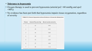
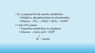
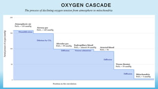
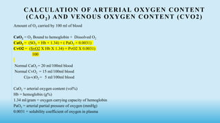
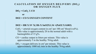
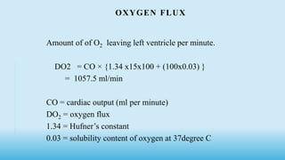
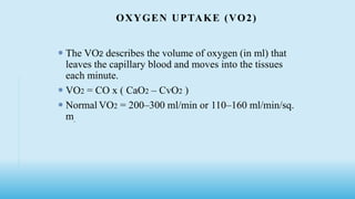
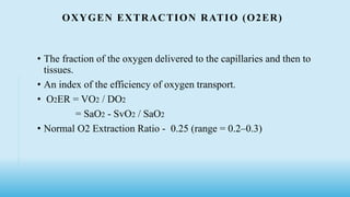
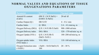
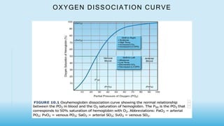
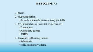
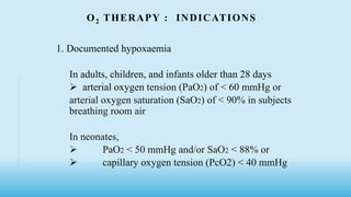
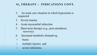
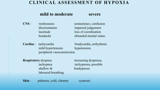
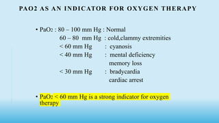
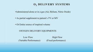
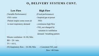
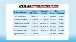
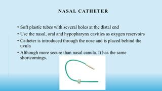
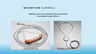
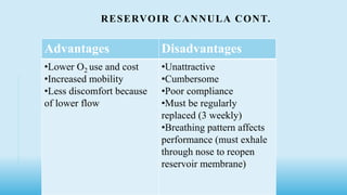
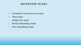
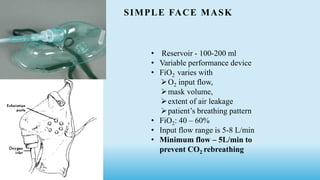
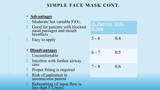
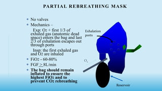
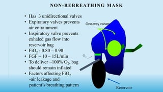
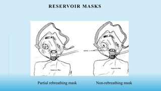
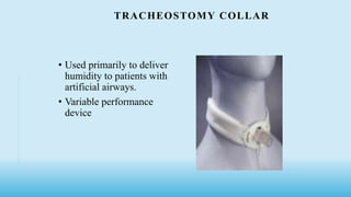
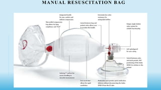
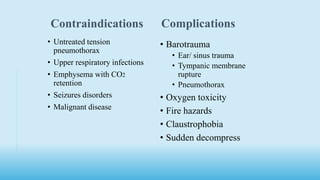
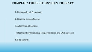
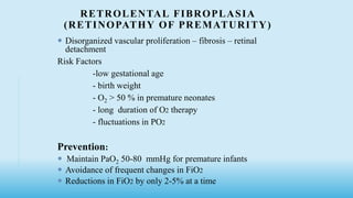
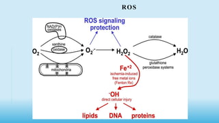
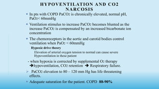
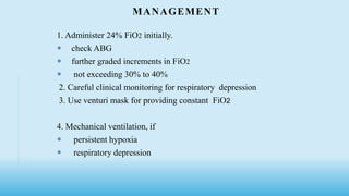
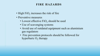
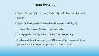
OXYGEN THERAPY
MODERATOR
Dr Ram
Assistant professor
Department of
Anaesthesiology
SMIMS
PRESENTED BY
Dr Gandam Raju
Junior resident
Department of
Anaesthesiology
SMIMS
INTRODUCTION
• Colorless, odorless, tasteless gas
• Discovered by Priestly in 1772
• Antoine Lavoisier coined the word oxygen 1779
• Oxygen therapy was introduced by Beddoes 1794
• Boiling point -183 C
• Melting point -216.6C
• Critical temp -118.6
• Constitutes about 20.95% of atmosphere
• Used at cellular level as the final electron acceptor in the electron
transport chain in the mitochondria of cell
• Though we depend on Oxygen for metabolic energy production, aerobic
metabolism is carried out in an oxygen restricted environment
• Oxygen requirement for basal metabolism at rest - 3.5mL/kg/minute
• humans as microaerophilic organism
3
OXYGEB
THERAPHY
• Tolerance to hypoxemia
• Oxygen therapy is used to prevent hypoxemia (arterial po2 <60 mmHg and spo2
<90%)
• No evidence has been put forth that hypoxemia impairs tissue oxygenation, regardless
of severity
4
OXYGEN
THERAPHY
O2 THERAPY AND AEROBIC METABOLISM
• Since hypoxemia doesn’t impair aerobic metabolism, then oxygen therapy
should not be necessary for preserving aerobic metabolism
5
OXYEGN
THERAPHY
• OXYGEN AND THE MICRO CIRCULATION
• Oxygen therapy produces systemic vasoconstriction, mainly in small arterioles
that control capillary blood flow
• At higher concentration of O2, arteriolar constriction is accompanied by
decrease in capillary density, resulting in decrease O2 availability in capillaries
• This is influences reduction of VO2 that occurs during normobaric hyperoxia
• It may be protective mechanism for protecting the tissue from oxygen induced
injury during periods of hyperoxia
6
OXYGEN
THERAPHY
OXYGEN THERAPY
Administration of O2 in concentration more than in
ambient air
↑Partial Pressure of O2 in inspiratory Gas (Pi o2)
↑Partial Pressure of O2 in alveoli (PAo2)
↑Partial Pressures of O2 in arterial blood (Pao2)
PAO2 = FiO2 (PB –PH2O ) – PaCO2/R
PAO2- partial pres of alveolar O2, FiO2 - fraction of inspired O2 ,
PB - Barometric pres , PH2O- pres of water , PaCO2- pres of arterial CO2,
R – Respiratory exchange ratio = 0.8
• O2 is required for the aerobic metabolism
• Oxidative phosphorylation in mitochondria
• Glucose + 6O2 → 6H2O + 6CO2 + 36ATP
• Lack of O2 causes
• Anaerobic metabolism in cytoplasm
• Glucose → lactic acid + 2ATP
↓
H+ + lactate-
OXYGEN CASCADE
The process of declining oxygen tension from atmosphere to mitochondria
CALCULATION OF ARTERIAL OXYGEN CONTENT
(CAO2) AND VENOUS OXYGEN CONTENT (CVO2)
Amount of O2 carried by 100 ml of blood
CaO2 = O2 Bound to hemoglobin + Dissolved O2
CaO2 = (SO2 × Hb × 1.34) + ( PaO2 × 0.0031)
CvO2 = (SvO2 X Hb X 1.34) + PvO2 X 0.0031)
100
Normal CaO2 = 20 ml/100ml blood
Normal CvO2 = 15 ml/100ml blood
C(a-v)O2 = 5 ml/100ml blood
CaO2 = arterial oxygen content (vol%)
Hb = hemoglobin (g%)
1.34 ml/gram = oxygen carrying capacity of hemoglobin
PaO2 = arterial partial pressure of oxygen (mmHg)
0.0031 = solubility coefficient of oxygen in plasma
CALCULATION OF OXYGEN DELIVERY (DO 2)
OR OXYGEN FLUX
DO2 = CaO2 X CO
or
DO2 = CO X OXYGEN CONTENT
DO2 = HR X SV X( Hb X SaO2X1.34 +(PaO2 X 0.003)
CaO2 = arterial oxygen content in cm3 per 100 cm3 blood (vol%).
This value is approximately 20 in the normal adult with a
haemoglobin of 15 g%.
CO = cardiac output in litters per minute. This value is
approximately 5 in the healthy 70-kg adult.
DO2 = oxygen delivery in cm3 per minute. This value is
approximately 1000 mL/min in the healthy 70-kg adult.
OXYGEN FLUX
Amount of of O2 leaving left ventricle per minute.
DO2 = CO × {1.34 x15x100 + (100x0.03) }
= 1057.5 ml/min
CO = cardiac output (ml per minute)
DO2 = oxygen flux
1.34 = Hufner’s constant
0.03 = solubility content of oxygen at 37degree C
OXYGEN UPTAKE (VO2)
 The VO2 describes the volume of oxygen (in ml) that
leaves the capillary blood and moves into the tissues
each minute.
 VO2 = CO x ( CaO2 – CvO2 )
 Normal VO2 = 200–300 ml/min or 110–160 ml/min/sq.
m.
OXYGEN EXTRACTION RATIO (O2ER)
• The fraction of the oxygen delivered to the capillaries and then to
tissues.
• An index of the efficiency of oxygen transport.
• O2ER = VO2 / DO2
= SaO2 - SvO2 / SaO2
• Normal O2 Extraction Ratio - 0.25 (range = 0.2–0.3)
NORMAL VALUES AND EQUATIONS OF TISSUE
OXYGENATIONS PARAMETERS
Parameter Equation Normal range
Arterial O2 content
(CaO2)
(Hb X 1.34 X SaO2) +
(0.0031 X PaO2)
20 ml/ dl
Cardiac Output (Q ) HR X SV 4 – 8 L/ min
Cardiac Index Q / BSA 2.4 – 4 L/min/sq. m
Oxygen Delivery (DO2) Q X 1.34 X Hb X SaO2 900 -1000 ml/min
Oxygen Delivery index DO2 /BSA 520 – 570 ml/min/ sq. m
Oxygen Uptake (VO2 ) Q X 1.34 X Hb X
(SaO2- SvO2 )
180 -280 ml/min/sq. m
Oxygen uptake index
(VO2 Index )
VO2 /BSA 110 -160 ml/min/sq. m
Oxygen Extraction ratio
( O2ER )
( SaO2 – SvO2/SaO2) X
100
20 – 30 %
OXYGEN DISSOCIATION CURVE
Reference ranges Arterial blood Venous blood
pH 7.35- 7.45 7.35- 7.43
pCO2 35 – 45 mm of Hg 38 -50 mm of Hg
pO2 80 – 100 mm of Hg 30 – 50 mm of Hg
HCO3- 22 – 26 mmol 23 – 27 mmol
O2 Saturation 95 – 100 % 60 -85 %
CLINICAL OBJECTIVES OF OXYGEN THERAPY
1. Treat hypoxemia
2. Decrease work of breathing (WOB)
3. Decrease myocardial Work
HYPOXIA: INADEQUATE DELIVERY OF O2 TO TISSUE
1. Hypoxaemia hypoxia ( decrease diffusion of O2 across the alveolar-capillary
membrane )
-low inspired FiO2
-V/Q inequalities
-increased shunt(eg cardiac anomalies)
2. Stagnant hypoxia (decreased cardiac output resulting in increased systemic transit
time )
-Shock
-Vasoconstriction
3. Anaemic hypoxia ( decreased O2 carrying capacity in the blood)
-Anaemia
-Carbon monoxide poisoning
4. Histotoxic hypoxia ( inability the tissue utilize available O2)
- Cyanide poisoning
5. Demand hypoxia ( increase O2 consumption)
- fever, seizures
HYPOXEMIA:
1. Shunt
2. Hypoventilation
• As carbon dioxide increases oxygen falls
3. V/Q mismatching (ventilation/perfusion)
• Pneumonia
• Pulmonary edema
• ARDS
4. Increased diffusion gradient
• Asbestosis
• Early pulmonary edema
O2 THERAPY : INDICATIONS
1. Documented hypoxaemia
In adults, children, and infants older than 28 days
 arterial oxygen tension (PaO2) of < 60 mmHg or
arterial oxygen saturation (SaO2) of < 90% in subjects
breathing room air
In neonates,
 PaO2 < 50 mmHg and/or SaO2 < 88% or
 capillary oxygen tension (PcO2) < 40 mmHg
O2 THERAPY : INDICATIONS CONT.
2 . An acute care situation in which hypoxemia is
suspected
3. Severe trauma
4. Acute myocardial infarction
5. Short-term therapy (e.g., post-anesthesia
recovery)
6. Increased metabolic demand eg.
 burns
 multiple injuries and
 severe infections.
ASSESSMENT
• Physical examination for C/F of hypoxemia
• Pulse oximetry
• ABG analysis
• pH
• pO2
• pCO2
• Mixed venous blood oxygenation
CLINICAL ASSESSMENT OF HYPOXIA
mild to moderate severe
CNS : restlessness somnolence, confusion
disorientation impaired judgement
lassitude loss of coordination
headache obtunded mental status
Cardiac : tachycardia bradycardia, arrhythmia
mild hypertension hypotension
peripheral vasoconstriction
Respiratory:dyspnea increasing dyspnoea,
tachypnea tachypnoea, possible
shallow & bradypnoea
laboured breathing
Skin : paleness, cold, clammy cyanosis
PAO2 AS AN INDICATOR FOR OXYGEN THERAPY
• PaO2 : 80 – 100 mm Hg : Normal
60 – 80 mm Hg : cold,clammy extremities
< 60 mm Hg : cyanosis
< 40 mm Hg : mental deficiency
memory loss
< 30 mm Hg : bradycardia
cardiac arrest
• PaO2 < 60 mm Hg is a strong indicator for oxygen
therapy
O2 DELIVERY SYSTEMS
•Administered alone or in a gas (Air, Helium, Nitric Oxide)
• As partial supplement to patient’s TV or MV
• Or Entire source of inspired volume
OXYGEN DELIVERY EQUIPMENTS
Low Flow High Flow
(Variable Performance) (Fixed performance)
O2 DELIVERY SYSTEMS CONT.
Low Flow High Flow
(Variable Performance) (Fixed performance)
-FiO2 varies - Inspired gas at preset
-Patient inspire some room air FIO2
to meet inspiratory demand - continuous high flow
- FiO2 not changed by
variation in ventilation
demand / breathing pattern
Minute ventilation <8-10L/Min.
RR < 20 / min.
TV < 0.8 L
(N) Inspiratory flow – 10-30L/Min - Consistent FiO2 and
flow >40 l/min
2 8
PRESENTATION
TITLE
TYPES OF FLOW DEVICES
Low flow devices High flow devices
1. Nasal cannulas
2. Simple face masks
3. Partial rebreathing masks
4. Non-rebreathing masks
5. Tracheostomy collars
1. Venturi mask
2. Face tent
3. Aerosol mask
4. T-piece
2 9
NASAL CANNULA
• Simple plastic tubing + prongs
• Flow from 1-6 L/min. of O2
• FiO2 ranges from 24-44% of O2
1 - 24%
2 - 28%
3 - 32%
4 - 36%
5 - 40%
6 - 44%
Advantages Disadvantages
• Inexpensive Pressure sores
• Well tolerated, comfortable Crusting of secretion
• Easy to eat, drink Drying of mucosa
• Used in patient with COPD Epistaxis
• Used with humidifier
NASAL CATHETER
• Soft plastic tubes with several holes at the distal end
• Use the nasal, oral and hypopharynx cavities as oxygen reservoirs
• Catheter is introduced through the nose and is placed behind the
uvula
• Although more secure than nasal canula. It has the same
shortcomings.
RESERVOIR CANNULA
Nasal reservoir Pendant reservoir
miniature reservoir is attached to the nasal canula
to economize oxygen delivery
RESERVOIR CANNULA CONT.
Advantages Disadvantages
•Lower O2 use and cost
•Increased mobility
•Less discomfort because
of lower flow
•Unattractive
•Cumbersome
•Poor compliance
•Must be regularly
replaced (3 weekly)
•Breathing pattern affects
performance (must exhale
through nose to reopen
reservoir membrane)
RESERVOIR MASKS
 Commonly used reservoir system
 Three types
1. Simple face mask
2. Partial rebreathing masks
3. Non rebreathing masks
SIMPLE FACE MASK
• Reservoir - 100-200 ml
• Variable performance device
• FiO2 varies with
O2 input flow,
mask volume,
extent of air leakage
patient’s breathing pattern
• FiO2: 40 – 60%
• Input flow range is 5-8 L/min
• Minimum flow – 5L/min to
prevent CO2 rebreathing
SIMPLE FACE MASK CONT.
 Advantages
1. Moderate but variable FiO2.
2. Good for patients with blocked
nasal passages and mouth
breathers
3. Easy to apply
 Disadvantages
1. Uncomfortable
2. Interfere with further airway
care
3. Proper fitting is required
4. Risk of aspiration in
unconscious patient
5. Rebreathing (if input flow is
less than 5 L/min)
O2 Flowrate
(L/min
Fi O2
5 - 6 0.4
6 - 7 0.5
7 - 8 0.6
PARTIAL REBREATHING MASK
 No valves
 Mechanics –
Exp: O2 + first 1/3 of
exhaled gas (anatomic dead
space) enters the bag and last
2/3 of exhalation escapes out
through ports
Insp: the first exhaled gas
and O2 are inhaled
 FiO2 - 60-80%
 FGF > 8L/min
 The bag should remain
inflated to ensure the
highest FiO2 and to
prevent CO2 rebreathing
+
Exhalation
ports
O2
Reservoir
NON-REBREATHING MASK
• Has 3 unidirectional valves
• Expiratory valves prevents
air entrainment
• Inspiratory valve prevents
exhaled gas flow into
reservoir bag
• FiO2 - 0.80 – 0.90
• FGF – 10 – 15L/min
• To deliver ~100% O2, bag
should remain inflated
• Factors affecting FiO2
-air leakage and
patient’s breathing pattern
Reservoir
O2
One-way valves
RESERVOIR MASKS
Partial rebreathing mask Non-rebreathing mask
TRACHEOSTOMY COLLAR
• Used primarily to deliver
humidity to patients with
artificial airways.
• Variable performance
device
HIGH FLOW/FIXED PERFORMANCE EQUIPMENT'S
Manual resuscitation bags
• used primarily for resuscitation and manual ventilation of
ventilator-dependent patients.
• deliver an FiO2 above 0.90 and TV up to 800 mL when
oxygen flows to the bag are 10 to 15 mL/min.
• highest FiO2s - use of an oxygen reservoir, connection to
an oxygen source, and slow rates of ventilation that allow
the bag to refill completely.
• PEEP valve used for pts who require PEEP > 5cm H2O.
MANUAL RESUSCITATION BAG
VENTURI OR VENTI MASK
• High flow O2 mask
• provide complete control of inhaled gas mixture and deliver a
constant FIO2 regardless of changes in ventilatory pattern .
• O2 is delivered to the mask at low flow rate
but at the inlet of mask , O2 is passed through a narrow orifice and
create a high velocity which pulls room air inside the mask
•Venturi masks entrain air by the Bernoulli principle and constant
pressure-jet mixing
•This physical phenomenon is based on a rapid of gas (e.g., oxygen)
moving through a restricted orifice. This produces viscous shearing
forces, which create a decreased pressure gradient (subatmospheric)
downstream relative to the surrounding gases. This pressure gradient
causes RA to be entrained until the pressures are equalized
VENTURI OR VENTI MASK CONT.
• multiple color coded jets to set the desired FiO2
• color of the device reflects the delivered oxygen concentration
• blue = 24%; yellow = 28%; white = 31%; green = 35%; pink =
40%; orange = 50%.(flow rate 4 to 12lit/min)
Advantages :
-Ability to deliver constant FiO2
-Air tight fit not required
-No apparatus dead space
-Humidification not required
FiO2 more controllable – ideal for decompensated patients
with COPD with CO2 retention.
Limitations :
-Interruption of O2 supply leads to hypoxemia and
hypercarbia in unstable patient
-Obstruction of entrainment ports - ↑ FiO2
AIR ENTRAINMENT NEBULIZERS
-Pneumatic jet with adjustable orifice to vary entrained
air for various FiO2 levels
-Provide bland mist therapy with some control of FiO2
Use:
-after extubation for aerosol therapy
-applied to many devices – tracheostomy dome/collar
face tent
T piece adapter
- Mist like tracer determines adequacy of flow.
Disadvantage :
Excess water in tubing obstructs gas flow
Bronchospasm -sterile water for aerosol can be irritating
OXYGEN TENTS / HOODS
 Ideal for short term oxygen therapy for newborn and
inactive Infant
Clear plastic shell covering the baby's head
 Easy access to chest, trunk, and extremities
 O2 and air premixed by an air oxygen blending device
and passed through a heated humidifiers.
 Delivers 80-90% oxygen at 10-15 liter per minute
 Minimum>7L/min to remove CO2 .
Tents cover the whole body.
OXYGEN TENT/HOODS
FACE TENT
OXYGEN HOOD
OXYGEN TENT
HELIOX (HELIUM – O2 THERAPY)
Less dense compared with pure O2
Popular mixtures 80:20(1.8 density) and 70:30(1.6) (Helium
oxygen)
Available in large sized compressed gas cylinders
used with small volume nebulizers for bronchodilator therapy –
11 L/min
Advantage:
Relief in upper airway obstructing lesions like foreign body,
subglottic edema, tracheal tumors
 use in lower airways obstruction, COPD, acute asthma
pressure needed to ventilate patients with smaller ET tube is
reduced to half
- WOB is ↓ when delivered by mechanical ventilator.
HYPERBARIC OXYGEN THERAPY
• A mode of medical treatment wherein
the patient breathes 100% oxygen at a pressure greater
than one Atmosphere Absolute (1 ATA)
• 1 ATM is equal to 760 mm Hg at sea level
Basis of Hyperbaric O2 Therapy
Dissolved O2 in plasma :
0.003ml / 100ml of blood / mm PO2
(Henry’s Law -The concentration of any gas in solution is
proportional to its partial pressure.)
The basis is to increase the concentration of dissolved oxygen
OXYGEN CONTENTS OF THE BLOOD AT
DIFFERENT ATMOSPHERIC PRESSURE
WITH HB 14GMS/DL WHEN BREATHING 100
% OXYGEN
Pressure
(Atm )
PaO2
( mm Hg )
Dissolved O2
(%)
Total O2
content
(ml/dl )
1 670 11 18.7
2 1200-1300 18 20
3 1700-1800 25 22
PHYSIOLOGICAL EFFECTS OF HBO
 It increases PaO2 and PO2 to levels higher than those at
1 atm.
 promote healing, phagocytosis and antibiotic action
O2 is available to tissues without any increase in Hb
 There is an increase in ambient pressures which
increases interstitial pressure as well- used to treat
decompression sickness
 Neovascularization
 Vasoconstriction
 An increased Hb saturation of venous blood reduces its
ability to carry CO2 and results in CO2 accumulation.
INDICATIONS OF HBOT
ACUTE CONDITIONS CHRONIC CONDITIONS
• Decompression sickness
• Air embolism
• Carbon monoxide
poisoning
• Severe crush injuries
• Thermal burns
• Acute arterial insufficiency
• Clostridial gangrene
• Necrotizing soft-tissue
infection
• Ischemic skin graft or flap
•Radiation necrosis
•Diabetic wounds of lower
limbs
•Refractory osteomyelitis
•Actinomycosis (chronic
systemic abscesses)
METHODS OF
ADMINISTRATION OF HBOT
Contraindications Complications
• Untreated tension
pneumothorax
• Upper respiratory infections
• Emphysema with CO2
retention
• Seizures disorders
• Malignant disease
• Barotrauma
• Ear/ sinus trauma
• Tympanic membrane
rupture
• Pneumothorax
• Oxygen toxicity
• Fire hazards
• Claustrophobia
• Sudden decompress
COMPLICATIONS OF OXYGEN THERAPY
1. Retinopathy of Prematurity
2. Reactive oxygen Species
3. Adsorption atelectasis
4.Decreased hypoxic drive (Hypoventilation and CO2 narcosis)
5. Fire hazards
RETROLENTAL FIBROPLASIA
(RETINOPATHY OF PREMATURITY)
 Disorganized vascular proliferation – fibrosis – retinal
detachment
Risk Factors
-low gestational age
- birth weight
- O2 > 50 % in premature neonates
- long duration of O2 therapy
- fluctuations in PO2
Prevention:
 Maintain PaO2 50-80 mmHg for premature infants
 Avoidance of frequent changes in FiO2
 Reductions in FiO2 by only 2-5% at a time
ROS
ROS
Mechanism: intracellular generation of free radicals (highly reactive metabolites)
e.g.: superoxide, H2O2 react with cellular DNA
singlet oxygen,  sulphydryl proteins & lipids
haemorrhagic intracellular  cytotoxicity
alveolar oedema
pulmonary fibrosis  decrease in pulmonary compliance
resp. failure
PULMONARY TOXICITY
 Presents with one of the following three
- Tracheobronchitis
- Acute lung injury (ARDS)
- Bronchopulmonary dysplasia
 Cough, substernal pain and nasal stuffiness occur within 6 hrs on
100% O2.
 More prolonged exposure causes disruption of the endothelial
lining, leakage of proteinacious fluid and an ARDS like
syndrome
 Depends on
- partial pressure of O2 in Inspired air
-duration of exposure
 Goal should be to use lowest possible FiO2 compatible with
adequate tissue oxygenation
 Inhalation of high O2 tension for prolonged periods should be
avoided.
ANTI-OXIDANTS
• Glutathione
• N-acetyl cystine
• Selenium
• Vit-C
• Vit- E
• Ceruloplasmin and transferrin
6 3
PRESENTATION
TITLE
ADSORPTION ATELECTASIS
• Air: contains 79% Nitrogen
relatively insoluble
maintains residual volume in the alveoli
• In high FiO2 inhalation,
Nitrogen washout of alveoli and get filled with oxygen
areas with low V/Q- O2 absorption in pulmonary capillaries
Partial or total collapse of alveoli
• Alveoli in low V/Q areas are more prone
e.g.: COPD, Bronchial asthma
HYPOVENTILATION AND CO2
NARCOSIS
 In pts with COPD PaCO2 is chronically elevated, normal pH,
PaO2< 60mmHg
 Ventilation stimulus to increase PaCO2 becomes blunted as the
increase PaCO2 is compensated by an increased bicarbonate ion
concentration
 The chemoreceptors in the aortic and carotid bodies control
ventilation when PaO2 < 60mmHg
Hypoxic drive theory
Elevation of arterial oxygen tension to normal can cause severe
Hypoventilation in these patient
- when hypoxia is corrected by supplemental O2 therapy
hypoventilation, CO2 retention  Respiratory failure.
 PaCO2 elevation to 80 – 120 mm Hg has life threatening
effects.
 Adequate saturation for the patient. COPD 88-90%
MANAGEMENT
1. Administer 24% FiO2 initially.
 check ABG
 further graded increments in FiO2
 not exceeding 30% to 40%
2. Careful clinical monitoring for respiratory depression
3. Use venturi mask for providing constant FiO2
4. Mechanical ventilation, if
 persistent hypoxia
 respiratory depression
FIRE HAZARDS
• High FiO2 increases the risk of fire
• Preventive measures
• Lowest effective FiO2 should be used
• Use of scavenging systems
• Avoid use of outdated equipment such as aluminium
gas regulators
• Fire prevention protocols should be followed for
hyperbaric O2 therapy
• Liquid Oxygen (O2) is one of the physical form of elemental
oxygen.
• Liquid O2 is oxygen that’s cooled to -183 deg C (-297 deg F)
• It is pale blue in color & strongly paramagnetic
• It is cryogenic . Boiling point -183 deg C at 760 mm Hg .
• 1 volume of liquid oxygen yields 842 times of its volume of O2 in
gaseous form at 15 deg C temperature & 1 atm pressure.
LIQUID OXYGEN
THERAPEUTIC USES -
• Liquid oxygen is widely used in clinical practice to provide a
basis for most modern anaesthetic techniques .
• Used in Hospitals as a source of oxygen during pre / intra/ post-
operative period
• To restore tissue oxygen tension towards normal by improving
oxygen availability in variety of conditions like –
I. Cyanosis of recent origin
II. surgical trauma
III. shock , severe hemorrhage
IV. In CPR
V. CO poisoning , etc ..
• Used in Home medical oxygen supply device .
• Liquid oxygen containers are used for transfer of patients , to
provide anaesthesia out side health care facility ( eg- by army
services )
• Also Used in SUBMARINE & in Overhead emergency O2
devices in aircrafts.
SUMMARY
• Therapeutic effectiveness of oxygen therapy is limited
to 25% - 50%
• Low V/Q hypoxemia is reversed with less than 50%
• Pulmonary oxygen toxicity is a potential risk factor
with FiO2 more than 50%
• Treat like any other drug by proper documentation:
• Date and time oxygen started.
• Method of delivery.
• Oxygen concentration and flow rate.
• Patient observation.
REFERENCES
• PAUL MARINO – THE ICU BOOK 4TH EDITION
• PAUL G BARASH CLINICAL ANESTHESIA
• MORGAN & MIKHAIL’S CLINICAL ANAESTHEIOLOGY
• STANDARD INTERNET SOURCES
7 2
PRESENTATION
TITLE
THANK YOU
TYPES OF HYPOXIA
DIFFUSION HYPOXIA/FINK EFFECT
OXYGEN CASCADE
ANAESTHETIC IMPLICATION
ODC
EFFECTS OF OXYGEN THERAPY

OXYGEN THERAPHY.pptx

  • 1.
    OXYGEN THERAPY MODERATOR Dr Ram Assistantprofessor Department of Anaesthesiology SMIMS PRESENTED BY Dr Gandam Raju Junior resident Department of Anaesthesiology SMIMS
  • 2.
    INTRODUCTION • Colorless, odorless,tasteless gas • Discovered by Priestly in 1772 • Antoine Lavoisier coined the word oxygen 1779 • Oxygen therapy was introduced by Beddoes 1794 • Boiling point -183 C • Melting point -216.6C • Critical temp -118.6 • Constitutes about 20.95% of atmosphere • Used at cellular level as the final electron acceptor in the electron transport chain in the mitochondria of cell
  • 3.
    • Though wedepend on Oxygen for metabolic energy production, aerobic metabolism is carried out in an oxygen restricted environment • Oxygen requirement for basal metabolism at rest - 3.5mL/kg/minute • humans as microaerophilic organism 3 OXYGEB THERAPHY
  • 4.
    • Tolerance tohypoxemia • Oxygen therapy is used to prevent hypoxemia (arterial po2 <60 mmHg and spo2 <90%) • No evidence has been put forth that hypoxemia impairs tissue oxygenation, regardless of severity 4 OXYGEN THERAPHY
  • 5.
    O2 THERAPY ANDAEROBIC METABOLISM • Since hypoxemia doesn’t impair aerobic metabolism, then oxygen therapy should not be necessary for preserving aerobic metabolism 5 OXYEGN THERAPHY
  • 6.
    • OXYGEN ANDTHE MICRO CIRCULATION • Oxygen therapy produces systemic vasoconstriction, mainly in small arterioles that control capillary blood flow • At higher concentration of O2, arteriolar constriction is accompanied by decrease in capillary density, resulting in decrease O2 availability in capillaries • This is influences reduction of VO2 that occurs during normobaric hyperoxia • It may be protective mechanism for protecting the tissue from oxygen induced injury during periods of hyperoxia 6 OXYGEN THERAPHY
  • 7.
    OXYGEN THERAPY Administration ofO2 in concentration more than in ambient air ↑Partial Pressure of O2 in inspiratory Gas (Pi o2) ↑Partial Pressure of O2 in alveoli (PAo2) ↑Partial Pressures of O2 in arterial blood (Pao2) PAO2 = FiO2 (PB –PH2O ) – PaCO2/R PAO2- partial pres of alveolar O2, FiO2 - fraction of inspired O2 , PB - Barometric pres , PH2O- pres of water , PaCO2- pres of arterial CO2, R – Respiratory exchange ratio = 0.8
  • 8.
    • O2 isrequired for the aerobic metabolism • Oxidative phosphorylation in mitochondria • Glucose + 6O2 → 6H2O + 6CO2 + 36ATP • Lack of O2 causes • Anaerobic metabolism in cytoplasm • Glucose → lactic acid + 2ATP ↓ H+ + lactate-
  • 9.
    OXYGEN CASCADE The processof declining oxygen tension from atmosphere to mitochondria
  • 10.
    CALCULATION OF ARTERIALOXYGEN CONTENT (CAO2) AND VENOUS OXYGEN CONTENT (CVO2) Amount of O2 carried by 100 ml of blood CaO2 = O2 Bound to hemoglobin + Dissolved O2 CaO2 = (SO2 × Hb × 1.34) + ( PaO2 × 0.0031) CvO2 = (SvO2 X Hb X 1.34) + PvO2 X 0.0031) 100 Normal CaO2 = 20 ml/100ml blood Normal CvO2 = 15 ml/100ml blood C(a-v)O2 = 5 ml/100ml blood CaO2 = arterial oxygen content (vol%) Hb = hemoglobin (g%) 1.34 ml/gram = oxygen carrying capacity of hemoglobin PaO2 = arterial partial pressure of oxygen (mmHg) 0.0031 = solubility coefficient of oxygen in plasma
  • 11.
    CALCULATION OF OXYGENDELIVERY (DO 2) OR OXYGEN FLUX DO2 = CaO2 X CO or DO2 = CO X OXYGEN CONTENT DO2 = HR X SV X( Hb X SaO2X1.34 +(PaO2 X 0.003) CaO2 = arterial oxygen content in cm3 per 100 cm3 blood (vol%). This value is approximately 20 in the normal adult with a haemoglobin of 15 g%. CO = cardiac output in litters per minute. This value is approximately 5 in the healthy 70-kg adult. DO2 = oxygen delivery in cm3 per minute. This value is approximately 1000 mL/min in the healthy 70-kg adult.
  • 12.
    OXYGEN FLUX Amount ofof O2 leaving left ventricle per minute. DO2 = CO × {1.34 x15x100 + (100x0.03) } = 1057.5 ml/min CO = cardiac output (ml per minute) DO2 = oxygen flux 1.34 = Hufner’s constant 0.03 = solubility content of oxygen at 37degree C
  • 13.
    OXYGEN UPTAKE (VO2) The VO2 describes the volume of oxygen (in ml) that leaves the capillary blood and moves into the tissues each minute.  VO2 = CO x ( CaO2 – CvO2 )  Normal VO2 = 200–300 ml/min or 110–160 ml/min/sq. m.
  • 14.
    OXYGEN EXTRACTION RATIO(O2ER) • The fraction of the oxygen delivered to the capillaries and then to tissues. • An index of the efficiency of oxygen transport. • O2ER = VO2 / DO2 = SaO2 - SvO2 / SaO2 • Normal O2 Extraction Ratio - 0.25 (range = 0.2–0.3)
  • 15.
    NORMAL VALUES ANDEQUATIONS OF TISSUE OXYGENATIONS PARAMETERS Parameter Equation Normal range Arterial O2 content (CaO2) (Hb X 1.34 X SaO2) + (0.0031 X PaO2) 20 ml/ dl Cardiac Output (Q ) HR X SV 4 – 8 L/ min Cardiac Index Q / BSA 2.4 – 4 L/min/sq. m Oxygen Delivery (DO2) Q X 1.34 X Hb X SaO2 900 -1000 ml/min Oxygen Delivery index DO2 /BSA 520 – 570 ml/min/ sq. m Oxygen Uptake (VO2 ) Q X 1.34 X Hb X (SaO2- SvO2 ) 180 -280 ml/min/sq. m Oxygen uptake index (VO2 Index ) VO2 /BSA 110 -160 ml/min/sq. m Oxygen Extraction ratio ( O2ER ) ( SaO2 – SvO2/SaO2) X 100 20 – 30 %
  • 16.
  • 17.
    Reference ranges Arterialblood Venous blood pH 7.35- 7.45 7.35- 7.43 pCO2 35 – 45 mm of Hg 38 -50 mm of Hg pO2 80 – 100 mm of Hg 30 – 50 mm of Hg HCO3- 22 – 26 mmol 23 – 27 mmol O2 Saturation 95 – 100 % 60 -85 %
  • 18.
    CLINICAL OBJECTIVES OFOXYGEN THERAPY 1. Treat hypoxemia 2. Decrease work of breathing (WOB) 3. Decrease myocardial Work
  • 19.
    HYPOXIA: INADEQUATE DELIVERYOF O2 TO TISSUE 1. Hypoxaemia hypoxia ( decrease diffusion of O2 across the alveolar-capillary membrane ) -low inspired FiO2 -V/Q inequalities -increased shunt(eg cardiac anomalies) 2. Stagnant hypoxia (decreased cardiac output resulting in increased systemic transit time ) -Shock -Vasoconstriction 3. Anaemic hypoxia ( decreased O2 carrying capacity in the blood) -Anaemia -Carbon monoxide poisoning 4. Histotoxic hypoxia ( inability the tissue utilize available O2) - Cyanide poisoning 5. Demand hypoxia ( increase O2 consumption) - fever, seizures
  • 20.
    HYPOXEMIA: 1. Shunt 2. Hypoventilation •As carbon dioxide increases oxygen falls 3. V/Q mismatching (ventilation/perfusion) • Pneumonia • Pulmonary edema • ARDS 4. Increased diffusion gradient • Asbestosis • Early pulmonary edema
  • 21.
    O2 THERAPY :INDICATIONS 1. Documented hypoxaemia In adults, children, and infants older than 28 days  arterial oxygen tension (PaO2) of < 60 mmHg or arterial oxygen saturation (SaO2) of < 90% in subjects breathing room air In neonates,  PaO2 < 50 mmHg and/or SaO2 < 88% or  capillary oxygen tension (PcO2) < 40 mmHg
  • 22.
    O2 THERAPY :INDICATIONS CONT. 2 . An acute care situation in which hypoxemia is suspected 3. Severe trauma 4. Acute myocardial infarction 5. Short-term therapy (e.g., post-anesthesia recovery) 6. Increased metabolic demand eg.  burns  multiple injuries and  severe infections.
  • 23.
    ASSESSMENT • Physical examinationfor C/F of hypoxemia • Pulse oximetry • ABG analysis • pH • pO2 • pCO2 • Mixed venous blood oxygenation
  • 24.
    CLINICAL ASSESSMENT OFHYPOXIA mild to moderate severe CNS : restlessness somnolence, confusion disorientation impaired judgement lassitude loss of coordination headache obtunded mental status Cardiac : tachycardia bradycardia, arrhythmia mild hypertension hypotension peripheral vasoconstriction Respiratory:dyspnea increasing dyspnoea, tachypnea tachypnoea, possible shallow & bradypnoea laboured breathing Skin : paleness, cold, clammy cyanosis
  • 25.
    PAO2 AS ANINDICATOR FOR OXYGEN THERAPY • PaO2 : 80 – 100 mm Hg : Normal 60 – 80 mm Hg : cold,clammy extremities < 60 mm Hg : cyanosis < 40 mm Hg : mental deficiency memory loss < 30 mm Hg : bradycardia cardiac arrest • PaO2 < 60 mm Hg is a strong indicator for oxygen therapy
  • 26.
    O2 DELIVERY SYSTEMS •Administeredalone or in a gas (Air, Helium, Nitric Oxide) • As partial supplement to patient’s TV or MV • Or Entire source of inspired volume OXYGEN DELIVERY EQUIPMENTS Low Flow High Flow (Variable Performance) (Fixed performance)
  • 27.
    O2 DELIVERY SYSTEMSCONT. Low Flow High Flow (Variable Performance) (Fixed performance) -FiO2 varies - Inspired gas at preset -Patient inspire some room air FIO2 to meet inspiratory demand - continuous high flow - FiO2 not changed by variation in ventilation demand / breathing pattern Minute ventilation <8-10L/Min. RR < 20 / min. TV < 0.8 L (N) Inspiratory flow – 10-30L/Min - Consistent FiO2 and flow >40 l/min
  • 28.
  • 29.
    TYPES OF FLOWDEVICES Low flow devices High flow devices 1. Nasal cannulas 2. Simple face masks 3. Partial rebreathing masks 4. Non-rebreathing masks 5. Tracheostomy collars 1. Venturi mask 2. Face tent 3. Aerosol mask 4. T-piece 2 9
  • 30.
    NASAL CANNULA • Simpleplastic tubing + prongs • Flow from 1-6 L/min. of O2 • FiO2 ranges from 24-44% of O2 1 - 24% 2 - 28% 3 - 32% 4 - 36% 5 - 40% 6 - 44%
  • 31.
    Advantages Disadvantages • InexpensivePressure sores • Well tolerated, comfortable Crusting of secretion • Easy to eat, drink Drying of mucosa • Used in patient with COPD Epistaxis • Used with humidifier
  • 32.
    NASAL CATHETER • Softplastic tubes with several holes at the distal end • Use the nasal, oral and hypopharynx cavities as oxygen reservoirs • Catheter is introduced through the nose and is placed behind the uvula • Although more secure than nasal canula. It has the same shortcomings.
  • 33.
    RESERVOIR CANNULA Nasal reservoirPendant reservoir miniature reservoir is attached to the nasal canula to economize oxygen delivery
  • 34.
    RESERVOIR CANNULA CONT. AdvantagesDisadvantages •Lower O2 use and cost •Increased mobility •Less discomfort because of lower flow •Unattractive •Cumbersome •Poor compliance •Must be regularly replaced (3 weekly) •Breathing pattern affects performance (must exhale through nose to reopen reservoir membrane)
  • 35.
    RESERVOIR MASKS  Commonlyused reservoir system  Three types 1. Simple face mask 2. Partial rebreathing masks 3. Non rebreathing masks
  • 36.
    SIMPLE FACE MASK •Reservoir - 100-200 ml • Variable performance device • FiO2 varies with O2 input flow, mask volume, extent of air leakage patient’s breathing pattern • FiO2: 40 – 60% • Input flow range is 5-8 L/min • Minimum flow – 5L/min to prevent CO2 rebreathing
  • 37.
    SIMPLE FACE MASKCONT.  Advantages 1. Moderate but variable FiO2. 2. Good for patients with blocked nasal passages and mouth breathers 3. Easy to apply  Disadvantages 1. Uncomfortable 2. Interfere with further airway care 3. Proper fitting is required 4. Risk of aspiration in unconscious patient 5. Rebreathing (if input flow is less than 5 L/min) O2 Flowrate (L/min Fi O2 5 - 6 0.4 6 - 7 0.5 7 - 8 0.6
  • 38.
    PARTIAL REBREATHING MASK No valves  Mechanics – Exp: O2 + first 1/3 of exhaled gas (anatomic dead space) enters the bag and last 2/3 of exhalation escapes out through ports Insp: the first exhaled gas and O2 are inhaled  FiO2 - 60-80%  FGF > 8L/min  The bag should remain inflated to ensure the highest FiO2 and to prevent CO2 rebreathing + Exhalation ports O2 Reservoir
  • 39.
    NON-REBREATHING MASK • Has3 unidirectional valves • Expiratory valves prevents air entrainment • Inspiratory valve prevents exhaled gas flow into reservoir bag • FiO2 - 0.80 – 0.90 • FGF – 10 – 15L/min • To deliver ~100% O2, bag should remain inflated • Factors affecting FiO2 -air leakage and patient’s breathing pattern Reservoir O2 One-way valves
  • 40.
    RESERVOIR MASKS Partial rebreathingmask Non-rebreathing mask
  • 41.
    TRACHEOSTOMY COLLAR • Usedprimarily to deliver humidity to patients with artificial airways. • Variable performance device
  • 42.
    HIGH FLOW/FIXED PERFORMANCEEQUIPMENT'S Manual resuscitation bags • used primarily for resuscitation and manual ventilation of ventilator-dependent patients. • deliver an FiO2 above 0.90 and TV up to 800 mL when oxygen flows to the bag are 10 to 15 mL/min. • highest FiO2s - use of an oxygen reservoir, connection to an oxygen source, and slow rates of ventilation that allow the bag to refill completely. • PEEP valve used for pts who require PEEP > 5cm H2O.
  • 43.
  • 44.
    VENTURI OR VENTIMASK • High flow O2 mask • provide complete control of inhaled gas mixture and deliver a constant FIO2 regardless of changes in ventilatory pattern . • O2 is delivered to the mask at low flow rate but at the inlet of mask , O2 is passed through a narrow orifice and create a high velocity which pulls room air inside the mask •Venturi masks entrain air by the Bernoulli principle and constant pressure-jet mixing •This physical phenomenon is based on a rapid of gas (e.g., oxygen) moving through a restricted orifice. This produces viscous shearing forces, which create a decreased pressure gradient (subatmospheric) downstream relative to the surrounding gases. This pressure gradient causes RA to be entrained until the pressures are equalized
  • 45.
    VENTURI OR VENTIMASK CONT.
  • 46.
    VENTURI OR VENTIMASK CONT. • multiple color coded jets to set the desired FiO2 • color of the device reflects the delivered oxygen concentration • blue = 24%; yellow = 28%; white = 31%; green = 35%; pink = 40%; orange = 50%.(flow rate 4 to 12lit/min)
  • 47.
    Advantages : -Ability todeliver constant FiO2 -Air tight fit not required -No apparatus dead space -Humidification not required FiO2 more controllable – ideal for decompensated patients with COPD with CO2 retention. Limitations : -Interruption of O2 supply leads to hypoxemia and hypercarbia in unstable patient -Obstruction of entrainment ports - ↑ FiO2
  • 48.
    AIR ENTRAINMENT NEBULIZERS -Pneumaticjet with adjustable orifice to vary entrained air for various FiO2 levels -Provide bland mist therapy with some control of FiO2 Use: -after extubation for aerosol therapy -applied to many devices – tracheostomy dome/collar face tent T piece adapter - Mist like tracer determines adequacy of flow. Disadvantage : Excess water in tubing obstructs gas flow Bronchospasm -sterile water for aerosol can be irritating
  • 49.
    OXYGEN TENTS /HOODS  Ideal for short term oxygen therapy for newborn and inactive Infant Clear plastic shell covering the baby's head  Easy access to chest, trunk, and extremities  O2 and air premixed by an air oxygen blending device and passed through a heated humidifiers.  Delivers 80-90% oxygen at 10-15 liter per minute  Minimum>7L/min to remove CO2 . Tents cover the whole body.
  • 50.
  • 51.
    HELIOX (HELIUM –O2 THERAPY) Less dense compared with pure O2 Popular mixtures 80:20(1.8 density) and 70:30(1.6) (Helium oxygen) Available in large sized compressed gas cylinders used with small volume nebulizers for bronchodilator therapy – 11 L/min Advantage: Relief in upper airway obstructing lesions like foreign body, subglottic edema, tracheal tumors  use in lower airways obstruction, COPD, acute asthma pressure needed to ventilate patients with smaller ET tube is reduced to half - WOB is ↓ when delivered by mechanical ventilator.
  • 52.
    HYPERBARIC OXYGEN THERAPY •A mode of medical treatment wherein the patient breathes 100% oxygen at a pressure greater than one Atmosphere Absolute (1 ATA) • 1 ATM is equal to 760 mm Hg at sea level Basis of Hyperbaric O2 Therapy Dissolved O2 in plasma : 0.003ml / 100ml of blood / mm PO2 (Henry’s Law -The concentration of any gas in solution is proportional to its partial pressure.) The basis is to increase the concentration of dissolved oxygen
  • 53.
    OXYGEN CONTENTS OFTHE BLOOD AT DIFFERENT ATMOSPHERIC PRESSURE WITH HB 14GMS/DL WHEN BREATHING 100 % OXYGEN Pressure (Atm ) PaO2 ( mm Hg ) Dissolved O2 (%) Total O2 content (ml/dl ) 1 670 11 18.7 2 1200-1300 18 20 3 1700-1800 25 22
  • 54.
    PHYSIOLOGICAL EFFECTS OFHBO  It increases PaO2 and PO2 to levels higher than those at 1 atm.  promote healing, phagocytosis and antibiotic action O2 is available to tissues without any increase in Hb  There is an increase in ambient pressures which increases interstitial pressure as well- used to treat decompression sickness  Neovascularization  Vasoconstriction  An increased Hb saturation of venous blood reduces its ability to carry CO2 and results in CO2 accumulation.
  • 55.
    INDICATIONS OF HBOT ACUTECONDITIONS CHRONIC CONDITIONS • Decompression sickness • Air embolism • Carbon monoxide poisoning • Severe crush injuries • Thermal burns • Acute arterial insufficiency • Clostridial gangrene • Necrotizing soft-tissue infection • Ischemic skin graft or flap •Radiation necrosis •Diabetic wounds of lower limbs •Refractory osteomyelitis •Actinomycosis (chronic systemic abscesses)
  • 56.
  • 57.
    Contraindications Complications • Untreatedtension pneumothorax • Upper respiratory infections • Emphysema with CO2 retention • Seizures disorders • Malignant disease • Barotrauma • Ear/ sinus trauma • Tympanic membrane rupture • Pneumothorax • Oxygen toxicity • Fire hazards • Claustrophobia • Sudden decompress
  • 58.
    COMPLICATIONS OF OXYGENTHERAPY 1. Retinopathy of Prematurity 2. Reactive oxygen Species 3. Adsorption atelectasis 4.Decreased hypoxic drive (Hypoventilation and CO2 narcosis) 5. Fire hazards
  • 59.
    RETROLENTAL FIBROPLASIA (RETINOPATHY OFPREMATURITY)  Disorganized vascular proliferation – fibrosis – retinal detachment Risk Factors -low gestational age - birth weight - O2 > 50 % in premature neonates - long duration of O2 therapy - fluctuations in PO2 Prevention:  Maintain PaO2 50-80 mmHg for premature infants  Avoidance of frequent changes in FiO2  Reductions in FiO2 by only 2-5% at a time
  • 60.
  • 61.
    ROS Mechanism: intracellular generationof free radicals (highly reactive metabolites) e.g.: superoxide, H2O2 react with cellular DNA singlet oxygen,  sulphydryl proteins & lipids haemorrhagic intracellular  cytotoxicity alveolar oedema pulmonary fibrosis  decrease in pulmonary compliance resp. failure
  • 62.
    PULMONARY TOXICITY  Presentswith one of the following three - Tracheobronchitis - Acute lung injury (ARDS) - Bronchopulmonary dysplasia  Cough, substernal pain and nasal stuffiness occur within 6 hrs on 100% O2.  More prolonged exposure causes disruption of the endothelial lining, leakage of proteinacious fluid and an ARDS like syndrome  Depends on - partial pressure of O2 in Inspired air -duration of exposure  Goal should be to use lowest possible FiO2 compatible with adequate tissue oxygenation  Inhalation of high O2 tension for prolonged periods should be avoided.
  • 63.
    ANTI-OXIDANTS • Glutathione • N-acetylcystine • Selenium • Vit-C • Vit- E • Ceruloplasmin and transferrin 6 3 PRESENTATION TITLE
  • 64.
    ADSORPTION ATELECTASIS • Air:contains 79% Nitrogen relatively insoluble maintains residual volume in the alveoli • In high FiO2 inhalation, Nitrogen washout of alveoli and get filled with oxygen areas with low V/Q- O2 absorption in pulmonary capillaries Partial or total collapse of alveoli • Alveoli in low V/Q areas are more prone e.g.: COPD, Bronchial asthma
  • 65.
    HYPOVENTILATION AND CO2 NARCOSIS In pts with COPD PaCO2 is chronically elevated, normal pH, PaO2< 60mmHg  Ventilation stimulus to increase PaCO2 becomes blunted as the increase PaCO2 is compensated by an increased bicarbonate ion concentration  The chemoreceptors in the aortic and carotid bodies control ventilation when PaO2 < 60mmHg Hypoxic drive theory Elevation of arterial oxygen tension to normal can cause severe Hypoventilation in these patient - when hypoxia is corrected by supplemental O2 therapy hypoventilation, CO2 retention  Respiratory failure.  PaCO2 elevation to 80 – 120 mm Hg has life threatening effects.  Adequate saturation for the patient. COPD 88-90%
  • 66.
    MANAGEMENT 1. Administer 24%FiO2 initially.  check ABG  further graded increments in FiO2  not exceeding 30% to 40% 2. Careful clinical monitoring for respiratory depression 3. Use venturi mask for providing constant FiO2 4. Mechanical ventilation, if  persistent hypoxia  respiratory depression
  • 67.
    FIRE HAZARDS • HighFiO2 increases the risk of fire • Preventive measures • Lowest effective FiO2 should be used • Use of scavenging systems • Avoid use of outdated equipment such as aluminium gas regulators • Fire prevention protocols should be followed for hyperbaric O2 therapy
  • 68.
    • Liquid Oxygen(O2) is one of the physical form of elemental oxygen. • Liquid O2 is oxygen that’s cooled to -183 deg C (-297 deg F) • It is pale blue in color & strongly paramagnetic • It is cryogenic . Boiling point -183 deg C at 760 mm Hg . • 1 volume of liquid oxygen yields 842 times of its volume of O2 in gaseous form at 15 deg C temperature & 1 atm pressure. LIQUID OXYGEN
  • 69.
    THERAPEUTIC USES - •Liquid oxygen is widely used in clinical practice to provide a basis for most modern anaesthetic techniques . • Used in Hospitals as a source of oxygen during pre / intra/ post- operative period • To restore tissue oxygen tension towards normal by improving oxygen availability in variety of conditions like – I. Cyanosis of recent origin II. surgical trauma III. shock , severe hemorrhage IV. In CPR V. CO poisoning , etc ..
  • 70.
    • Used inHome medical oxygen supply device . • Liquid oxygen containers are used for transfer of patients , to provide anaesthesia out side health care facility ( eg- by army services ) • Also Used in SUBMARINE & in Overhead emergency O2 devices in aircrafts.
  • 71.
    SUMMARY • Therapeutic effectivenessof oxygen therapy is limited to 25% - 50% • Low V/Q hypoxemia is reversed with less than 50% • Pulmonary oxygen toxicity is a potential risk factor with FiO2 more than 50% • Treat like any other drug by proper documentation: • Date and time oxygen started. • Method of delivery. • Oxygen concentration and flow rate. • Patient observation.
  • 72.
    REFERENCES • PAUL MARINO– THE ICU BOOK 4TH EDITION • PAUL G BARASH CLINICAL ANESTHESIA • MORGAN & MIKHAIL’S CLINICAL ANAESTHEIOLOGY • STANDARD INTERNET SOURCES 7 2 PRESENTATION TITLE
  • 73.
  • 74.
    TYPES OF HYPOXIA DIFFUSIONHYPOXIA/FINK EFFECT OXYGEN CASCADE ANAESTHETIC IMPLICATION ODC EFFECTS OF OXYGEN THERAPY