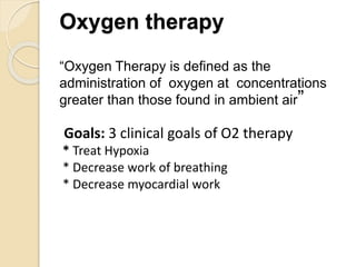
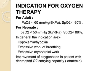
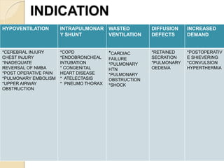
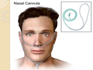
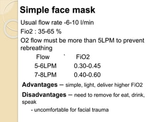
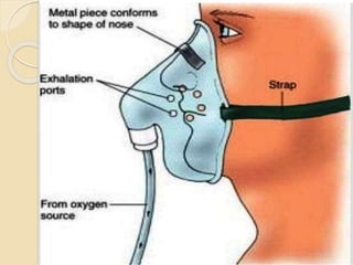
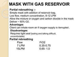
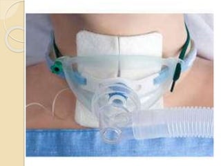
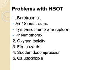
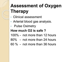
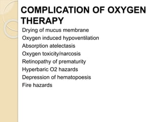
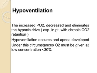
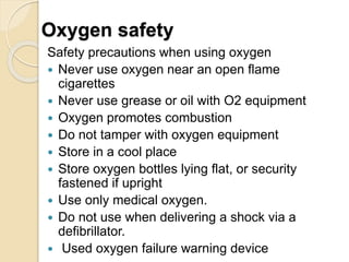
Oxygen therapy involves administering oxygen at concentrations greater than in ambient air to treat hypoxia and reduce work of breathing and myocardial work. It can be delivered via low-flow nasal cannulas or high-flow face masks, hoods, and tents. Precautions must be taken when using oxygen to avoid drying tissues, hypoventilation, absorption atelectasis, oxygen toxicity, and fire hazards. Clinical assessment and arterial blood gases are used to monitor patients and determine appropriate oxygen concentrations and durations of therapy.
This document defines and discusses the pathophysiology of different types of shock: cardiogenic, obstructive, hypovolemic, and distributive. It notes that shock occurs when there is inadequate perfusion and oxygenation of cells, leading to cellular and organ dysfunction. The key signs of shock include tachycardia, hypotension, altered mental status, and decreased urine output. Early goal-directed resuscitation is important to prevent end organ damage and death, and should focus on airway management, oxygenation, fluid resuscitation, and treating the underlying cause.
Diabetic Ketoacidosis/Hyperosmolar Coma in ESRD
• Clinical Picture of hyperglycemia is modified (due to absence of renal function).
o The absence of polyuria and glycosuria “safety valve” severe hyperglycemia (serum glucose level >1,000 mg/dL)
o Alteration of mental status is unusual (Due to absence of water loss induced by osmotic diuresis).
o Asymptomatic mostly in spite of severe hyperglycemia
o Thirst, weight gain, and may be pulmonary edema or coma
o Severe hyperkalemia in DKA in insulin-dependent dialysis patients.
• Diagnosis in the ESKD patient is based on hyperglycemia, positive serum ketones, metabolic acidemia, and an increased anion gap.
o Which is not easy due to the plasma reaction for ketones may be negative, the anion gap may not be affected and the clinical presentation itself of severe hyperglycemia and ketoacidosis are atypical.
• Management of hyperglycemia with or without ketoacidosis differs from that in patients without renal failure in that administration of large amounts of fluid is unnecessary and generally contraindicated.
o Insulin is the only treatment needed can correct all clinical and laboratory abnormalities of hyperglycemia.
o Can administer a continuous infusion of low-dose regular insulin (starting at 2 units/hr) with close clinical monitoring and measurement of serum glucose and potassium concentrations at 2- to 3-hour intervals.
o Urgent dialysis if pulmonary edema and hyperkalemia.
• IV bicarbonate is not indicated may exacerbate volume overload.
• No phosphate replacement is generally needed.
• Hypophosphatemia is not expected.
• Magnesium deficiency is absent.
The document provides information on pathophysiology fundamentals including the general adaptation model of stress response, characteristics of the hematologic system, disseminated intravascular coagulation (DIC), acute coronary syndromes, myocardial infarction, and hypertension. It describes the body's physical response to stress in stages of alarm, resistance, and exhaustion. It also summarizes pathophysiologic changes, causes, signs and symptoms, diagnosis, and treatment of various conditions.
The document discusses pulmonary embolism, which is the blockage of pulmonary arteries by blood clots or other materials. It defines pulmonary embolism and discusses its incidence, risk factors including deep vein thrombosis, clinical features such as chest pain and dyspnea, pathophysiology involving right heart strain, diagnostic studies, and treatment including anticoagulation with heparin and warfarin as well as surgical interventions in severe cases.
This document discusses bronchiectasis, including its definition, etiology, clinical features, diagnosis, management, and complications. Some key points:
- Bronchiectasis is irreversible dilation of the airways caused by infection or other insults that damages the airways and impairs mucus clearance.
- It has various etiologies including infection, immunodeficiency, genetic disorders, and aspiration. Recurrent infections lead to a vicious cycle of inflammation and further airway damage.
- Symptoms include chronic productive cough and sputum. Investigations include chest CT, which shows characteristic findings like airway dilation.
- Management focuses on airway clearance, antibiotics for infections, and
This document provides information on asthma, including its definition, history, burden, types (atopic, non-atopic, drug-induced, occupational, exercise-induced), pathogenesis, genetic and environmental factors, clinical features, and methods of diagnosis. Asthma is a chronic airway disorder marked by episodic bronchoconstriction due to increased airway sensitivity and inflammation. It can be triggered by allergens, irritants, infections, and other stimuli and is characterized by symptoms like wheezing, coughing, and shortness of breath.
Atherosclerosis is a condition where the artery walls thicken due to a build-up of fatty materials like cholesterol. It starts when damage occurs to the inner layer of the artery from issues like high blood pressure, smoking, or high cholesterol. This damage allows plaque to form and build up in the artery wall over time from substances like macrophages, cholesterol crystals, and calcium deposits. Left untreated, it can block blood flow and potentially cause heart attacks or strokes since many people do not notice symptoms until a serious event occurs. Risk factors include age, family history, and lifestyle habits, and it can be treated through medication, procedures like stenting and bypass surgery, and lifestyle changes.
Respiratory acidosis and alkalosis are acid-base disorders caused by problems with ventilation.
Respiratory acidosis occurs when Paco2 is elevated due to conditions that decrease ventilation like lung disease or muscle fatigue. It causes a decrease in pH but HCO3 rises in compensation. Chronic respiratory acidosis is treated by gradually lowering Paco2.
Respiratory alkalosis is caused by excessive ventilation lowering Paco2, seen in anxiety, pain, or drug effects. It increases pH but HCO3 falls as the kidneys compensate. Severe acute respiratory alkalosis can reduce blood flow and cause arrhythmias.
Edema is an abnormal accumulation of fluid in the interstitial spaces of the body that can occur for various reasons. It is classified as either generalized, affecting multiple body areas, or localized affecting a single area. Edema can be caused by increased hydrostatic pressure, reduced plasma oncotic pressure, increased vessel permeability, obstruction of lymphatic fluid clearance, or changes in tissue water retention. Common symptoms include weight gain, swollen limbs, distended veins, and crackles in the lungs. Treatment involves addressing the underlying cause, restricting sodium intake, using diuretics, elevating limbs, and preventing complications. Nurses monitor for worsening symptoms, ensure treatment adherence, and provide skin care to prevent issues.
Crystalloids are electrolyte solutions that can freely diffuse throughout the extracellular space. The principal crystalloid is isotonic saline (0.9% NaCl), which expands the interstitial space rather than plasma volume. Ringer's lactate is also commonly used as it more closely matches plasma composition. Dextrose 5% in water (D5W) is hypotonic and expands both intra and extracellular spaces, providing calories but not electrolytes. Each crystalloid has different indications and disadvantages to consider when selecting the appropriate fluid for treatment.
This document discusses eosinophilic pneumonias, which are characterized by infiltration of the lungs with eosinophils. It begins by providing a brief history and classification, dividing causes into those of known cause (such as parasites, drugs, tropical pulmonary eosinophilia) and unknown cause (idiopathic acute eosinophilic pneumonia, chronic eosinophilic pneumonia, Churg-Strauss syndrome, idiopathic hypereosinophilic syndrome). It then discusses several types of eosinophilic pneumonia in more detail, including their presentations, investigations, treatments, and key distinguishing features.
Cardiomyopathy refers to diseases of the heart muscle that weaken the heart's ability to pump blood effectively. The three main types are dilated, hypertrophic, and restrictive cardiomyopathy. Dilated cardiomyopathy causes the left ventricle to enlarge and weaken, impairing its ability to pump blood. Causes include viral infections, toxins, genetic factors, and hypertension. Symptoms include fatigue, shortness of breath, and fluid retention. Diagnosis involves echocardiograms, electrocardiograms, and cardiac catheterization. Treatment focuses on managing symptoms through medications, lifestyle changes, and potentially surgery or transplantation.
Acute respiratory distress syndrome (ARDS) occurs when fluid builds up in the tiny, elastic air sacs (alveoli) in your lungs. The fluid keeps your lungs from filling with enough air, which means less oxygen reaches your bloodstream. This deprives your organs of the oxygen they need to function.
This document discusses the treatment of acute exacerbations of asthma and COPD. For asthma, treatment involves high doses of inhaled bronchodilators, systemic corticosteroids, oxygen therapy, and intravenous fluids. For COPD, treatment focuses on oxygenation, ventilation support using non-invasive ventilation if needed, nebulized short-acting bronchodilators, corticosteroids, and antibiotics for bacterial infections. Lung volumes measured via spirometry can differentiate obstructive disorders like COPD and asthma from restrictive disorders. Obstructive disorders show reduced airflow and FEV1/FVC ratio, while restrictive disorders have normal ratio but reduced lung volumes. Emergent tests for diagnosis include ECG, arterial blood gases,
Pulmonary edema is a condition caused by excessive fluid in the lungs, making it difficult to breathe. There are two main types: cardiogenic, caused by issues with the heart like weakened pumping from heart disease or damage, and non-cardiogenic, caused by things like drug use, smoke inhalation, or viral infections which damage the lungs. Complications can include swelling in the legs and abdomen. Treatment focuses on removing excess fluid with diuretics, relieving symptoms, and addressing the underlying cause.
Respiratory acidosis is a condition caused by a buildup of carbon dioxide in the body due to impaired lung function. This disrupts the body's acid-base balance, causing fluids like blood to become too acidic. Causes include lung diseases like asthma, obesity, and drugs that suppress breathing. Symptoms include confusion, fatigue, and shortness of breath. Treatment focuses on the underlying lung condition through medications, ventilation support, and oxygen if needed. The prognosis depends on the severity and cause of the respiratory acidosis.
This document defines pneumoconiosis as a permanent alteration of lung structure caused by inhaling mineral dust. Not all dusts are pathogenic, with silica being highly fibrogenic. Pneumoconiosis resulted in 260,000 deaths globally in 2013. It can lead to serious complications like cor pulmonale and pulmonary hypertension. The tendency for pneumoconiosis to develop into carcinomas depends on factors like particle size, composition, and amount retained in the lungs. There are three main types classified by tissue response: fibrotic nodules, interstitial fibrosis, and hypersensitivity reactions. Coal worker's pneumoconiosis is examined in detail, including pathogenesis, radiological features, gross depiction, and histological
This document provides guidelines for the management of acute severe asthma. Initial assessment involves measuring peak expiratory flow, vital signs, and arterial blood gases. Treatment includes high doses of inhaled bronchodilators, systemic corticosteroids, oxygen, intravenous fluids and magnesium. Mechanical ventilation may be required if the patient deteriorates or is unable to be adequately ventilated. Close monitoring of peak expiratory flow, oxygen saturation, and serum aminophylline levels is important.
1. The document discusses several obstructive lung diseases including emphysema, chronic bronchitis, asthma, and bronchiectasis.
2. Emphysema is characterized by destruction of alveolar walls without significant fibrosis leading to enlarged air spaces. Chronic bronchitis involves thickening of bronchial walls and excess mucus production.
3. Asthma is a chronic inflammatory disease involving recurrent airway obstruction, inflammation, and hyperresponsiveness triggered by various stimuli. Bronchiectasis permanently dilates the bronchi and bronchioles due to destruction of muscles and tissues often caused by infection or obstruction.
This document discusses pulmonary embolism (PE), which refers to obstruction of the pulmonary artery or its branches by a thrombus (blood clot). PE can be caused by factors that increase clotting like surgery, trauma, or heart failure. When a thrombus blocks a pulmonary vessel, it impairs gas exchange and increases pulmonary vascular resistance, raising pressure in the pulmonary artery and overworking the right ventricle. Diagnosis involves tests like ventilation-perfusion scans and treatment focuses on anticoagulation, thrombolytic drugs, or occasionally surgery to remove clots. Nursing care aims to prevent clots, monitor for complications, manage pain and oxygen therapy.
Glomerulonephritis (GN) refers to a group of kidney diseases characterized by damage to the glomeruli. There are many potential causes of GN, including both immune-mediated mechanisms like deposition of immune complexes or antibodies, and non-immune factors like hypertension. This can lead to inflammation in the glomeruli and injury to the filtration barrier. Pathological features are classified based on patterns of involvement seen on biopsy. Treatment involves controlling risk factors like blood pressure and use of corticosteroids, immunosuppressants, or plasmapheresis depending on the underlying etiology and severity.
Chronic gastritis is long-term inflammation of the stomach lining that can be caused by factors like H. pylori infection, medications, alcohol, or diet. It is classified based on etiology (cause), morphology (appearance), and location in the stomach. The OLGA staging system scores and stages gastritis severity based on the degree of atrophy in the antrum and corpus as seen histologically. Treatment involves eliminating the cause, such as treating an H. pylori infection, and managing symptoms.
Acute myocardial infarction, or heart attack, results from prolonged ischemia due to a blockage in a coronary artery that supplies blood to heart muscle. Risk factors include increasing age, male sex, hypertension, dyslipidemia, diabetes, smoking, obesity, physical inactivity, excessive alcohol intake, and family history. Diagnosis involves electrocardiogram changes, elevated cardiac biomarkers, and symptoms like chest pain. Management focuses on oxygen, pain relief, antiplatelet/anticoagulant drugs, revascularization, and lifestyle changes to prevent future heart attacks.
H. pylori plays a crucial role in the pathogenesis of peptic ulcers. It has been found in 90% of patients with chronic gastritis, 95% with duodenal ulcer disease, and 70% with gastric ulcer. Peptic ulcers are caused by a loss of balance between protective and hostile factors in the stomach and duodenum. Factors like H. pylori infection, NSAIDs, and excess acid secretion can damage the mucosal lining, while factors like mucus, bicarbonate, and blood flow work to protect it. Chronic peptic ulcers form in areas of the stomach and duodenum with reduced acid levels. They appear as round or oval lesions that penetrate
This document provides information on bronchial asthma including its definition, classification, pathophysiology, and treatment approaches. It discusses the different types of asthma such as atopic, non-atopic, and drug-induced asthma. It describes the cells and mediators involved in asthma inflammation. It covers the mechanisms and classes of drugs used to treat asthma, including bronchodilators, leukotriene antagonists, mast cell stabilizers, corticosteroids, and anti-IgE antibody. It provides details on the mechanisms of action and side effects of various bronchodilators and corticosteroids. It also discusses inhalational drug delivery systems and the treatment of acute asthma attacks.
oxygen is a medication. oxygen therapy must be known to all health professionals for optimum management of patient and optimum use of resourses. even more oxygen can cause oxygen toxicity and can harm the patient in many ways. There are various methods for giving oxygen,varieties of face masks, cylinders. also there is criteria when to give oxygen ,how to give oxygen,what are the benefits and mechanism of oxygen therapy.
Oxygen therapy involves delivering oxygen concentrations above 21% to address hypoxemia or hypoxia. Various oxygen delivery systems can be used, ranging from low-flow nasal cannulas to high-flow venturi masks and non-rebreathing masks. Complications of prolonged high-concentration oxygen therapy include hypoventilation, absorption atelectasis, pulmonary toxicity, and retrolental fibroplasia in premature infants. The appropriate oxygen concentration and delivery method depends on careful evaluation of each patient's condition and needs.
Oxygen therapy involves delivering oxygen concentrations higher than 21% to treat hypoxemia or hypoxia. There are various oxygen delivery systems ranging from low-flow nasal cannulas to high-flow venturi masks and non-rebreathing masks. Proper evaluation is needed to determine if a patient requires oxygen therapy. While effective for certain conditions, oxygen therapy can cause complications if used improperly such as absorption atelectasis, hypoventilation, fire hazards, and oxygen toxicity in neonates. Careful consideration of each patient's needs is important when selecting the appropriate oxygen delivery method.
Oxygen therapy requires understanding oxygen delivery and toxicity risks. The presentation covered:
1. Indications for oxygen therapy include hypoxia, dyspnea, and low blood oxygen levels.
2. Oxygen delivery devices include nasal cannulas, masks, and venti-masks which provide varying levels of oxygen concentration depending on flow rates and patient breathing.
3. High oxygen concentrations over long periods risk toxicity including hypoventilation, absorption atelectasis, and pulmonary damage. Careful monitoring is needed to avoid risks while meeting patient needs.
portable hyperbaric
chamber, are used for
altitude sickness and
decompression illness
in remote areas.
This document discusses the history, basics, indications, contraindications, complications, and applications of hyperbaric oxygen therapy (HBOT). It begins with definitions and the initial discovery of HBOT in the 1600s. It then covers the physics and physiology behind how increased pressure and oxygen concentration improves oxygen delivery to tissues. Common indications for HBOT include carbon monoxide poisoning, decompression sickness, gas embolism, and infections like clostridial myonecrosis. Complications can include barotrauma, seizures from oxygen toxicity, and fire hazards. Applications of HBOT include wound
Oxygen therapy is the administration of oxygen to increase oxygen saturation in tissues. There are various methods of oxygen delivery including nasal cannula, face masks, and venturi masks. The type of delivery system used depends on the needed oxygen concentration and flow rate, ranging from 24-100% oxygen. Proper monitoring is important to ensure safe and effective oxygen therapy.
Oxygen therapy involves administering supplemental oxygen to increase oxygen levels in the blood. It is used when a patient has hypoxemia or low blood oxygen levels. Different devices are used to deliver oxygen depending on the patient's condition and oxygen needs. The main devices discussed are nasal cannulas, masks, venturi masks, and high flow devices. Potential complications of oxygen therapy include oxygen toxicity if delivered at too high of concentrations for too long, depression of ventilation, retinopathy of prematurity in infants, and fire hazards. Care must be taken to closely monitor patients on oxygen therapy.
This document provides information about oxygen therapy, including:
1. It discusses the anatomy and physiology of respiration and defines oxygen therapy as the administration of oxygen at a higher concentration than in the atmosphere.
2. The types of oxygen delivery discussed are nasal cannula, face masks, face tents, and transtracheal oxygen delivery. Humidification is also addressed.
3. Nursing responsibilities in administering oxygen therapy are outlined, such as explaining the therapy to patients and demonstrating safe oxygen use. Monitoring patients' response to therapy and pulse oximetry readings are also covered.
Oxygen therapy involves administering oxygen at concentrations greater than in room air to treat hypoxemia. The document defines oxygen therapy and discusses various types including low-flow systems like nasal cannulas and masks, and high-flow systems like Venturi masks and T-pieces. It covers indications for oxygen therapy, how to assess a patient's oxygen needs, goals of therapy, factors in choosing a delivery method, and complications of therapy like oxygen toxicity.
The document discusses oxygen therapy and administration, including the types of oxygen delivery systems like nasal cannulas, simple face masks, and reservoir masks. It covers indications for oxygen therapy when hypoxemia is present based on arterial blood gas values. Equations are provided for calculating oxygen content, delivery, uptake, and extraction from the blood under normal conditions.
Oxygen therapy involves administering oxygen at concentrations greater than in room air to treat hypoxemia. It works by increasing oxygen saturation in tissues where levels are too low. Oxygen can be delivered through various devices at low or high flows, with the goal of maintaining adequate oxygenation without toxicity. Long-term oxygen therapy improves outcomes for conditions like COPD but requires monitoring to prevent carbon dioxide retention and acidemia.
Oxygen therapy provides oxygen to tissues through various delivery methods. It is indicated for patients with hypoxemia or critical illness putting them at risk of tissue hypoxia. Low-flow devices like nasal cannulas or face masks deliver oxygen at rates lower than patient demand, mixing it with room air. High-flow devices exceed patient demand to maintain a constant oxygen concentration. Humidification is important to prevent drying of airways from medical gases. Potential complications include carbon dioxide narcosis, atelectasis, oxygen toxicity, and retrolental fibroplasia from extended high oxygen use.
Oxygen therapy involves administering oxygen at concentrations greater than room air to treat hypoxemia. The purpose is to increase oxygen saturation in tissues where it is too low due to illness or injury. Oxygen can be delivered via various low or high flow devices like nasal cannulas, masks, tents or venturi masks to maintain adequate oxygen saturation. Close monitoring of oxygen saturation levels via pulse oximetry or arterial blood gases is needed to properly titrate oxygen therapy.
Oxygen therapy involves prescribing supplemental oxygen to ensure adequate oxygen delivery to tissues. It is considered a medical treatment and requires a doctor's order specifying the oxygen concentration, flow rate, and duration. Oxygen therapy is used to treat hypoxemia in both acute and chronic conditions. Different devices are used to deliver oxygen at either low or high concentrations, with masks being variable performance devices and venturi masks or high flow cannulas providing fixed performance. Proper use and settings are important for safety and effectiveness.
Oxygen therapy aims to maintain adequate tissue oxygenation while minimizing strain on the cardiopulmonary system. It is indicated for documented hypoxemia defined as PaO2 < 60 mmHg or SaO2 < 90% on room air, or below desirable levels for a specific condition. Various oxygen delivery devices include nasal cannula, masks, and ventilators, which provide concentrations from 24-100% at flows of 2-10 LPM. Potential complications include oxygen toxicity, respiratory depression, retinopathy, and fire hazards from long term high concentration use without proper monitoring and control of flow rates.
The document discusses three basic essentials for life: oxygen, water, and food. It then focuses on oxygen, defining it as an element, gas, and drug. It describes oxygen therapy as administering oxygen at concentrations greater than room air to treat hypoxemia. Hypoxemia and different types of hypoxia are defined. Common signs and symptoms as well as indications for oxygen therapy are listed. Various oxygen delivery devices, their uses, advantages, and disadvantages are outlined.
Seminar Presentation on Oxygen Administration.pptxRebiraWorkineh
Oxygen therapy is used to treat conditions caused by low oxygen levels in the blood. It works by delivering oxygen through devices like nasal cannulas, masks, and ventilators at higher percentages than is found in regular air. The goals are to increase oxygen in the tissues and relieve symptoms. Different devices are used depending on the needed oxygen concentration and flow rate, ranging from low flow nasal cannulas for lower amounts to venturi masks or ventilators for higher amounts. Nurses must carefully monitor patients on oxygen therapy and watch for side effects like drying of tissues or oxygen toxicity from levels that are too high.
This document discusses hypoxia and oxygen therapy. It begins by providing historical background on the discovery of oxygen. It then describes the oxygen cascade and factors that can affect oxygen levels at different points. Various methods of oxygen delivery are presented, including low-flow and high-flow systems. Key goals and indications for oxygen therapy are outlined. Precautions for oxygen toxicity are discussed. The document concludes by providing guidance on selecting an appropriate oxygen delivery system based on the patient and clinical situation.
Nano-gold for Cancer Therapy chemistry investigatory projectSIVAVINAYAKPK
chemistry investigatory project
The development of nanogold-based cancer therapy could revolutionize oncology by providing a more targeted, less invasive treatment option. This project contributes to the growing body of research aimed at harnessing nanotechnology for medical applications, paving the way for future clinical trials and potential commercial applications.
Cancer remains one of the leading causes of death worldwide, prompting the need for innovative treatment methods. Nanotechnology offers promising new approaches, including the use of gold nanoparticles (nanogold) for targeted cancer therapy. Nanogold particles possess unique physical and chemical properties that make them suitable for drug delivery, imaging, and photothermal therapy.
Pictorial and detailed description of patellar instability with sign and symptoms and how to diagnose , what investigations you should go with and how to approach with treatment options . I have presented this slide in my 2nd year junior residency in orthopedics at LLRM medical college Meerut and got good reviews for it
After getting it read you will definitely understand the topic.
“Psychiatry and the Humanities”: An Innovative Course at the University of Mo...Université de Montréal
“Psychiatry and the Humanities”: An Innovative Course at the University of Montreal Expanding the medical model to embrace the humanities. Link: https://www.psychiatrictimes.com/view/-psychiatry-and-the-humanities-an-innovative-course-at-the-university-of-montreal
The Children are very vulnerable to get affected with respiratory disease.
In our country, the respiratory Disease conditions are consider as major cause for mortality and Morbidity in Child.
The biomechanics of running involves the study of the mechanical principles underlying running movements. It includes the analysis of the running gait cycle, which consists of the stance phase (foot contact to push-off) and the swing phase (foot lift-off to next contact). Key aspects include kinematics (joint angles and movements, stride length and frequency) and kinetics (forces involved in running, including ground reaction and muscle forces). Understanding these factors helps in improving running performance, optimizing technique, and preventing injuries.
Breast cancer: Post menopausal endocrine therapyDr. Sumit KUMAR
Breast cancer in postmenopausal women with hormone receptor-positive (HR+) status is a common and complex condition that necessitates a multifaceted approach to management. HR+ breast cancer means that the cancer cells grow in response to hormones such as estrogen and progesterone. This subtype is prevalent among postmenopausal women and typically exhibits a more indolent course compared to other forms of breast cancer, which allows for a variety of treatment options.
Diagnosis and Staging
The diagnosis of HR+ breast cancer begins with clinical evaluation, imaging, and biopsy. Imaging modalities such as mammography, ultrasound, and MRI help in assessing the extent of the disease. Histopathological examination and immunohistochemical staining of the biopsy sample confirm the diagnosis and hormone receptor status by identifying the presence of estrogen receptors (ER) and progesterone receptors (PR) on the tumor cells.
Staging involves determining the size of the tumor (T), the involvement of regional lymph nodes (N), and the presence of distant metastasis (M). The American Joint Committee on Cancer (AJCC) staging system is commonly used. Accurate staging is critical as it guides treatment decisions.
Treatment Options
Endocrine Therapy
Endocrine therapy is the cornerstone of treatment for HR+ breast cancer in postmenopausal women. The primary goal is to reduce the levels of estrogen or block its effects on cancer cells. Commonly used agents include:
Selective Estrogen Receptor Modulators (SERMs): Tamoxifen is a SERM that binds to estrogen receptors, blocking estrogen from stimulating breast cancer cells. It is effective but may have side effects such as increased risk of endometrial cancer and thromboembolic events.
Aromatase Inhibitors (AIs): These drugs, including anastrozole, letrozole, and exemestane, lower estrogen levels by inhibiting the aromatase enzyme, which converts androgens to estrogen in peripheral tissues. AIs are generally preferred in postmenopausal women due to their efficacy and safety profile compared to tamoxifen.
Selective Estrogen Receptor Downregulators (SERDs): Fulvestrant is a SERD that degrades estrogen receptors and is used in cases where resistance to other endocrine therapies develops.
Combination Therapies
Combining endocrine therapy with other treatments enhances efficacy. Examples include:
Endocrine Therapy with CDK4/6 Inhibitors: Palbociclib, ribociclib, and abemaciclib are CDK4/6 inhibitors that, when combined with endocrine therapy, significantly improve progression-free survival in advanced HR+ breast cancer.
Endocrine Therapy with mTOR Inhibitors: Everolimus, an mTOR inhibitor, can be added to endocrine therapy for patients who have developed resistance to aromatase inhibitors.
Chemotherapy
Chemotherapy is generally reserved for patients with high-risk features, such as large tumor size, high-grade histology, or extensive lymph node involvement. Regimens often include anthracyclines and taxanes.
STUDIES IN SUPPORT OF SPECIAL POPULATIONS: GERIATRICS E7shruti jagirdar
Unit 4: MRA 103T Regulatory affairs
This guideline is directed principally toward new Molecular Entities that are
likely to have significant use in the elderly, either because the disease intended
to be treated is characteristically a disease of aging ( e.g., Alzheimer's disease) or
because the population to be treated is known to include substantial numbers of
geriatric patients (e.g., hypertension).
Travel Clinic Cardiff: Health Advice for International TravelersNX Healthcare
Travel Clinic Cardiff offers comprehensive travel health services, including vaccinations, travel advice, and preventive care for international travelers. Our expert team ensures you are well-prepared and protected for your journey, providing personalized consultations tailored to your destination. Conveniently located in Cardiff, we help you travel with confidence and peace of mind. Visit us: www.nxhealthcare.co.uk
pharmacy exam preparation for undergradute students.pptx
Oxygen therapy
1.
2. Oxygen therapy
“Oxygen Therapy is defined as the
administration of oxygen at concentrations
greater than those found in ambient air”
Goals: 3 clinical goals of O2 therapy
* Treat Hypoxia
* Decrease work of breathing
* Decrease myocardial work
3. Physical properties of oxygen
It was first recognized as a distinct gas by Joseph
Priestley in 1774 .
Is a colourless , odourless non-flammable gas.
Molecular weight 32 & specific gravity 1.105
Makes up 20.9% of air by volume and 23% air by
weight.
Boiling point -183 C
Melting point –216.6C
Critical temp. –118.4C , Critical pressure 736.9psi
4. Preparation & storage
O2 manufactured by fractional distillation of
liquid air .
Before liquefaction ,co2 removed by filter.
O2 & N2 separated by means of their
boiling points. O2 = -183 C, N2= -195 C
5. O2 can be store either
1) Cylinder
Oxygen can be stored under pressure in cylinders
made of molybdenum steel.
Cylinders are black with white shoulders.
The pressure inside at 15°C is 137 bar.
Pin index 2,5
Oxygen cylinder content estimated Boyle’s law
Capacity: E cylinder =643.9L/1900 psi
M cylinder = 3450L/2200psi
H cyinder= 6900L/2200 psi
2) Oxygen concentrators
3)Vacuum Insulated Evaporator (VIE).
6. INDICATION FOR OXYGEN
THERAPY
For Adult :
PaO2 < 60 mmHg(8KPa), SpO2< 90% .
For Neonate :
paO2 < 50mmHg (6.7KPa), SpO2< 88%.
In general the indication are:-
Hypoxemia/hypoxia
Excessive work of breathing
Excessive myocardial work
Improvement of oxygenation in patient with
decreased O2 carrying capacity ( anaemia)
13. Oxygen Transport in the body
1. Diffusion of O2 from alveolar air to pulmonary
capillary
PO2 in alveolus = 105 mm Hg
PO2 in pulmonary capillaries at arterial end = 40
mm Hg
The pressure difference of O2 = 65 mm Hg
2. Transport of O2 in Blood :
Dissolved state :
Approximatly (0.27-0.3) mL/100 mL of Blood .
In the form of O2-Hb complex :
About 97% of O2 is arrived in the form of O2-Hb .
15. Oxygen content
Arterial Oxygen content(CaO2)= SpO2 x Hb%
x 1.34 mL/dL + 0.003 x PaO2~20ml
Venous Oxygen content(CvO2)= SpO2 xHb%
x 1.34 mL/dL + 0.003 x PvO2 ~15ml
O2 delivery = CaO2 x cardiac output
O2 consumption = (CaO2-CvO2) x cardiac
output 25%
16. OXYGEN DELIVERY SYSTEM
Classification
1. Low-Flow Systems/Variable performance
2. High Flow / fixed performance equipment .
Low flow/variable
performance devices
High flow /fixed
performance devices
* Flow rate less than
patients inspiratory flow
rate.
* FiO2 varies with
RR&TV
* FiO2 not predictable
*Flow rate more than patients
inspiratory flow rate
* Not influenced by RR/TV
* FiO2 fixed & accurate
17. Low-Flow Systems/Variable performance :
Is adequate for patients with
Minute ventilation < ~8-10L/min
Breathing frequencies < 20 breath/min
Tidal volumes(VT)< ~0.8L
Normal inspiratory flow (10-30L/min)
Low flow devices:
No capacity system : Nasal cannula , Nasal catheter
Low capacity system(<100ml) : Simple mask of children
Medium capacity system(100-250ml) : Adult Simple face
mask
nebulizer mask
High capacity system(250-1500ml): facemask with reservoir
bag
*Partial rebreathing
* Non rebreathing
Very high capacity system(>1500ml): oxygen hood ,oxygen
tents
18. Nasal cannula (prongs)
Capable to deliver an FiO2 ranging from 0.24-0.44
Maximum of 6LPM : causes crusting of the secretion ,
drying of the nasal mucosa & epistaxis
Advantages – inexpensive , well tolerated, comfortable
patient can eat and drink
Disadvantages – pressure sore, irritant to the mucosal
Flow L/M FiO2
LPM, 1 0.24
LPM , 2 0.28
LPM, 3 0.34
LPM, 4 0.38
LPM, 5-6 0.44
19.
20. Simple face mask
Usual flow rate -6-10 l/min
Fio2 : 35-65 %
O2 flow must be more than 5LPM to prevent
rebreathing
Flow ` FiO2
5-6LPM 0.30-0.45
7-8LPM 0.40-0.60
Advantages – simple, light, deliver higher FiO2
Disadvantages – need to remove for eat, drink,
speak
- uncomfortable for facial trauma
21.
22. MASK WITH GAS RESERVOIR
Partial rebreathing :-
Simple mask with addition of reservoir bag.
Low flow, medium concentration, 8-12 LPM
Allow the mixture or oxygen and carbon dioxide in the mask
Deliver ~ 60% O2.
Advantages
Client can inhale room air if oxygen supply is iterrupted .
Disadvantages
Requires tight seal (eating and talking difficult,
uncomfortable)
Partial rebreathing
Flow FiO2
7 LPM 0.35-0.75
15LPM 0.65- 1.0
23. Non rebreathing mask :–
one way valve to prevent rebreathing
Advantages
Delivers the highest possible oxygen Conc (95-100%)
at a
flow rate 8-15 L/min.
Disadvantages
1.Impractical for long term Therapy
2.suffocation
3.Expensive 4.Uncomfortable
Non rebreathing
Flow FiO2
7-15 0.40-1.0
24.
25.
26. Oxygen Hood
An oxygen hood is used for babies
who can breathe on their own but still
need extra oxygen.
A hood is a plastic dome or box with
warm, moist oxygen inside.
The hood is placed over the baby's
head
27.
28. Oxygen tents
An oxygen tent is a bendable piece of
clear
plastic held over the child's bed or crib
by a
frame.
29. Oxygen tents
An oxygen tent is a bendable piece of clear plastic held
over the child's bed or crib by a frame.
Oxygen tents
31. Venturi mask
• Operate on Bernoulli principle
• Is designed with wide- bore tubing and various color -
coded jet adapters
• Each color code correspond to a precise .
• The amount of air determined by the size of the orifice
(jet adapter).
• Oxygen from 24 - 50% . flow of 4 to 15 L/min.
32. Venturi mask
Advantages –
Delivers most precise oxygen Conc.
Doesn’t dry mucous membranes
Disadvantages –
Uncomfortable
Risk for skin irritation
produce respiratory depression in COPD
33.
34. AMBU BAG
AMBU- Artificial Manual Breathing Unit
(or)
Bag Valve Mask Ventilation is a hand-
held device commonly used to provide
positive pressure ventilation to patients
who are not breathing or not breathing
adequately.
35.
36. T-PIECE
Used on end of ET tube when
weaning from ventilator
Provides accurate FIO2
Provides good humidity
38. Tracheostomy Collar/ Mask
Inserted directed into trachea
Is indicated for chronic O2 therapy
need
O2 flow rate 8 to 10L
Provides accurate FIO2
Provides good humidity.
Comfortable ,more efficient .
39.
40. Hyperbaric Oxygen Therapy
(HBOT)
Hyperbaric O2 therapy use a pressurized
chamber greater than one atmosphere combined
with the delivery of 100% oxygen (FiO2 = 1.0),
drives the diffusion of oxygen into the blood
plasma at up to 10 times normal concentration.
42. Indication
1. Air or Gas Embolism
2. Carbon Monoxide Poisoning
3. Gas Gangrene
4. Crush Injury, Compartment Syndrome and
Other Acute Traumatic Ischemias
5. Decompression Sickness
6. Arterial Insufficiencies: • Central Retinal
Artery Occlusion
7. Severe Anemia
8. Intracranial Abscess
9. Necrotizing Soft Tissue Infections
43. Problems with HBOT
1. Barotrauma .
• Air / Sinus trauma
• Tympanic membrane rupture
• Pneumothorax
2. Oxygen toxicity
3. Fire hazards
4. Sudden decompression
5. Calutrophobia
44. Assessment of Oxygen
Therapy
• Clinical assessment
• Arterial blood gas analysis.
• Pulse Oximetry
How much O2 is safe ?
100% - not more than 12 hours
80% - not more than 24 hours
60 % - not more than 36 hours
45. COMPLICATION OF OXYGEN
THERAPY
Drying of mucus membrane
Oxygen induced hypoventilation
Absorption atelectasis
Oxygen toxicity/narcosis
Retinopathy of prematurity
Hyperbaric O2 hazards
Depression of hematopoesis
Fire hazards
46. Hypoventilation
The increased PO2, decreased and eliminates
the hypoxic drive ( esp. in pt. with chronic CO2
retention )
Hypoventilation occures and apnea developed
Under this circumstances O2 must be given at
low concentration <30%
47. Absorption Atelectasis
During 100% oxygen delivery, nitrogen in
alveoli is washed out and replaced by
oxygen. In contrast to nitrogen, oxygen is
extremely soluble in blood and diffuses
very quickly into the pulmonary
vasculature, so that not enough gas is left
in the alveoli to maintain patency, and the
alveolus collapses; this is known as
absorption atelectasis .
48.
49. Pulmonary Oxygen Toxicity
High FiO2 for prolonged time
Generation of free radical
React with cellular DNA
Cytotoxicity ,damage capillary endothelium , Interstitial
edema
Thickened alveolar capillary membrane
Pulmonary fibrosis and hypertension
50.
51. Retrolental Fibroplasia
Excessive O2 to pre-mature infants may
result in constriction of immature retinal
vessels, endothelial damage, retinal
detachment and possible blindness
Recommended that PO2 be maintained
between 60-90 mmHg range in neonate
52. Oxygen safety
Safety precautions when using oxygen
Never use oxygen near an open flame
cigarettes
Never use grease or oil with O2 equipment
Oxygen promotes combustion
Do not tamper with oxygen equipment
Store in a cool place
Store oxygen bottles lying flat, or security
fastened if upright
Use only medical oxygen.
Do not use when delivering a shock via a
defibrillator.
Used oxygen failure warning device