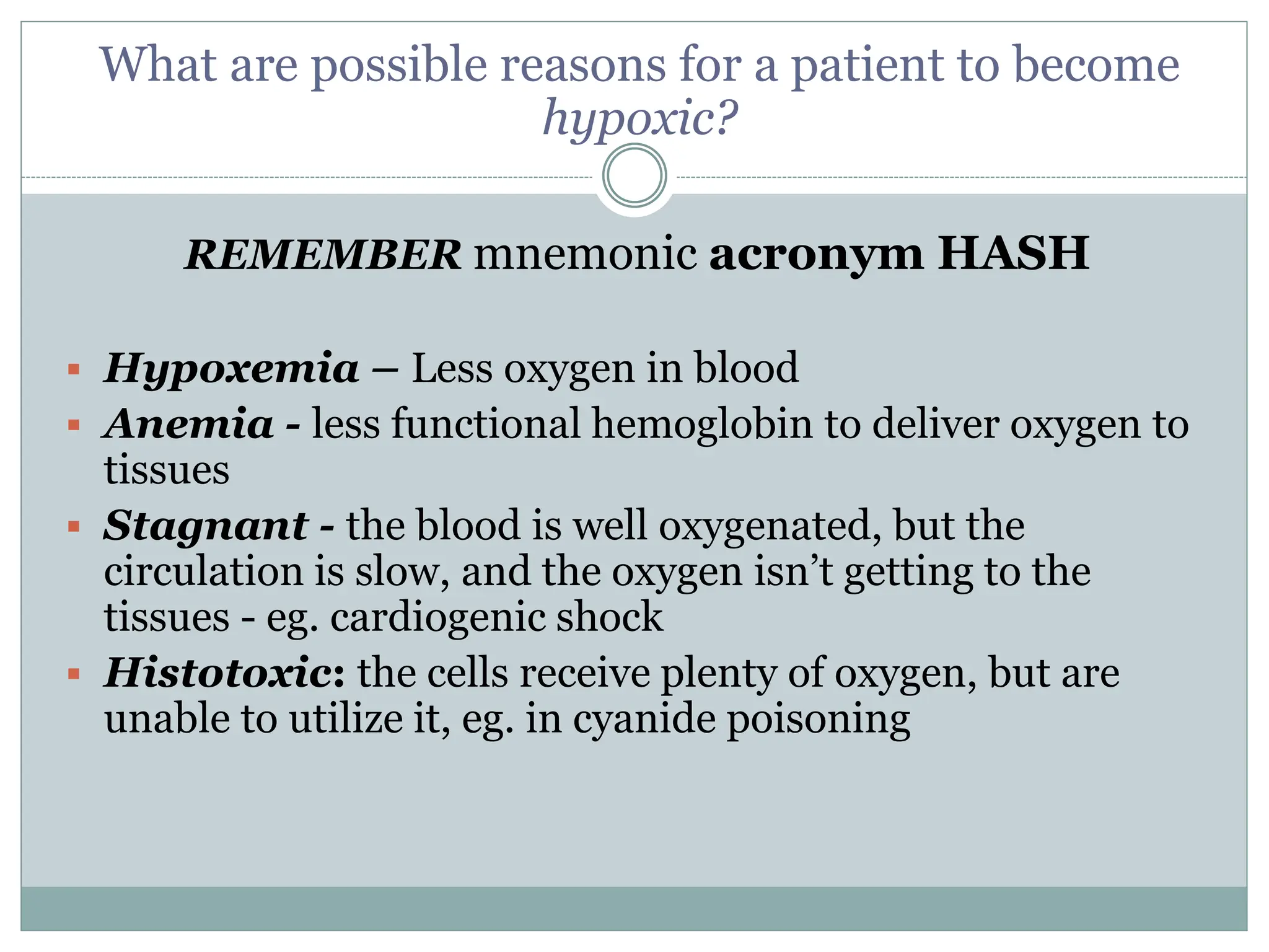
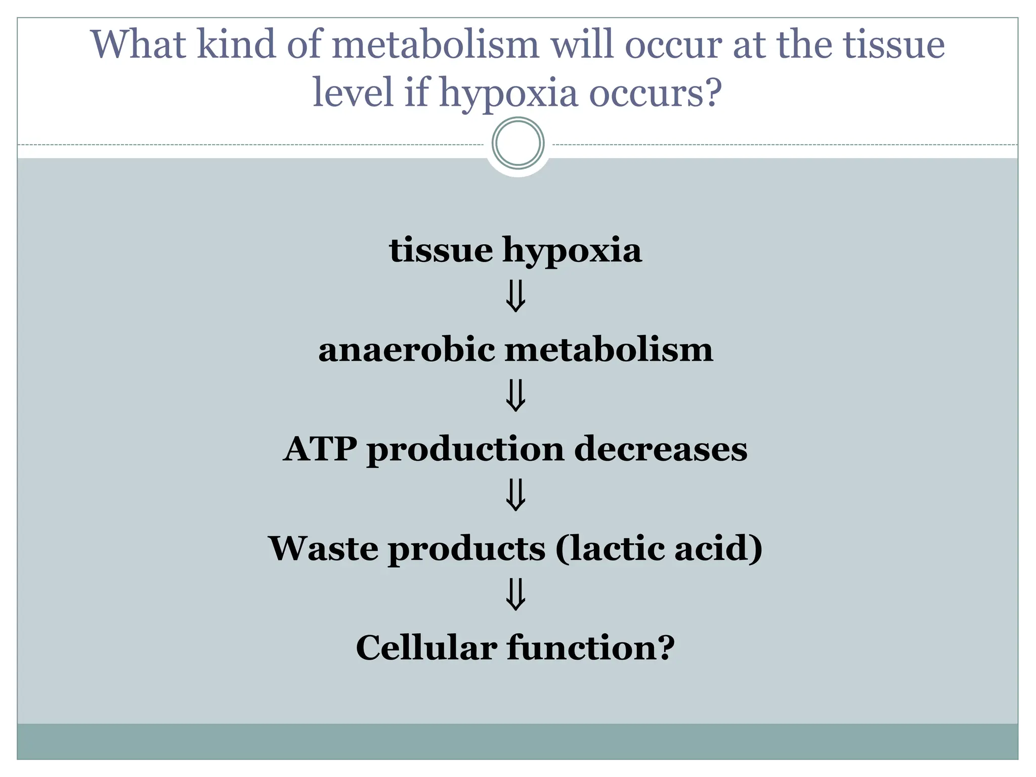
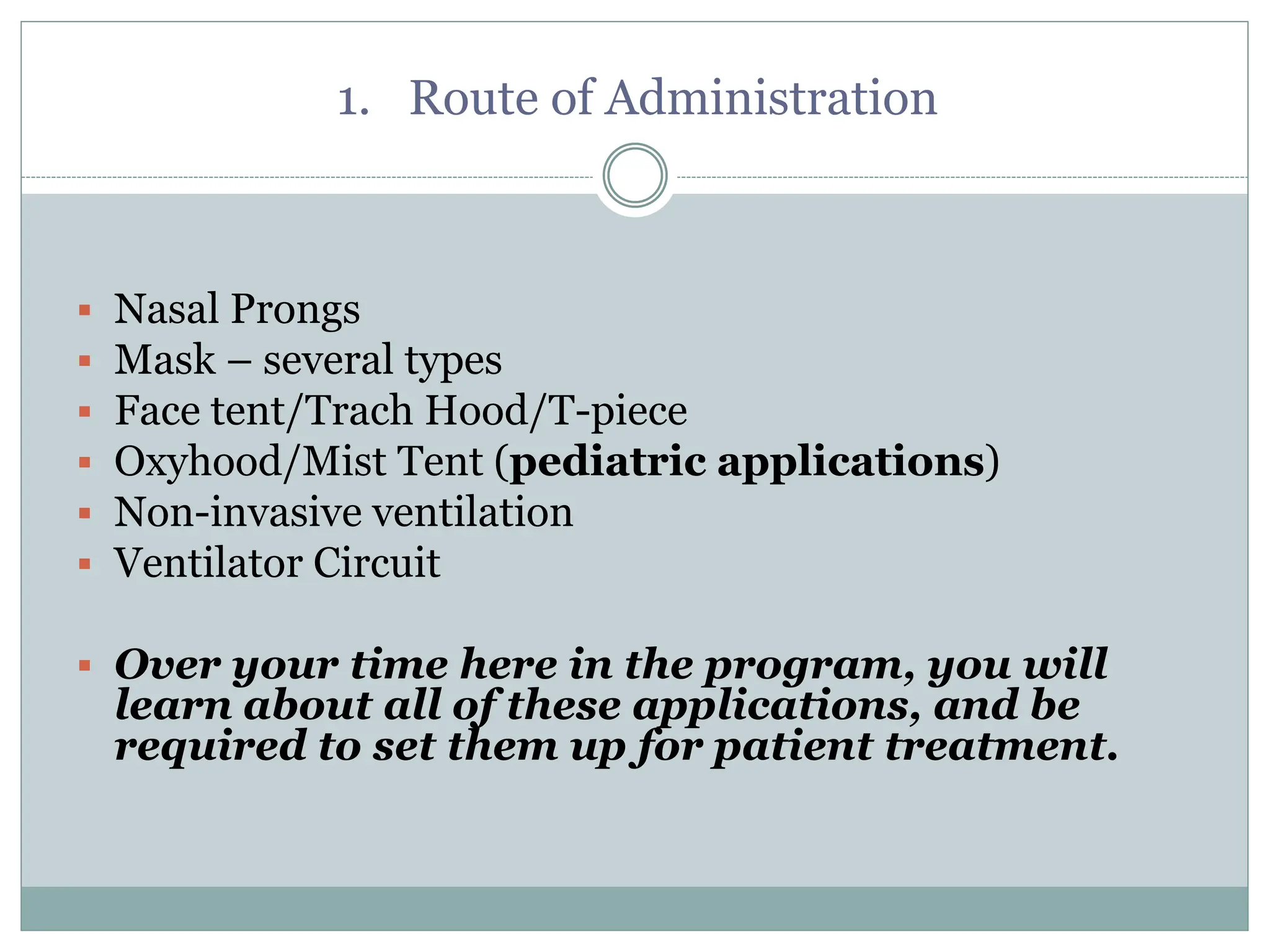
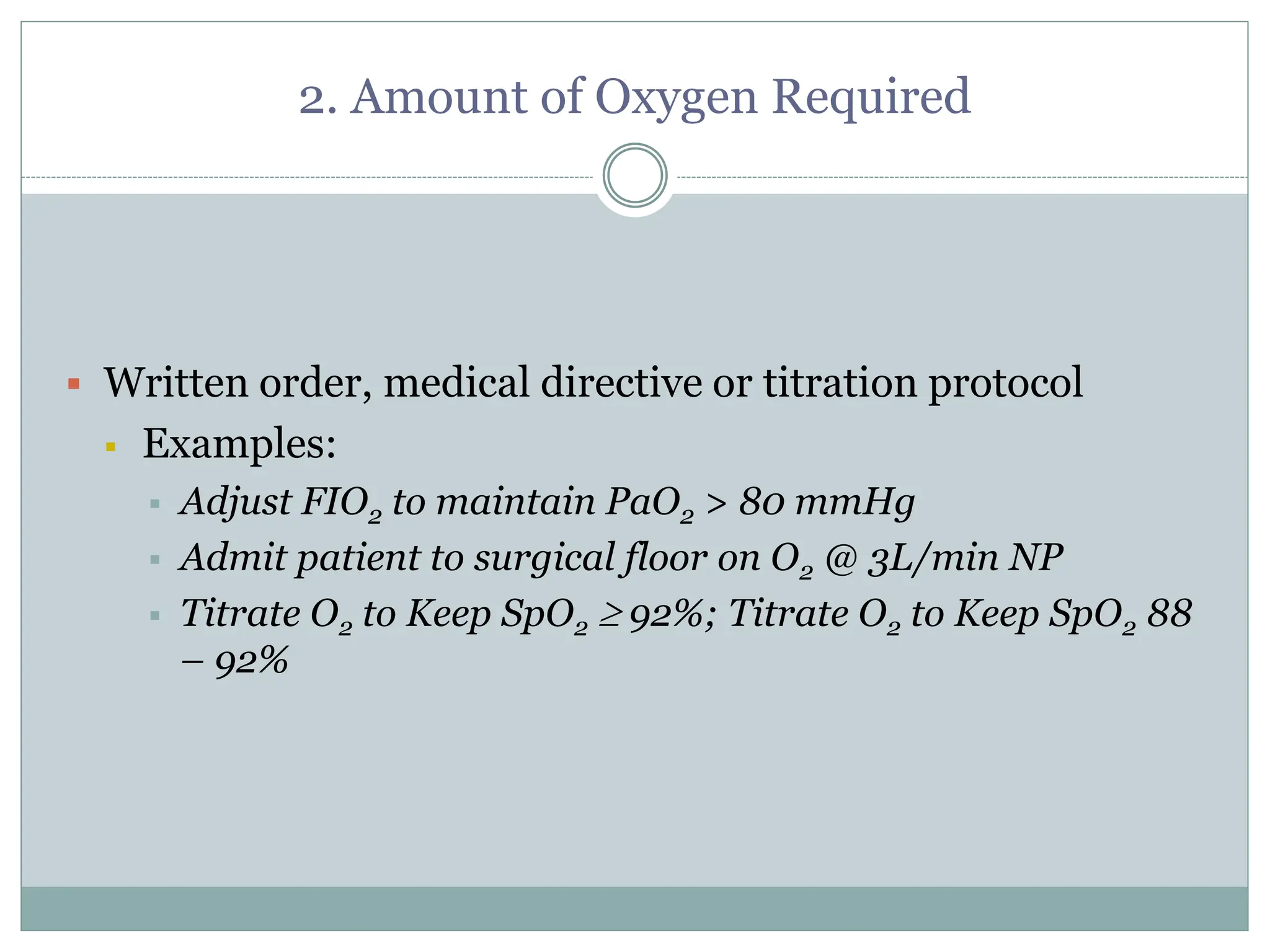
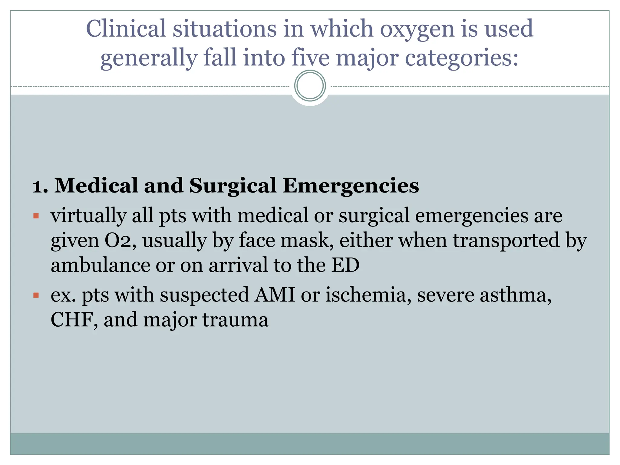
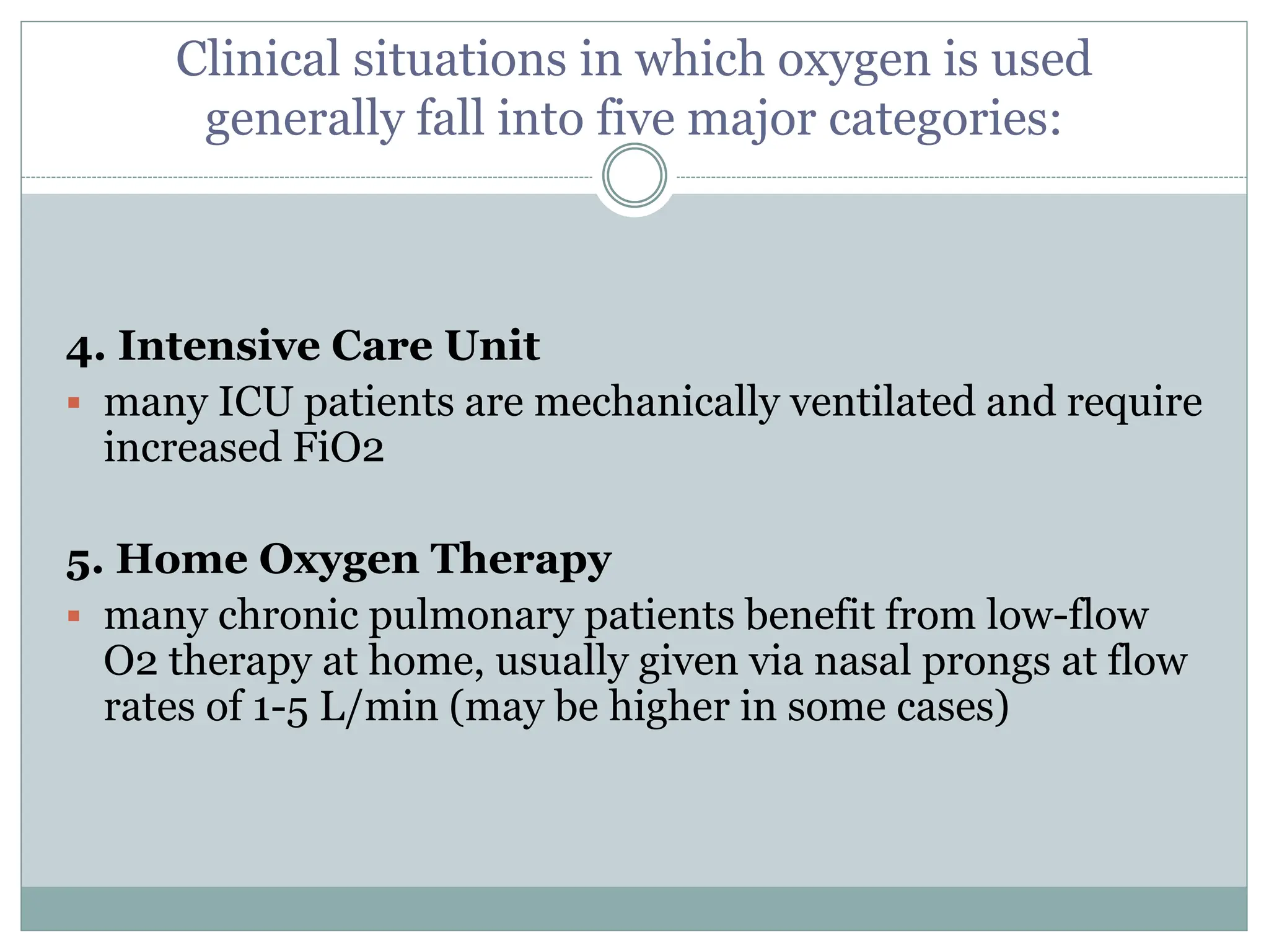
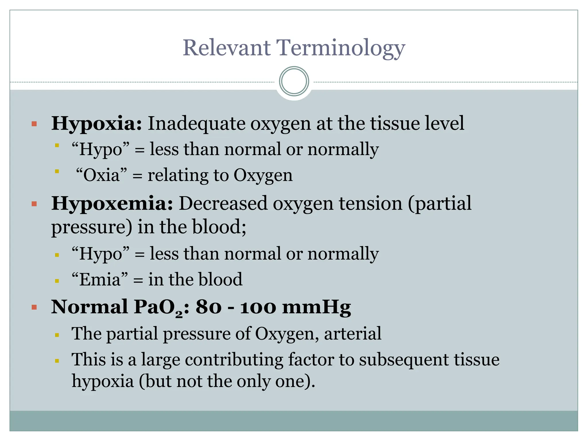
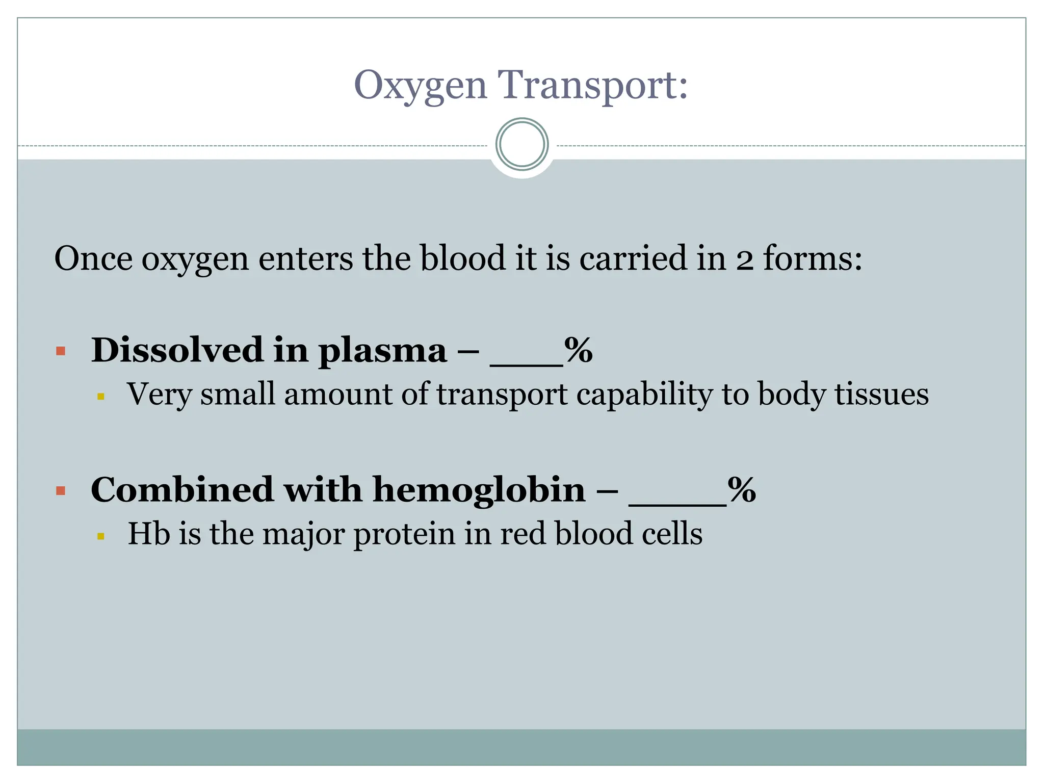
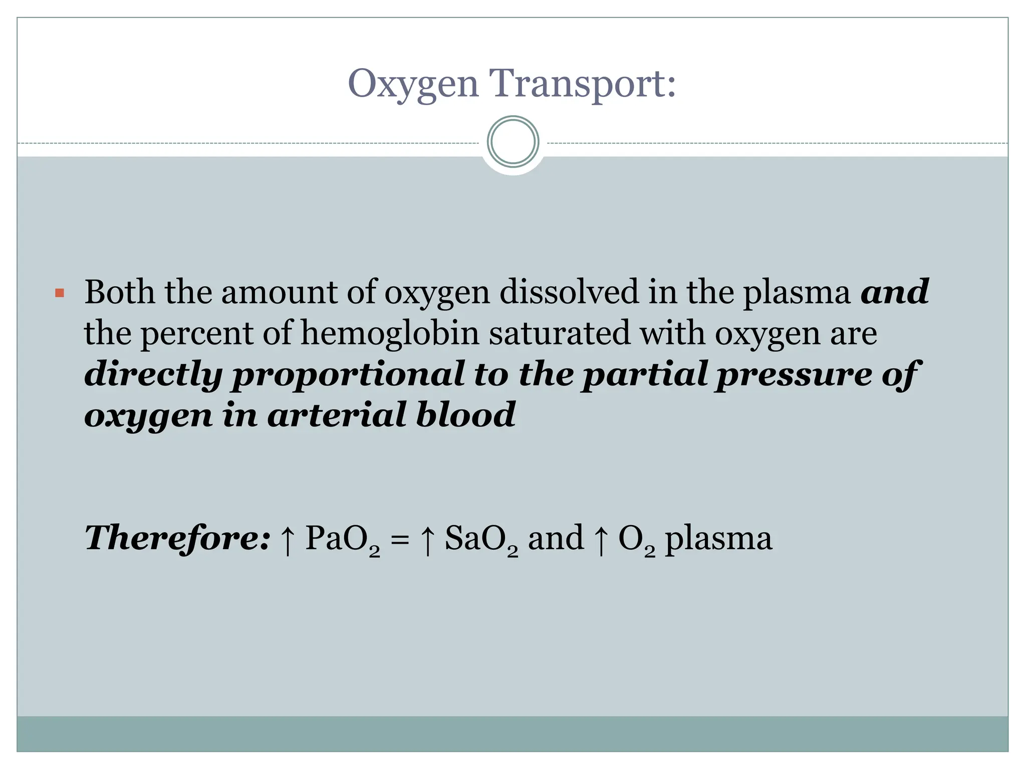
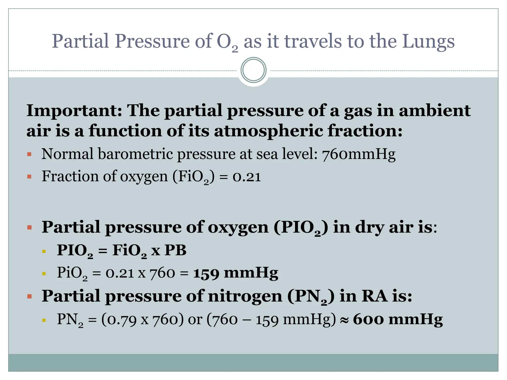
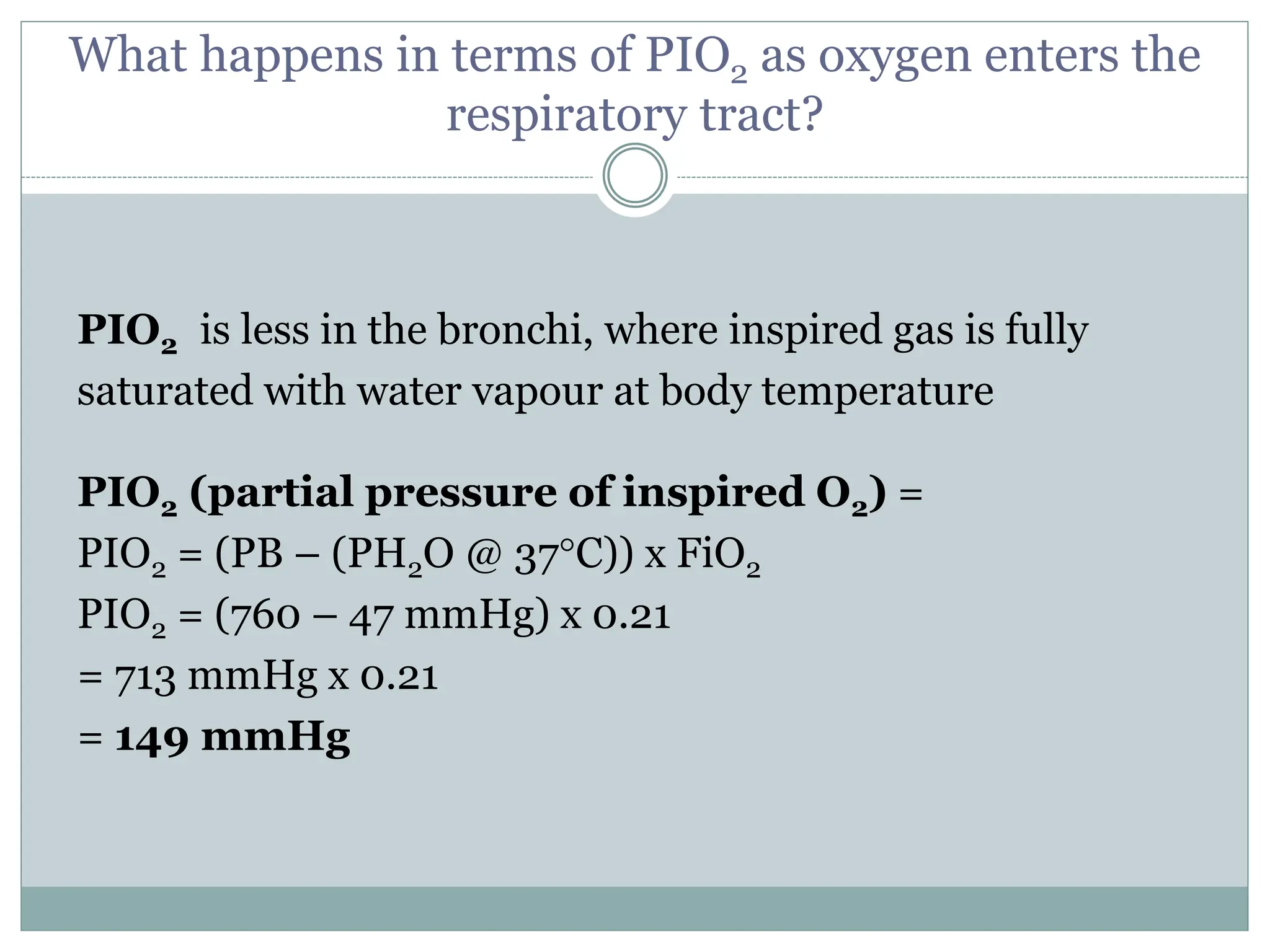
This document provides an overview of oxygen therapy, including relevant terminology, oxygen transport and monitoring, causes of hypoxemia and hypoxia, administration of oxygen therapy, and clinical situations where oxygen is commonly used. Key points covered include the partial pressure of oxygen in the lungs and blood, how supplemental oxygen increases this pressure gradient and oxygen saturation, and the five major categories where oxygen therapy is generally applied: medical emergencies, pulmonary disease, the perioperative period, the ICU, and home oxygen therapy.

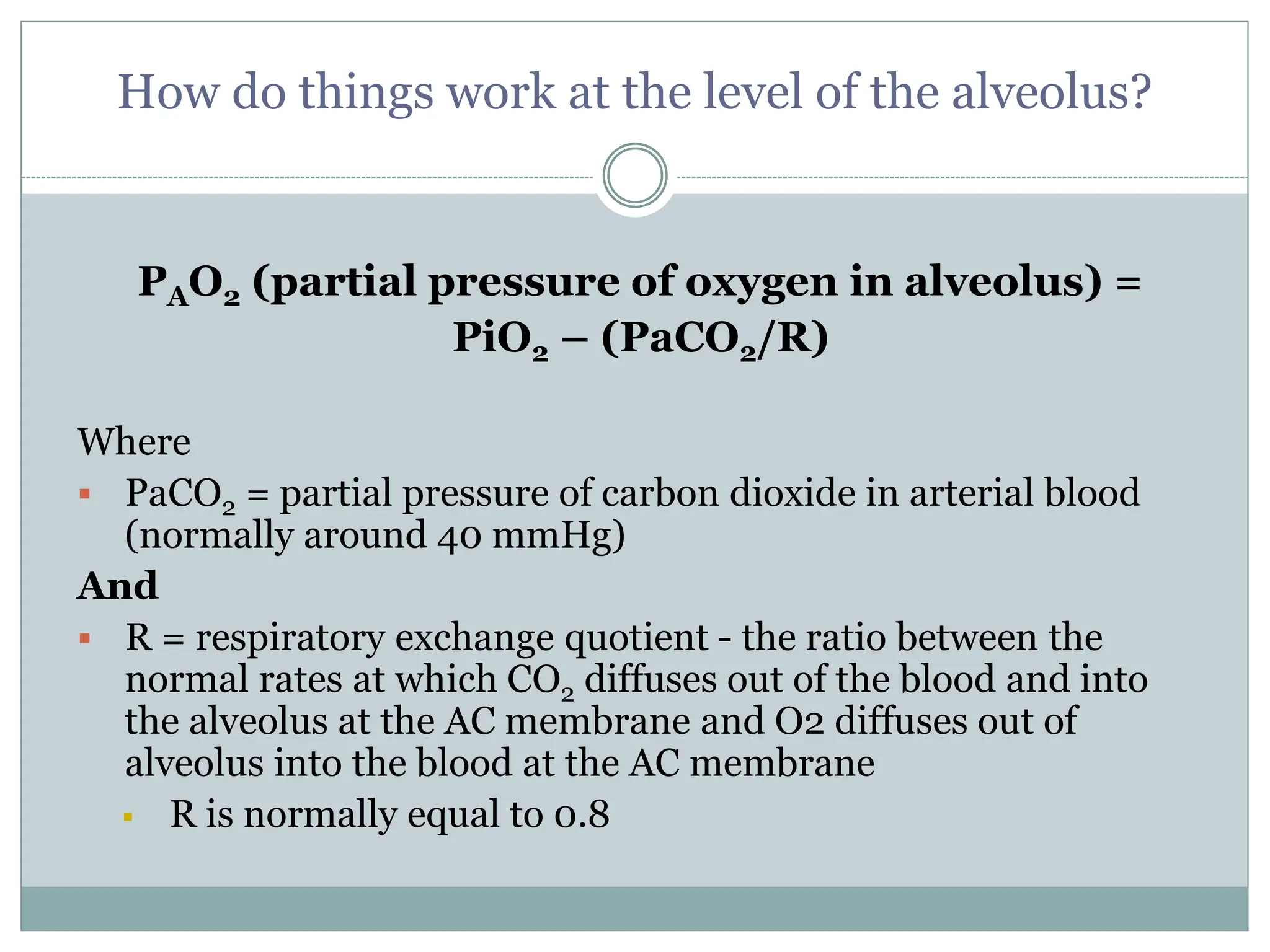
![Therefore; The ALVEOLAR AIR EQUATION
PAO2=[(PB – PH2O) x FiO2] – (PaCO2/0.8)
PAO2 = 149.73 or 150 mmHg – (40 mmHg/0.8)
100 mmHg
Airway Alveoli](https://image.slidesharecdn.com/sc-lecture5-introtooxygentherapy-240124064554-67ff2a1b/75/Introduction-to-Oxygen-Therapy-ppt-8-2048.jpg)