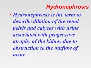
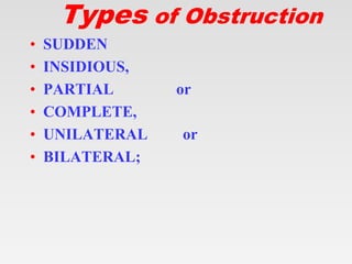
Tips on using my ppt.
1. You can freely download, edit, modify and put your
name etc.
2. Don’t be concerned about number of slides. Half the
slides are blanks except for the title.
3. First show the blank slides (eg. Aetiology ) > Ask
students what they already know about ethology of
today's topic. > Then show next slide which enumerates
aetiologies.
4. At the end rerun the show – show blank> ask questions >
show next slide.
5. This will be an ACTIVE LEARNING SESSION x
three revisions.
6. Good for self study also. Good for self study also.
Display blank slide> Think what you already know about
this > Read next slide.
Hydronephrosis.
Hydronephrosis.
• Hydronephrosis is the term to
describe dilation of the renal
pelvis and calyces with urine
associated with progressive
atrophy of the kidney due to
obstruction to the outflow of
urine.
Types of Obstruction
• SUDDEN
• INSIDIOUS,
• PARTIAL or
• COMPLETE,
• UNILATERAL or
• BILATERAL;
Epidemiology
Epidemiology
• Autopsy 3.1%.
• Differences based on sex did not become
apparent until age 20 years.
• At age 20-60 years, hydronephrosis was
more common in women, which was
suggested to be due to pregnancy and
gynecologic malignancy.
• In men, prostatic diseases were indicated as
the cause of the rise in prevalence after age
60 years.
Epidemiology
• Autopsy studies also indicate that
hydronephrosis is present in 2-2.5% of
children.
• The prevalence is slightly increased in boys,
most of whom in the study were younger
than 1 year.
• These occurrence rates likely underestimate
the prevalence because conditions such as
temporary obstruction due to prior
pregnancy or calculi were not included.
Types of Lesions
Types of Lesions
It can be caused by lesions that are
• Intrinsic to the urinary tract or
• Extrinsic lesions that compress
the ureter.
Obstructive lesions of the urinary tract
Etiology
Etiology
• 1. Congenital anomalies
• 2. Urinary calculi
• 3. Benign prostatic hypertrophy
• 4. Tumors
• 5. Inflammation
• 6. Sloughed papillae or blood clots
• 7. Pregnancy
• 8. Uterine prolapse and cystocele
• 9. Functional disorders
Etiology
• Anatomic abnormalities (including urethral
valves or stricture, and stenosis at the
ureterovesical or ureteropelvic junction)
account for the majority of cases in
children.
• Calculi are most common in young adults,
• Prostatic hypertrophy or carcinoma,
retroperitoneal or pelvic neoplasms, and
calculi are the primary causes in older
patients.
Physiological
Physiological
• Hydronephrosis or hydroureter is a normal
finding in pregnant women.
• progesterone effects and mechanical
compression of the ureters at the pelvic
brim.
• More prominent on the right
• These changes can be visualized on
ultrasound examination by the second
trimester, and they may not resolve until 6-
12 weeks post partum.
Acute Vs. Chronic
Acute Vs. Chronic
• We may consider acute as hydronephrosis
that, when corrected, allows full recovery of
renal function.
• Chronic hydronephrosis is a situation in
which the loss of function is irreversible
even with correction of the obstruction.
• Early experiments with dogs showed that if
acute unilateral obstruction is corrected
within 2 weeks, full recovery of renal
function is possible. However, after 6
weeks of obstruction, function is
When the obstruction is SUDDEN and
COMPLETE,
GLOMERULAR FILTRATION IS
REDUCED.
• It leads to
• MILD DILATION OF THE PELVIS and
CALYCES
and sometimes to
• ATROPHY OF THE RENAL
PARENCHYMA.
• When the obstruction is
•SUBTOTAL or
INTERMITTENT,
GLOMERULAR
FILTRATION IS NOT
SUPPRESSED, and
PROGRESSIVE
DILATION ENSUES
The kidney may be slightly to
massively ENLARGED,
depending on
the degree and the duration of
the obstruction.
• In far-advanced cases the kidney may
become transformed into
• A THIN-WALLED CYSTIC
STRUCTURE having a diameter of up to
15 to 20 cm
• with striking PARENCHYMAL
ATROPHY,
• TOTAL OBLITERATION OF THE
PYRAMIDS, and
• THINNING OF THE CORTEX.
Pathology
•
Pathology
• Compression of the papillae, thinning of
the parenchyma around the calyces, and
coalescence of the septa between calyces.
• Dilation of the tubular lumen and flattening
of the tubular epithelium.
• Fibrotic changes and increased collagen
deposition are observed in the peritubular
interstitium.
Complications
Complications
• Urinary stasis may result in
• Infection
• Renal scarring
• Calculus formation
• Sepsis
• Renal failure
• Hypertension
Clinical Features
Clinical Features
I. Acute obstruction –
• PAIN due to distention of the
collecting system or renal capsule.
• The site of obstruction determines the
location of pain. Upper ureteral or
renal pelvic lesions lead to flank pain
or tenderness, whereas lower ureteral
obstruction causes pain that may
radiate to the ipsilateral testicle or
labia.
Clinical Features
• II Unilateral complete or partial
hyd ronephrosis may remain SILENT
FOR LONG PERIODS, since the
unaffected kidney can maintain
adequate renal function.
• Sometimes its existence first becomes
apparent in the course of USG or
INTRAVENOUS PYELOGRAPHY.
Clinical Features
III. In bilateral partial
obstruction the earliest
manifestation is inability to
concentrate the urine,
reflected by
POLYURIA AND
NOCTURIA.
.
Clinical Features
Other symptoms
• Anuria
• Hematuria
• Fever
• Antenatal USG.
Clinical Features:Physical Exam
• With severe hydronephrosis, the kidney
may be palpable.
• With bilateral hydronephrosis, lower
extremity edema may occur.
• Costovertebral angle tenderness on the
affected side is common.
• A palpably distended bladder
Diagnostic Studies
Diagnostic Studies
• Laboratory Tests
• Imaging
– X-ray + Contrast studies.
– USG
– CT
– MRI
– Nuclear Scan
• Other Tests
Laboratory Studies
Laboratory Studies
• Urinalysis-
– Pyuria suggests the presence of infection.
– Microscopic hematuria may indicate the
presence of a stone or tumor.
• CBC leukocytosis, which may indicate
acute infection.
• Serum Creatinine levels
• Hypercalemia can be a life-threatening
condition.
Imaging studies
Imaging studies
• Renal ultrasonography is the test of choice
• Intravenous Urography
• CT Indications :
– Ultrasonography results are equivocal
– The kidneys cannot be well visualized
– The cause of the obstruction cannot be
identified.
Imaging studies
• Diuretic Renography with technetium Tc
99m-mercaptoacetyltriglycine
(Tc99mMAG3),
• A perfusion pressure flow study
• Diffusion-weighted magnetic resonance
imaging (MRI)
• Antegrade or retrograde pyelography is
usually used to relieve, rather than
diagnose, urinary tract obstruction.
Imaging in children
Imaging in children
• Detection of antenatal hydronephrosis by
ultrasound usually occurs in the second
trimester with a renal pelvic dilation (RPD)
cutoff of greater than or equal to 4 mm.
• Postnatal ultrasound.
• A voiding cystourethrography (VCUG) is
performed to detect VUR and, in boys, to
evaluate the posterior urethra.
• Diuretic renography
• Magnetic resonance urography (MRU)
Differential Diagnosis
• Other problems to consider in the
differential diagnosis include the following:
• Peripelvic cyst
• Congenital megacalices
• Calyceal diverticula
• Capacious extrarenal pelvis<>
• High urine flow
• Pyelonephritis
Non Operative Therapy
•
Non Operative Therapy
• Pain control
• Treatment or prevention of infection
• Oral alkalinization therapy for uric acid
stones and
• Steroid therapy for retroperitoneal fibrosis.
Minimally invasive Therapy
• Extracorporeal shockwave lithotripsy
• Urethral Catheterisation.
• SPC
• Ureteral catheterisation +- stent
• Nephrostomy
• Post. Urethral Valve fulguration.
Operative Therapy:Indications
• Any signs of infection Fever, leucocytosis
• The potential for loss of renal function also
adds to the urgency (eg, hydronephrosis or
hydroureter bilaterally or in a solitary
kidney).
• Refractory pain in a patient with an
obstructing ureteral calculus necessitates
intervention,
• Intractable nausea and vomiting.
Operative Therapy
Mostly extrinsic
• Retroperitoneal fibrosis,
• Retroperitoneal tumors
• Aortic aneurysms.
• Some stones that cannot be treated
endoscopically or with extracorporeal
shockwave lithotripsy
• Ureteral tumors,
On The Horizon
• Fetal Surgery VAS –VesicoAmniotic
Shunting
Get this ppt in mobile
1. Download Microsoft
PowerPoint from play
store.
2. Open Google assistant
3. Open Google lens.
4. Scan qr code from
next slide.
Get my ppt collection
• https://www.slideshare.net/drpradeeppande/
edit_my_uploads
• https://www.dropbox.com/sh/x600md3cvj8
5woy/AACVMHuQtvHvl_K8ehc3ltkEa?dl
=0
• https://www.facebook.com/doctorpradeeppa
nde/?ref=pages_you_manage

Hydronephrosis.pptx

  • 1.
    Tips on usingmy ppt. 1. You can freely download, edit, modify and put your name etc. 2. Don’t be concerned about number of slides. Half the slides are blanks except for the title. 3. First show the blank slides (eg. Aetiology ) > Ask students what they already know about ethology of today's topic. > Then show next slide which enumerates aetiologies. 4. At the end rerun the show – show blank> ask questions > show next slide. 5. This will be an ACTIVE LEARNING SESSION x three revisions. 6. Good for self study also. Good for self study also. Display blank slide> Think what you already know about this > Read next slide.
  • 2.
  • 3.
    Hydronephrosis. • Hydronephrosis isthe term to describe dilation of the renal pelvis and calyces with urine associated with progressive atrophy of the kidney due to obstruction to the outflow of urine.
  • 4.
  • 5.
    Types of Obstruction •SUDDEN • INSIDIOUS, • PARTIAL or • COMPLETE, • UNILATERAL or • BILATERAL;
  • 6.
  • 7.
    Epidemiology • Autopsy 3.1%. •Differences based on sex did not become apparent until age 20 years. • At age 20-60 years, hydronephrosis was more common in women, which was suggested to be due to pregnancy and gynecologic malignancy. • In men, prostatic diseases were indicated as the cause of the rise in prevalence after age 60 years.
  • 8.
    Epidemiology • Autopsy studiesalso indicate that hydronephrosis is present in 2-2.5% of children. • The prevalence is slightly increased in boys, most of whom in the study were younger than 1 year. • These occurrence rates likely underestimate the prevalence because conditions such as temporary obstruction due to prior pregnancy or calculi were not included.
  • 10.
  • 11.
    Types of Lesions Itcan be caused by lesions that are • Intrinsic to the urinary tract or • Extrinsic lesions that compress the ureter.
  • 12.
    Obstructive lesions ofthe urinary tract
  • 13.
  • 14.
    Etiology • 1. Congenitalanomalies • 2. Urinary calculi • 3. Benign prostatic hypertrophy • 4. Tumors • 5. Inflammation • 6. Sloughed papillae or blood clots • 7. Pregnancy • 8. Uterine prolapse and cystocele • 9. Functional disorders
  • 15.
    Etiology • Anatomic abnormalities(including urethral valves or stricture, and stenosis at the ureterovesical or ureteropelvic junction) account for the majority of cases in children. • Calculi are most common in young adults, • Prostatic hypertrophy or carcinoma, retroperitoneal or pelvic neoplasms, and calculi are the primary causes in older patients.
  • 16.
  • 17.
    Physiological • Hydronephrosis orhydroureter is a normal finding in pregnant women. • progesterone effects and mechanical compression of the ureters at the pelvic brim. • More prominent on the right • These changes can be visualized on ultrasound examination by the second trimester, and they may not resolve until 6- 12 weeks post partum.
  • 20.
  • 21.
    Acute Vs. Chronic •We may consider acute as hydronephrosis that, when corrected, allows full recovery of renal function. • Chronic hydronephrosis is a situation in which the loss of function is irreversible even with correction of the obstruction. • Early experiments with dogs showed that if acute unilateral obstruction is corrected within 2 weeks, full recovery of renal function is possible. However, after 6 weeks of obstruction, function is
  • 22.
    When the obstructionis SUDDEN and COMPLETE, GLOMERULAR FILTRATION IS REDUCED. • It leads to • MILD DILATION OF THE PELVIS and CALYCES and sometimes to • ATROPHY OF THE RENAL PARENCHYMA.
  • 23.
    • When theobstruction is •SUBTOTAL or INTERMITTENT, GLOMERULAR FILTRATION IS NOT SUPPRESSED, and PROGRESSIVE DILATION ENSUES
  • 24.
    The kidney maybe slightly to massively ENLARGED, depending on the degree and the duration of the obstruction.
  • 25.
    • In far-advancedcases the kidney may become transformed into • A THIN-WALLED CYSTIC STRUCTURE having a diameter of up to 15 to 20 cm • with striking PARENCHYMAL ATROPHY, • TOTAL OBLITERATION OF THE PYRAMIDS, and • THINNING OF THE CORTEX.
  • 26.
  • 27.
    Pathology • Compression ofthe papillae, thinning of the parenchyma around the calyces, and coalescence of the septa between calyces. • Dilation of the tubular lumen and flattening of the tubular epithelium. • Fibrotic changes and increased collagen deposition are observed in the peritubular interstitium.
  • 28.
  • 29.
    Complications • Urinary stasismay result in • Infection • Renal scarring • Calculus formation • Sepsis • Renal failure • Hypertension
  • 30.
  • 31.
    Clinical Features I. Acuteobstruction – • PAIN due to distention of the collecting system or renal capsule. • The site of obstruction determines the location of pain. Upper ureteral or renal pelvic lesions lead to flank pain or tenderness, whereas lower ureteral obstruction causes pain that may radiate to the ipsilateral testicle or labia.
  • 32.
    Clinical Features • IIUnilateral complete or partial hyd ronephrosis may remain SILENT FOR LONG PERIODS, since the unaffected kidney can maintain adequate renal function. • Sometimes its existence first becomes apparent in the course of USG or INTRAVENOUS PYELOGRAPHY.
  • 33.
    Clinical Features III. Inbilateral partial obstruction the earliest manifestation is inability to concentrate the urine, reflected by POLYURIA AND NOCTURIA. .
  • 34.
    Clinical Features Other symptoms •Anuria • Hematuria • Fever • Antenatal USG.
  • 35.
    Clinical Features:Physical Exam •With severe hydronephrosis, the kidney may be palpable. • With bilateral hydronephrosis, lower extremity edema may occur. • Costovertebral angle tenderness on the affected side is common. • A palpably distended bladder
  • 36.
  • 37.
    Diagnostic Studies • LaboratoryTests • Imaging – X-ray + Contrast studies. – USG – CT – MRI – Nuclear Scan • Other Tests
  • 38.
  • 39.
    Laboratory Studies • Urinalysis- –Pyuria suggests the presence of infection. – Microscopic hematuria may indicate the presence of a stone or tumor. • CBC leukocytosis, which may indicate acute infection. • Serum Creatinine levels • Hypercalemia can be a life-threatening condition.
  • 40.
  • 41.
    Imaging studies • Renalultrasonography is the test of choice • Intravenous Urography • CT Indications : – Ultrasonography results are equivocal – The kidneys cannot be well visualized – The cause of the obstruction cannot be identified.
  • 42.
    Imaging studies • DiureticRenography with technetium Tc 99m-mercaptoacetyltriglycine (Tc99mMAG3), • A perfusion pressure flow study • Diffusion-weighted magnetic resonance imaging (MRI) • Antegrade or retrograde pyelography is usually used to relieve, rather than diagnose, urinary tract obstruction.
  • 43.
  • 44.
    Imaging in children •Detection of antenatal hydronephrosis by ultrasound usually occurs in the second trimester with a renal pelvic dilation (RPD) cutoff of greater than or equal to 4 mm. • Postnatal ultrasound. • A voiding cystourethrography (VCUG) is performed to detect VUR and, in boys, to evaluate the posterior urethra. • Diuretic renography • Magnetic resonance urography (MRU)
  • 45.
  • 46.
    Differential Diagnosis • Otherproblems to consider in the differential diagnosis include the following: • Peripelvic cyst • Congenital megacalices • Calyceal diverticula • Capacious extrarenal pelvis<> • High urine flow • Pyelonephritis
  • 47.
  • 48.
    Non Operative Therapy •Pain control • Treatment or prevention of infection • Oral alkalinization therapy for uric acid stones and • Steroid therapy for retroperitoneal fibrosis.
  • 49.
  • 50.
    Minimally invasive Therapy •Extracorporeal shockwave lithotripsy • Urethral Catheterisation. • SPC • Ureteral catheterisation +- stent • Nephrostomy • Post. Urethral Valve fulguration.
  • 51.
  • 52.
    Operative Therapy:Indications • Anysigns of infection Fever, leucocytosis • The potential for loss of renal function also adds to the urgency (eg, hydronephrosis or hydroureter bilaterally or in a solitary kidney). • Refractory pain in a patient with an obstructing ureteral calculus necessitates intervention, • Intractable nausea and vomiting.
  • 53.
    Operative Therapy Mostly extrinsic •Retroperitoneal fibrosis, • Retroperitoneal tumors • Aortic aneurysms. • Some stones that cannot be treated endoscopically or with extracorporeal shockwave lithotripsy • Ureteral tumors,
  • 54.
    On The Horizon •Fetal Surgery VAS –VesicoAmniotic Shunting
  • 55.
    Get this pptin mobile 1. Download Microsoft PowerPoint from play store. 2. Open Google assistant 3. Open Google lens. 4. Scan qr code from next slide.
  • 58.
    Get my pptcollection • https://www.slideshare.net/drpradeeppande/ edit_my_uploads • https://www.dropbox.com/sh/x600md3cvj8 5woy/AACVMHuQtvHvl_K8ehc3ltkEa?dl =0 • https://www.facebook.com/doctorpradeeppa nde/?ref=pages_you_manage

Editor's Notes

  • #2 drpradeeppande@gmail.com 7697305442
  • #58 drpradeeppande@gmail.com 7697305442