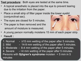
Body fluid analysis 
(excluding body cavity fluids & urine) 
Feb 5th, 2013 
By Dr. Niraj Gupta, 
MD (Pathology)
Contents 
Introduction 
Amniotic fluid 
Semen analysis 
Synovial fluid 
Gastric fluid 
Other body fluids: Sweat,saliva,tear
FLUID 
• A fluid is a non solid substance such as liquid or 
gas that tends to flow or conform to the shape of 
the container. Includes urine,fluids in the body 
cavity, joint fluid,sweat, semen etc. 
• They are generally involved with the process of 
transport ,excretion and lubrication. They allow 
the distribution of oxygen and nutrients to the 
tissues and organs and the transport of waste 
products from the tissues, enabling their 
elemination from body.
Total body 
water 
Extracellular 
fluid(33%) 
Intracellular 
fluid(66%) 
Interstitial fluid 
(25%) 
Plasma 
(7%) 
Transcellular 
fluid(~1%) 
50-70%
• Transcellular fluid is a small compartment 
that represents all those body fluids which are 
formed from the transport activities of cells. It is 
contained within the epithelial lined space. 
• It includes CSF, synovial fluid, GIT fluids , 
aqueous humor and serous fluid of bodycavity 
like pleural, peritoneal and pericardial fluid. 
• It is important because of specialized function 
involved. It is the smallest component of 
extracellular fluid.
ACCUMULATION OF EXCESS 
BODY FLUIDS 
• Body fluids are necessary for lubrication of the body 
cavity/organ interface during movement. 
• A delicate equilibrium is maintained by the 
capillaries and the lymphatic vessels. Any 
obstruction or altered pressure in these vessels can 
affect the amount of fluid and its constituents. 
• Several forces, within and outside of the capillaries, 
work together to maintain fluid equilibrium.
• The tissue’s colloidal osmotic pressure (interstitial fluid 
pressure), along with the capillary’s hydrostatic pressure 
(filtration pressure),regulates the outward flow of fluid from 
the capillary. 
• The colloidal osmotic pressure of the capillary and the 
tissue’s hydrostatic pressure regulate the inward flow of 
fluid into the capillary from the tissue. 
• However, an imbalance in pressures causes excess egress 
of fluid into tissue spaces and can lead to accumulation of 
fluid.
AMNIOTIC FLUID 
• Amniotic fluid is found around the developing 
fetus,inside a membranous sac, called the amnion. 
• This fluid serves to cushion and protect the 
developing fetus and also serves a key role in the 
exchange of water and molecules between the fetus 
and the maternal circulation. 
• The fluid has a composition similar to that of the 
maternal plasma and contains a small amount of 
sloughed fetal cells. 
• These cells provide the basis for cytogenetic 
analysis.
• The volume of amniotic fluid increases steadily 
throughout the pregnancy up to a maximum of 
1000–1200 mL at 36 weeks of gestation. 
• Measurement of amniotic fluid creatinine has 
been used to determine fetal age. 
• Prior to 36 weeks’ gestation,the amniotic fluid 
creatinine level ranges between 1.5 and 2.0 
mg/dL. 
• After 36 weeks rises to >2.0 mg/dL.
• The laboratory performs several crucial tests on 
amniotic fluid to assess the status of the fetus. 
• These tests can be divided into these groups: 
(a) tests to diagnose genetic and congenital 
disorders before birth, 
(b) tests to detect fetal distress from hemolytic 
disease of the newborn (HDN) or from infection, 
(c) tests to assess fetal lung maturity
Specimen Collection 
• Amniotic fluid is obtained by needle aspiration into the amniotic 
sac, usually transabdominally with simultaneous use of 
ultrasound,a procedure called amniocentesis. 
• Amniocentesis is generally performed between 15 and 18 
weeks of gestation for genetic studies,whereas tests for fetal 
distress and maturity are performed later in the third trimester. 
•A maximum of 30 mL of 
amniotic fluid is collected in 
sterile syringes. 
•The first 2 or 3 mL collected 
can be contaminated by 
maternal blood, tissue fluid, 
and cells and are discarded.
• In case of possible premature membrane rupture 
or maternal bladder puncture or rupture, it may be 
necessary to differentiate amniotic fluid from 
maternal urine. 
• To differentiate these two fluids,chemical levels of 
creatinine, urea, glucose, and protein can be of 
assistance. 
• Levels of creatinine and urea are much higher 
in urine than in amniotic fluid. 
• Glucose and protein levels tend to be higher in 
amniotic fluid than in urine.
• Normal amniotic fluid is colorless to pale 
yellow and slightly cloudy.
TESTING AMNIOTIC FLUID FOR GENETIC 
AND CONGENITAL DISORDERS 
• Valuable cytogenetic information related to the sex of 
the fetus and to genetic abnormalities can be obtained 
via amniocentesis. 
• Amniocentesis is often performed to detect Down 
syndrome and anencephaly prior to birth. 
• Fetal neural tube defects such as anencephaly and 
spina bifida cause elevated alpha fetoprotein (AFP) 
in amniotic fluid and in the maternal circulation. 
• In normal fetal development, AFP peaks at about 
16 weeks of gestation and then declines gradually to 
term.
• Measurement of amniotic fluid AFP levels 
is indicated when maternal serum levels 
are elevated or a family history of previous 
neural tube defects exists. 
• Both serum and amniotic fluid AFP levels 
are reported in terms of multiples of the 
median (MoM). 
• The median is the laboratory’s reference 
level for a given week of gestation.
A value two times the median value is 
considered abnormal (>2 MoM) for both maternal 
serum and amniotic fluid. 
• Elevated amniotic fluid AFP levels are followed 
by measurement of amniotic 
acetylcholinesterase (AChE). 
• The test is more specific for neural tube 
disorders than AFP, provided it is not 
performed on a bloody specimen, because 
blood contains AChE. 
Amniotic fluid AchE = > 5 U/L in NTD
Fetal karyotype: 
• Retrieval of fetal somatic cells can be used to identify 
genetic constitution of fetus. 
• The problem of karyotyping is that it take 3 or more 
weeks. 
• Nowadays ,numerical abnormalities in the 
chromosomes 21,18,13,X & Y can be detected more 
rapidly by using chromosome specific DNA probes 
tagged with fluorescent dyes,known as FISH.(90% 
specificity & gives results in 2days.)
FETAL LUNG MATURITY 
• Respiratory distress is the most frequent complication of early 
delivery. 
• Therefore, laboratory tests must be performed to determine the 
maturity of the fetal lungs. 
• Lecithin:Sphingomyelin Ratio and Phosphatidylglycerol- 
• Fetal lung surfactants include these three phospholipids:lecithin 
(also known as phosphatidylcholine), sphingomyelin, and 
phosphatidyl glycerol. It is produced by type II pneumocytes in 
the form of lamellar bodies. 
• The ratio of lecithin to sphingomyelin is used to assess fetal 
lung maturity. Up until the 33rd week of gestation,the levels of 
these two phospholipids are relatively equal.
• After 34 weeks of gestation,the level of 
sphingomyelin decreases, whereas the level of lecithin 
increases significantly. 
L/S ratio ≥ 2.0 usually indicate maturity, and 
ratio <1.5 indicate immaturity. 
• This is done by centrifugation and acetone 
precipitation. 
• Amniotic fluid surfactant lipids are chromatographed 
on thin-layer silica . 
• The phospholipids are made visible by heat charring 
or staining. 
• Densitometric quantification determines the L/S ratio.
• Many laboratories have replaced the L/S ratio with 
the more cost-effective phosphatidyl glycerol 
immunoassays, fluorescence polarization, and 
lamellar body density procedures. 
• Development of an immunologic agglutination test 
for phosphatidyl glycerol has provided a more rapid 
method for assessment of fetal maturity that does 
not require a laboratory to be equipped to perform 
thin-layer chromatography. 
• The Aminostat-FLM (Irving Scientific, Santa 
Ana,CA) uses antisera specific for phosphatidyl 
glycerol and is not affected by specimen 
contamination with blood and 
meconium.
Phosphatidylglycerol measurement in AF 
• Thin-layer chromatography 
– Absent: Fetal lung immaturity 
– Present: Fetal lung maturity 
• Slide-agglutination test 
– Negative: <0.5 mg/L (immature) 
– Positive : > 0.5 mg/L (mature) 
• Enzymatic assay: 19-83 mg/L 
< 19 = Immature lung 
> 83 = Mature lung
Fluorescence Polarization Assay (Microviscosity) 
• This assay provides a fluorescence polarization (P) 
surfactant:albumin ratio. 
• The microviscosity of lipid aggregates in the amniotic fluid may 
be assayed by mixing the fluid with a specific fluorescent dye 
that becomes incorporated in the lipids of surfactant. 
• The intensity of fluorescence is measured by polarized light. 
• Dye bound to surfactant has a longer fluorescence lifetime and 
exhibits a low polarization. 
P value is high with low levels of surfactant , 
P value is low with high levels of surfactant. 
• The degree of fluorescence polarization is inversely 
proportional to the quantity of pulmonary surfactant present.
FOAM STABILITY (SHAKE TEST) 
1. Mix equal parts of amniotic fluid with 95% ethanol. 
2. Vigorously shake for 15 seconds. 
3. Allow to sit undisturbed for 15 minutes. 
4. Observe for the presence of a continuous line of bubbles 
around the outside edge. 
• The presence of bubbles indicates that a sufficient amount of 
phospholipid is available to reduce the surface tension. 
bubbles present -- POSITIVE 
bubbles absent -- NEGATIVE 
• The highest concentration of 95% ethanol that is able to 
support a ring of foam is known as the foam stability index. 
Foam stability index= ≥0.47 indicate fetal lung maturity
Lamellar Bodies 
• Fetal lung surfactants are produced by fetal type II 
pneumocytes of the fetal lung and are stored as 
lamellar bodies after about 20 weeks of gestation. 
• Lamellar bodies are about the size of small platelets. 
• Lamellar bodies are storage forms of lung 
phospholipids and they enter the fetal lungs and the 
amniotic fluid at about 20–24 weeks of gestation. 
• Lamellar bodies affect the optical density of amniotic 
fluid and a measurement of the optical density of 
0.150 at 650 nm has been shown to correlate with an 
L/S ratio of 2.0 and to correlate with the presence of 
phosphatidyl glycerol.
• Lamellar body counts provide a reliable estimate of fetal 
• lung maturity. Lamellar body counts can be performed 
• easily with many hematology analyzers using the platelet 
• count channel. 
• Lamellar body counts of approximately 35,000 per microliter 
correspond to adequate fetal lung surfactant levels. 
Fig: Lamellar bodies in 
amniotic fluid on 
hematology counters
HEMOLYTIC DISEASE OF THE NEWBORN 
• HDN, also known as erythroblastosis fetalis, is caused when 
mother develops antibodies to an antigen on the fetal 
erythrocytes and these maternal antibodies cross the placenta 
to destroy many fetal red blood cells (RBCs). 
• Most frequently,HDN is caused by the sensitization of an Rh-negative 
mother to fetal Rh antigen. 
• The destruction of these fetal RBCs results in the appearance 
of elevated level of bilirubin in the amniotic fluid. 
• The measurement of amniotic fluid bilirubin is performed by 
spectrophotometric analysis. 
• the optical density (OD) of the fluid is measured in intervals 
between 365 nm and 550 nm and the readings plotted on 
semilogarithmic graph paper.
• When bilirubin is present, a rise in OD will be seen at 
450 nm because this is the wavelength of maximum 
bilirubin absorption. 
• The amount that the curve deviates from a straight 
line at 450 nm (the ΔA450) is directly proportional to 
the amount of bilirubin in the amniotic fluid. 
Fig: 
Spectrophotometric 
bilirubin scan showing 
bilirubin and 
oxyhemoglobin peaks.
• This difference in OD, referred to as the absorbance difference 
at 450 nm(A450), is then plotted on a Liley graph to determine 
the severity of the hemolytic disease. 
• The Liley graph plots the A450 against gestational age and is 
divided into three zones that represent the degree of hemolytic 
severity.
Infection 
• Evidence is mounting of the importance of 
microorganisms in the amniotic fluid contributing 
to the incidence of preterm delivery and 
spontaneous abortion. 
• Herpes,candidiasis, trichomoniasis and even 
bacterial vaginosis have been linked to preterm 
birth. 
• Gram stain, wet mount, culture, and molecular 
tests may be used on amniotic fluid to look for 
potential infectious agents.
Semen Analysis 
• The semen is a fluid that is ejaculated 
at the time of orgasm,contains sperms 
and secretions of the seminal 
vesicle,prostate, cowper’s gland and 
bulbourethral glands.
indications for performing a semen 
analysis 
• Male infertility 
• Determining the effectiveness of a vasectomy, 
• Forensic cases, eg. rape-case 
• Paternity cases 
• Sperm donor evaluation for IVF
Specimen Collection 
• The preferred method of semen collection is by masturbation. 
• Specimens are collected following a period of sexual 
abstinence of at least 3 days and not longer than 5 days. 
• Two or three samples are usually tested at 2-week intervals 
with two abnormal samples considered significant. 
• Specimen collection containers should be clean glass or plastic 
and have a wide opening. 
• specimen should be delivered to the laboratory within 1 hour of 
collection. 
• Shortly after ejaculation, the semen coagulates because of the 
action of a clotting enzyme, formed in the prostate, on a 
fibrinogenlike precursor substance that is produced by the 
seminal vesicles. Fresh sample should liquefy within 30 to 60 
minutes after collection. 
Failure to liquefy indicate inadequate prostate secretion.
MACROSCOPIC EXAMINATION 
APPEARANCE: 
• Normal semen - Gray-white color, translucent 
• white turbidity - Presence of white blood cells 
(infection) 
• Red coloration - Red blood cells(infection,cancer) 
• Yellow coloration - Urine contamination, specimen 
collection following prolonged 
abstinence, and 
medications.
• Viscosity: 
Slightly viscous and easily drawn into a pipette. 
Incompletely liquefied specimens will be clumped and 
highly viscous. Increased viscosity and incomplete 
liquefaction will impede sperm motility. 
Fators affects semen viscosity (liqification time): 
Dehydration,Infection of prostate and seminal vesicle and drugs 
• pH: 
Normal pH:7.2 to 8.0 
Increased pH -- infection within the reproductive tract. 
decreased pH – increased prostatic fluid.
• Volume: N= 2-5 ml 
• Decreased volume is more frequently associated with 
infertility and may indicate improper functioning of one 
of the semen-producing organs. 
• Low volume ejaculates are caused by 3 disorders:. 
• Ejaculatory disorders: Neurological 
diseases,Diabetes,Prior surgeries & Medications; can 
cause retrograde ejaculation, aspermia or both. 
• Genital tract obstructions :Prostate 
cysts,inflammatory disorders,Infections,Scars from 
prior surgeries. 
• Congenital anomalies : Seminal vesical aplasia 
(absence),congenital absence of the vas deferens
MICROSCOPIC EXAMINATION 
1. Sperm count -- ≥20 million/ml(20-160million/ml) 
2. Total sperm count 
per ejaculate -- ≥ 40 million 
3. Motility -- > 60% motile 
class A ≥ 25% rapidly progressive 
class A & B ≥ 50% progressive 
3. Morphology -- > 30% normal morphology 
4. Viability -- > 75% live 
5. WBCs -- ≤ 1 million/ml
SPERM CONCENTRATION/COUNT 
• In the clinical laboratory, sperm concentration is 
usually performed using the Neubauer counting 
chamber,by diluting the specimen and counting the 
cells in the Neubauer chamber. 
• The amount of the dilution and the number of squares 
counted vary among laboratories. 
• Method : Semen is diluted 1:20 with sodium 
bicaronate-formalin diluting fluid (1ml liquified semen 
in graduated tube and fill with diluting fluid to 20ml 
mark & mix well). 
• Place a coverslip over Neubauer counting chamber 
and filled with well mixed diluted semen. 
• Spematozoa is counted in 4 large corner squares.
Fig; Inclusion criteria of counting cells. 
Count cells (sperm heads, not tails) that 
touch the upper and left boarders of the 
counting grid. 
Do not count cells (sperm heads, not tails) 
that touch the lower and 
right boarders of the counting grid. Count 
only complete sperm.
• Sperm count per ml is calculated as follows 
Sperms counted (N) X correction factor for dilution (D) 
Sperm conct(C) = --------------------------------------------------------------------------- 
X 1000 
No. of squares counted X volume of 1 square 
Sperms counted(N) X 20 
= --------------------------------------- X 1000 
4 X 0.1 
= sperms counted (N) X 50,000 
eg. 500 X 50,000 = 25,000,000/ml
• Aspermia : is the complete lack of semen 
• Oligospermia : is a sperm count less than 
10 million/ml 
• Azoospermia : is the complete absence of 
sperm. 
• Oligozoospermia : is a sperm count less 
than 20 million/ml
• Causes of Aspermia: 
• Retrograde ejaculation due to prostate surgery ,excessive drug 
use(tamsulosin & antidepressants),or benign prostatic 
hyperplasia 
• Ejaculatory duct obstruction 
• Lesion of presacral sympathetic nerve fibers 
• Causes of Oligospermia : 
• Pre-testicular causes: 
• Hypogonadism 
• Drugs, alcohol, smoking 
• Strenuous riding (bicycle riding,horseback riding) 
• Medications, including androgens.
• Testicular factors: 
• Age 
• Genetic defects on the Y chromosome 
Y chromosome microdeletions 
• Abnormal set of chromosomes 
Klinefelter syndrome 
• Neoplasm, e.g. seminoma 
• Cryptorchidism 
• Varicocele 
• Trauma 
• Hydrocele 
• Mumps 
• Radiation 
• Post-testicular causes: 
• Vas deferens obstruction 
• Infection, e.g. prostatitis 
• Ejaculatory duct obstruction
Causes of Azoospermia 
• Pretesticular azoospermia 
• Hypopituitarism 
• Hyperprolactinemia, 
• Testosterone 
• Testicular azoospermia: 
• Klinefelter syndrome 
• Cryptorchidism 
• Sertoli cell-only syndrome 
Infection (mumps orchitis), 
• Surgery/trauma, 
• Radiation 
• Posttesticular azoospermia; 
• Vasectomy 
• Agenesis of the vas deferen 
• Obstructive azoospermia
MOTILITY 
• The presence of sperm capable of forward, progressive 
movement is critical for fertility, because once presented to the 
cervix, the sperm must propel themselves through the cervical 
mucosa to the uterus, fallopian tubes, and ovum. 
• Motility should be evaluated within 1 hour of specimen 
collection. 
• To evaluate sperm motility, place a small drop of liquefied 
semen on a prewarmed slide and coverslipped. 
• Atleast 200 spermatazoa counted (under 40X). 
• Normally ≥ 60% sperm should be motile. 
• “Asthenospermia" or Asthenozoospermia is the medical term 
for reduced sperm motility. 
• The movement of sperm is evaluated and may be subjectively 
estimated or counted into four categories
A. Rapidly progressive spermatazoa: Moving fast forward in a 
straight line. 
B. Slowly progressive spermatazoa: Slow linear or nonlinear, 
eg. Crooked/curved movement. 
C. Non progressive spermatazoa: Movement of tails,but with 
no forward progress. 
D. Immotile spermatazoa: No movement at all.
Causes of Asthenospermia 
• Infections: like mumps, tuberculosis, brucellosis, 
gonorrhea and Chlamydia, typhoid, influenza, 
smallpox, and syphilis. 
• Epididymitis 
• Prostatitis 
• Nutrition: 
• Vitamin deficiencies (vitamin C,Vit B12 & E, selenium, 
zinc and folate, specifically) can cause low motility. 
• Anabolic steroids ,cigarettes, alcohol and marijuana 
also have a negative impact on motility.
SPERM MORPHOLOGY 
• A smear is prepared by spreading a drop of seminal 
fluid on a glass slide,stained and percentage of 
normal & abnormal spermatozoa are counted. 
• 200 spermatozoa should be counted under oil 
immersion. 
• Normal spermatozoa consists of : head,neck & tail. 
• 
Principal 
piece 
End piece 
• A normal spermatozoa has 
a flattened oval head and an 
elongated tailpiece
• Teratospermia or teratozoospermia is a condition characterised by the 
presence of sperm with abnormal morphology that affects fertility in males. 
Excessive 
cytoplasmic 
membrane
Causes of abnormal sperm morphology 
• Infections 
• High fever 
• Organic solvents causes coiled tails 
• Usage of illegal drugs (cannabis, heroin, cocaine and 
LSD) 
• Alcohol consumption 
• Congenital testicular abnormalities 
• Varicocele 
• Cigarette smoke
SPERM VIABILITY 
• Decreased sperm viability may be suspected when a specimen 
has a normal sperm concentration with markedly decreased 
motility. 
• Viability is evaluated by mixing the specimen with an eosin-nigrosin 
stain, preparing a smear, and counting the number of 
dead cells in 100 sperm. Living cells are not infiltrated by the 
dye and remain a bluishwhite color, whereas dead cells stain 
red against the purple background. 
• Normal viability requires 75% living cells and should correspond 
to the previously evaluated motility.
ANTISPERM ANTIBODIES 
• Antisperm antibodies can be present in both men 
and women. They may be detected in semen, 
cervical mucosa,or serum and are considered a 
possible cause of infertility. 
• Two frequently used tests to detect the presence of 
antibody- coated sperm are 
• Mixed agglutination reaction(MAR) test and 
• Immunobead test.
Mixed agglutination reaction(MAR) test 
• The MAR test is a screening procedure used primarily to detect 
the presence of IgG antibodies. 
• The semen sample containing motile sperm is incubated with 
IgG antihuman globulin (AHG) and a suspension of IgG coated-latex 
particles. 
• The bivalent AHG will bind simultaneously to both the antibody 
on the sperm and the antibody on the latex particles, forming 
microscopically visible clumps of sperm and particles. 
• Less than 10 percent of the motile sperm attached to the 
particles is considered normal.
The immunobead test 
• It is a more specific procedure 
• It can be used to detect the presence of IgG, 
IgM, and IgA antibodies and will demonstrate 
what area of the sperm (head, neck, or tail) the 
autoantibodies are affecting. 
• Sperm are mixed with polyacrylamide beads 
known to be coated with either anti-IgG, anti- 
IgM, or anti-IgA. 
• Microscopic examination of the sperm will 
show the beads attached to sperm at particular 
areas. 
• Depending on the type of beads used, the test 
could be reported as “IgM tail antibodies,” “IgG 
head antibodies,” and so forth. 
• The presence of beads on less than 20 percent 
of the sperm is considered normal.
Biochemical analysis 
Table :
SEMINAL FLUID FRUCTOSE 
• Specimens for fructose levels should be tested within 
2 hours or frozen to prevent fructolysis. 
• Procedure: 
1. Prepare reagent.(50 mg resorcinol dissolved in 33 mL 
concentrated HCl; diluted upto 100 mL with distilled 
water). 
2. Mix 1 mL semen with 9 mL reagent. 
3. Boil. 
4. Observe for orange-red color. 
• Absence of fructose indicates obstructed or 
absent vas deferens.
Sims-Huhner test (Postcoital test) 
• A Sims-Huhner test is done when a couple is 
experiencing difficulty conceiving, and is usually done 
after the man has had a normal semen analysis. 
• Consistency of cervical mucus as well as the ability of 
spermatozooa to penetrate the mucus and maintain 
activity are two useful indicators. 
• The PCT is scheduled close to ovulation(12-13th day) 
when mucus is abundant. 
• Following 2-3 hours after intercourse,vaginal and 
cervical mucus is removed and examined to 
determine how well the sperm are able to move 
through the mucus.
Interpretation of postcoital test
Hamster oocyte sperm penetration test 
• The test is done for unexplained infertility with normal 
semen. 
• Hamster oocytes are enzymatically treated to remove 
the outer layers(zona pellucida),hence zona-free. 
• Oocytes are incubated with sperms and observed for 
penetration rate compared with control. 
• Reported as 
a)No.of eggs penetrated 
penetration rate < 15% - low fertility 
b)No. of sperm penetration/egg 
Normal = ≥ 5 
• Test has high incidence of false negative results
Microbiology of semen specimens 
• Microorganisms can be detected in semen, including 
Enterobacteriaceae , 
• Gardnerella vaginalis , 
• Chlamydia trachomatis , 
• Mycoplasma genitalium , and 
• Ureaplasma urealyticum . 
• Semen quality was neither related to occurrence of 
microorganisms nor pyospermia. 
• However, pyospermia is associated with simultaneous 
growth of Gardnerella vaginalis and Ureaplasma 
urealyticum. 
• Culture can be done.
SYNOVIAL FLUID 
• Joint fluid is called as synovial fluid because of its 
resemblance to egg white.It is viscous, mucinous substance 
that lubricates most joints. It is an imperfect ultrafiltrate of 
plasma combined with hyaluronic acid produced by synovial 
cells. 
• It acts as a lubricant and adhesive and provides nutrients for 
avascular articular cartilage. 
• Specimen collection – Arthrocentesis is performed by 
physicians and the affected joint aspirated.Two step process in 
which 1st thrust made in skin and next into synovial capsule. 
specimen should be separated into 3 parts – 
1)5-10ml into sterile heparanized tube for microbiological studies. 
2) 2-5ml in an anticoagulant tube for microscopic examination. 
3)5ml fluid in plain tube and allowed to clot (normal SF doesn’t 
clot)
Laboratory testing 
Normal Synovial Fluid Values 
•Volume <3.5 mL 
•Color Pale yellow ,transparent 
•Clarity Clear 
•Viscosity Able to form a string 4–6 cm long 
•Erythrocyte count None 
•Leukocyte count <0-150/ml 
•Neutrophils <20% of the differential 
•Lymphocytes <15% of the differential 
•Monocytes and 65% of the differential 
macrophages 
•Crystals None present 
•Glucose <10 mg/dL lower than the blood glucose 
•Lactate <250 mg/dL 
•Total protein <3 g/dL 
•Uric acid Equal to blood value
 Volume –recorded at bed side. usually less, knee joint has upto 
4ml 
 Colour- normal colorless and clear. 
• Yellow/clear-non inflammatory effusions 
• Yellow/cloudy-inflammatory 
• White/cloudy-crystals 
• Red/brown/xanthochromatic-hemorrhage. 
 Inclusions- 
1)free floating aggregates of tissue 
Rice bodies-degenerated synovium with fibrin. Seen in RA. 
2) Ochronotic shards- debris from metal and plastic prosthesis 
(ground pepper appearance). 
 Viscosity – normal- high viscosity. 
String test- normally – app. 5 cm string formed or cling to the 
sides of tube. Abnormal – when <3cm/or fall to bottom. 
• ↓ viscosity in inflammatory conditions
Synovial fluid inclusions. 
A. “Ground pepper” ochronotic 
shards. 
B. “Rice bodies” fibrin-enriched 
synovium fragments 
String test showing normal 
synovial fluid viscosity
 Clotting – Clotting of synovial fluid can result when fibrinogen is 
present as a result of a traumatic tap. 
• Depositing part of the specimen into a heparin tube avoid 
clotting of synovial fluid. 
 Mucin clot test /Ropes TEST – indicates integrity of hyaluronic 
acid-protein complex (mucin). 
• . 
•Normally SF forms tight ropy 
clot on addition of acetic acid. 
•A good mucin clot indicates 
good integrity of the hyaluronate. 
•A poor mucin clot--breaks up 
easily, is associated with 
destruction or dilution of 
hyaluronate 
Fig.: Mucin clot test of 
normal synovial fluid.
Chemical examination 
 Proteins – normally 1-3 g/dL. All proteins present in plasma except high 
molecular weight. 
• increased protein level in Inflammatory contitios like ankylosing spondylitis 
and arthritis. 
 Glucose- normally synovial fluid glucose level is 10mg/dL lower than 
serum levels. 
• Infectious joint disorders demonstrate large decreases in synovial fluid 
glucose and can be as much as 20–100 mg/dL less than serum levels. 
 Uric acid- SF uric acid level = 6-8mg/dL . 
• Useful for diagnosing gout. Crystal identification is used for this 
determination, 
 Lactic acid- Normal level = < 25mg/dL . 
• In septic arthritis level = >1000mg/dL 
 Lactate dehydrogenase- levels increased in RA,infectious arthritis and 
gout. This is due to increased neutrophils. 
 Rheumatoid factor -RF is an antibody to immunoglobulin. 
• Found in serum and SF of patients of RA.
Synovial Fluid Findings by Disease Category
Microscopic examination 
• Synovial fluid cell counts,should be performed within 1 hour of 
collection. 
• It is done by hemacytometer and automated cell counter. 
• Normal WBC count = < 200 cells/μL (0-150 cells/μL 
 DIFFERENTIAL CELL COUNT- 
• NEUTROPHILS = 7% 
• LYMPHOCYTES = 24% 
• MONOCYTES = 48% 
• MACROPHAGE = 10% 
• SYNOVIAL LINING CELL = 4% 
• EOSINOPHILS = > 2% 
( ↑ed eosinophils in allergic diseases with arthritis and parasitic 
arthritis)
• Septic arthritis - high no. of neutrophil. In later stages have 
inclusions like IgG, IgM and RF. They appear to have dark 
cytoplasmic granules called as RA cells or RAGOCYTES. 
• LE cells -are also seen in patients with 
SLE and in some cases of RA. 
These are neutrophils that have engulfed 
nucleus of lymphocyte that has been 
altered by AntiNuclearAntibody(ANA). 
• Reiter cell- Macrophage that has 
phagocytosed 1 or more neutrophils. 
Not specific for Reiter’s Syndrome. 
LE cell
• Tart cell: a macrophage containing 
a phagocytized nucleus that retains 
some nuclear detail 
(Macrophage with engulfed nuclear 
material). 
• Lipophage :Lipids may be released from bone marrow after 
injury to the bone. 
Lipid-laden macrophage may be 
present in synovial fluid.
Crystals examination 
MATERIAL SHAPE/SIZE BIREFRINGENE CAUSE MONOSODIUM 
URATE 
Needle/rod. 
Strongly -ve GOUT 8-20 microm 
Calcium 
pyrophosphate 
dihydrate 
Rhomboid/rod/square 
Weakly +ve PSEUDOGOUT <10 micro m 
CALCIUM OXALATE bipyramidal strong Long term renal 
dialysis 
hydroxypatite Aggregates 
only/small round 
weak Acute/chronic 
arthritis 
CHOLESTROL Flat/plate corner 
notch,<100microm 
variable TB arthritis/RA 
CHARCOT LEYDEN spindle variable Eosinophilic 
synnovitis 
Crystalline 
corticosteroids 
Blunt, jagged edges +ve Intraarticular 
injection 
Lipid crystal Maltese cross app +ve Acute arthritis
Monosodium urate crystals. The needle-shaped 
crystals demonstrate negative 
birefringence, because they are yellow when 
aligned with the compensator. 
MSU 
Calcium pyrophosphate dihydrate crystals. 
The rhomboidal crystal demonstrates positive 
birefringence, because it is blue when 
aligned with the compensator filter. 
CPPD 
Cholesterol crystals in synovial fluid. 
Polarized light
Microbiologic Examination 
• Infectious agents can enter the synovial fluid,between them 
bacteria being the most common. 
• Bacteria and other microorganisms enter the synovial capsule 
through the bloodstream,deep penetrating wounds, and rupture 
of osteomyelitis into the joint. 
• Bacteria may be introduced during procedures such as 
arthroscopy, intra-articular steroid injections, and prosthetic 
joint surgery. 
• Gram stain is performed on synovial fluid smears prepared by 
centrifugation or cytocentrifugation. 
• Synovial fluid Gram stains are positive in only 50% of cases 
with joint sepsis 
• So, if Gram staining does not suggest the presence of 
infectious agents, both aerobic and anaerobic cultures should 
be performed.
Gastric fluid analysis 
• Gastric analysis consists of a series of tests used to analyze 
the contents of the stomach for acidity, appearance, and 
volume. 
• Gastric juice can be aspirated through an oral or nasogastric 
tube(Ryle’s tube) or during endoscopy. 
• In some diseases of Stomach alterations in gastric secretion 
occurs, thereby chemical examination of gastric contents has 
limited but specific value in diagnosis & assessment of 
disorders of upper GIT. 
• Hence to get complete data of gastric function, the contents of 
stomach should be examined - 
During resting period 
During digestion after meals 
After stimulation 
• Normal Gastric acid secretion = 1500-2000ml/24hr
Chief constituents of Gastric juice 
Renin / Chymosin: 
A milk clotting 
factor found in the 
stomach of young 
animals, 
but is probably 
absent in humans.
Abnormal constituents 
a) Blood :bleeding ulcer,malignancy 
b) Food remnants: after 6 hrs of eating(delayed 
emptying) 
c) Large quantity of mucus or bile 
d) Pyogenic bacteria,yeast cell,lactobacillus: mostly 
found in Achlorhydria 
e) Parasites(round worm), amoeba(cyst form) 
f) Organic acid,lactic acid :found in absence of Hcl 
g) Tubercular bacilli: through swallowed sputum 
h) Tissue fragments,large no. of epithelial cells from 
exfoliation 
i) Malignant cells: by exfoliation
Indications of Gastric Function Tests 
• To diagnose Gastric Ulcers 
• To exclude the diagnosis of Pernicious Anaemia & Peptic ulcer 
• For presumptive diagnosis of Zollinger Ellison Syndrome 
• To determine the completeness of Surgical Vagotomy. 
Contraindication 
• Esohageal strictures or varices 
• Active nasopharyngeal disease 
• Diverticula 
• Recent H/o severe gastric hemorrhage 
• Aortic aneurysm 
• Congestive cardiac failure 
• Cardiac arrythmias 
• Pyloric stenosis
• Basic acid output(BAO) : is the amount of Hcl 
secreted by the stomachin the absence of any 
external stimuli (visual/olfactory/auditory). 
• Maximum acid output(MAO) : is the amount of Hcl 
secreted by the stomach following stimulation by 
pentagastrin. 
• Peak aci output(PAO) : is calculated from the 2 
highest consecutive 15-minute samples following 
stimulationand multiplied by 2.It indicates greatest 
possible acid secretory capacity. 
• Acidity is estimated by titration
Estimation of BAO 
• Sample is collected in the morning after 12 hr overnight fast. 
• Gastric secretion that has accumulated overnight is aspirated and 
discarded.Followed by 
• Aspiration of gastric secretions at 15-minute interval for 1hr. 
• Total 4 consecutive samples are collected. 
• Each sample is titrated to pH 3.5 using 0.1N sodium hydroxide. 
• The acid output in 4 samples is totaled and result is expressed as 
concentration of acid in mEq/hr or mmol/hr. 
• Normal BAO=upto 5mEq/hr 
 MAO & PAO estimation : After determination of BAO pt is given a s/c or 
I/M injection of pentagastrin(6μg/kg). 
• Immediately afterwards gastric secretions are aspirated at 15-minute 
intervals for 1 hr. 
 MAO : is determined by titrating each of the four specimens and averaging 
the results. (Normal MAO= 12-26 mEq/hr) 
 PAO : is sum of two consecutive,15-minute samples showing highest 
acidity, multiplied by 2.(after stimulation) 
• Normal PAO = 1-20 mEq/hr.
Interpretation of results : 
1) Volume : Normal = 20-100 ml 
• Causes of Increased volume of gastric juice: 
• Delayed emptying: Pyloric stenosis 
• Increased secretions: Zollinger-Ellison syndrome,duodenal 
ulcer. 
2) Color : Normal gastric secretion – colorless 
• Red -- Fresh bleeding from ulcer, trauma, cancer 
• Brown -- Old haemorrhage 
• Yellow to green – Bile regurgitation 
3) pH : 
• Normal gastric pH= 1.5-3.5 
• Increased pH- pernicious anemia(>7)due to absence of Hcl.
4) Basic acid output (BAO): N= upto 5 mEq/hr 
• Duodenal ulcer = 5-15 mEq/hr 
• Zollinger-Ellison syndrome = > 20 mEq/hr 
5) Peak acid output (PAO): N = 1-20 mEq/hr 
• Duodenal ulcer = 20-60 meq/hr 
• Zollinger-Ellison syndrome = > 60 mEq/hr 
• Achlorhydria = 0 mEq/hr 
6) BAO/PAO : N = < 0.20 (or < 20%) 
• Gastric ulcer =0.20-0.40 (20-40%) 
• Duodenal ulcer = 0.40-0.60 (40-60%) 
• Zollinger-Ellison syndrome = > 0.60 (> 60%)
Other test for gastric acid secretions 
These tests are named after the stimulant used for MAO. 
Histamin: It was the first standard stimulant used for gastric acid 
secretion test.Subcutaneous injection of histamin phosphate 
(0.04mg/kg) is given to stimulate gastric acid. 
Pentagastrin: It is currently most preferred agent administered in 
dose of 6μg/kg. 
Tubeless analysis : A resin bound dye, Diagnex blue is given 
orally.The release of dye by the action of gastric acid and its 
appearance in urine indicates the presence of gastric acid. 
Fractional test meal : In the past,test meals(eg.Oat meal gruel, 
alcohol) were administered orally to stimulate gastric secretion 
and determine MAO or PAO.
Hollander’s test (insulin hypogastric analysis) 
• This test was used for confirmation of comleteness of vagotomy 
earlier but no longer recommended due to risk associated with 
hypoglycemia. 
• Principal : Hypoglycemia is a potent stimulus for gastric acid 
secretion and is mediated by vagus nerve.This response is 
abolished by vagotomy. 
• Procedure : After determining BAO,Insuline (0.2units/kg) is 
administered I/V. and acid output is estimated every 15-min for 
2hrs(8-post stimulation sample). 
• Result : Vagotomy is considered complete if after insulin-induced 
hypoglycemia (Blood glucose < 45mg/dl) , no acid 
output is observed within 45 minutes.
Tests for gastrin 
 S.Gastrin levels : Normal fasting value= 20-150 pg/ml by RIA 
• S.Gastrin levels Increased in: 
1. Atrophic gastritis (low gastric acid secretion) 
2. Zollinger-Ellison syndrome (High gastric acid secretion) 
 Gastrin Provocation test : 
1. Secretin test : I/V injection of secretin(1unit/kg) is given. 
if S.gastrin levels rise by > 50% of basal value in 5-15 
minutes,it is diagnostic of Zollinger-Ellison 
syndrome(Gastrinoma). 
2. Calcium infusion test : I/V infusion of calcium (5mg/kg/hr)is 
given for 3 hrs. 
Rise in S.gastrin level by > 50% of basal value is diagnostic of 
Zollinger-Ellison syndrome(Gastrinoma).
OTHER 
BODY FLUIDS
Sweat test 
• The sweat test measures the concentration of chloride that is 
excreted in sweat. It is used to screen for cystic fibrosis. 
• For normal salt reabsorption to occur, individual ions of sodium 
and chloride must be taken from the sweat and moved back 
into cells of the sweat duct. 
• These ions are moved by transporters called ion channels. For 
sodium, there is a sodium channel; for chloride, there is a 
chloride channel called CFTR. 
• For sweat to be produced with the proper concentrations of 
sodium and chloride, sodium channels and chloride channels 
(CFTRs) must work properly. 
• In cystic fibrosis(AR),the CFTR chloride channel is defective, 
and does not allow chloride to be reabsorbed into sweat duct 
cells. The concentration of chloride in sweat is therefore 
elevated in individuals with cystic fibrosis.
• The sweat test is usually done on the forearm, but may be done 
on the thigh of infants and children. 
• First Step - Stimulating Sweat: Place two electrodes 
containing a sweat-inducing drug called pilocarpine on the skin. 
A small electric current is delivered through the electrodes to 
help the pilocarpine stimulate sweat. 
• Second Step – Collecting Sweat: After about 10 minutes, 
stop the current and remove the electrodes.The test site is 
carefully cleaned and dried, then a piece of preweighed filter 
paper is placed over the test site and covered with parafilm to 
prevent evaporation. Sweat is collected for 30 minutes. The 
filter paper is retrieved and weighed to determine the weight of 
sweat collected. 
• People with cystic fibrosis have high levels of salt but the 
amount of chloride is the factor that determines the outcome of 
the test.
• Chloride Ranges in Infants: 
< 30 = normal 
30 – 59 = borderline 
≥ 60 = positive for CF 
• Chloride Ranges in Children and Adults: 
< 40 = normal 
40 – 59 = borderline 
≥ 60 = positive for CF in children, but could be 
normal for adults. 
• If an adult has a result close to 60 mmol/liter, genetic 
testing may be done to confirm the diagnosis.
Saliva 
• Saliva testing is a diagnostic technique that involves laboratory 
analysis of saliva to identify markers of endocrine, immunologic, 
inflammatory, infectious, and other types of conditions. 
• Saliva is a useful biological fluid for assaying steroid hormones 
such as cortisol, genetic material like RNA, proteins such as 
enzymes and antibodies. 
• Saliva testing is used to screen for or diagnose numerous 
conditions and disease states, including Cushing's disease, 
anovulation, HIV, cancer, parasites, hypogonadism, and 
allergies. 
• Most saliva testing is performed using enzyme-linked 
immunosorbent assay (ELISA), polymerase chain reaction 
(PCR), high-resolution mass spectrometry (HRMS), or newer 
technologies such as fiber-optic-based detection.
• late-night salivary cortisol testing is a suitable alternative to 
serum cortisol testing for diagnosing Cushing’s syndrome, 
reporting that both sensitivity and specificity exceeded 90%. 
• Early morning salivary cortisol to be “as good as serum” as an 
Addison’s disease screening technique. 
• Saliva also contains some antibody molecules that apparently 
derive from plasma. 
• Thus measurements are sometime performed on saliva to 
detect antibodies against infectious agents . 
• Testing of saliva for antibodies against HIV has been 
practiced widely in sexually transmitted disease clinics. 
• A 2011 study demonstrated that HBV surface antigen saliva 
testing using ELISA had a sensitivity of 93.6% and specificity of 
92.6%. 
• Genetic testing requires DNA from the patient that can be 
obtained from buccal cells in saliva or from swabs of the interior 
of the mouth.
Tears 
• Tear is the watery secretion of the lacrimal glands that 
clean and lubricate the eyes. 
• Average basal tear volume = 5-9 μL 
• Flow rate of tear =0.5-2.0 μL/minute 
• Test for assessment of tear secretion : 
• Schirmer's test : determines whether the eye 
produces enough tears to keep it moist. 
• Indication: 
• Dry eyes(keratoconjunctivitis sicca) 
• Excessive watering of the eyes(epiphora)
Test procedure: Both eyes are tested at the same time. 
• A topical anesthetic is placed into the eye to prevent tearing 
due to the irritation from the paper. 
• Place a small strip of filter paper inside the lower eyelid 
(conjunctival sac). 
• The eyes are closed for 5 minutes. 
• The paper is then removed and the 
amount of moisture is measured. 
• A young person normally moistens 15 mm of each paper strip. 
• Result : 
1. Normal - ≥15 mm wetting of the paper after 5 minutes. 
2. Mild - 14-9 mm wetting of the paper after 5 minutes. 
3. Moderate - 8-4 mm wetting of the paper after 5 minutes. 
4. Severe - <4 mm wetting of the paper after 5 minutes. 
Persons with Sjögren's syndrome moisten < 5 mm in 5 
minutes
Tear film breakup time(BUT) test 
• This test is done to determine dry eyes. 
• Take a glass rod dipped in the 2% fluorescein solution & apply 
on the inferior temporal bulbar conjunctiva.Patient is asked to 
blink 3-4 times in order to distribute the fluorescein evenly over 
the cornea.Examine the cornea under cobalt blue light on 
slit lamp. 
• The dry area is indicated by the 
presence of a black spot. 
• The time between the last blink and 
the first appearance of a dry spot 
should be recorded in seconds as 
the tear film breakup time. 
• Repeat 3 times & take average. 
• BUT ≥ 20 seconds is Normal
SUMMARY 
• Body fluids are diverse, with variation in 
physical appearance, properties, cell types, 
and cell counts. 
• In general, studies of body fluids are most 
helpful to assess inflammation,infection, 
malignancy, and hemorrhage.
• The study of body fluids presents challenges 
to the laboratory. 
• Analysis involves multiple departments of 
the 
laboratory and specialized knowledge of 
each type of body fluid. 
• Hematology is important in examining the 
cells and crystals found.
• Microbiology can help detect infectious agents in 
a nearby body cavity or membrane, and 
immunological tests and other miscellaneous 
tests can also provide the physician with critical 
information. 
• Further consultation with pathology required for 
the identification of tumor cells and other 
abnormal cells. 
• Thus it’s a muldisciplinary & multispeciality field 
of medical science.
References 
• Henry’s Clinical Diagnosis and Management by Laboratory 
Methods,22nd ed.,2011,Elsevier Saunders, Philadelphia. 
• Graff’s Textbook of Routine Urinalysis and Body Fluids,2nd ed., 
2011, Lippincott Williams & Wilkins,Wolters kluwer,Philadelphia. 
• Urinalysis and Body Fluids; Susan King Strasinger & Marjorie 
Schaub Di Lorenzo; 4th ed., 2001; F. A. Davis Company • 
Philadelphia. 
• Williams Obstetrics, 23rd Ed.,2010, The McGraw-Hill Companies 
• Delmar’s Manual of Laboratory and Diagnostic Tests, 
Rick Daniels; 2010, Delmar,Cengage ;USA 
• Various internet references
Body fluid analysis

Body fluid analysis

  • 1.
    Body fluid analysis (excluding body cavity fluids & urine) Feb 5th, 2013 By Dr. Niraj Gupta, MD (Pathology)
  • 2.
    Contents Introduction Amnioticfluid Semen analysis Synovial fluid Gastric fluid Other body fluids: Sweat,saliva,tear
  • 3.
    FLUID • Afluid is a non solid substance such as liquid or gas that tends to flow or conform to the shape of the container. Includes urine,fluids in the body cavity, joint fluid,sweat, semen etc. • They are generally involved with the process of transport ,excretion and lubrication. They allow the distribution of oxygen and nutrients to the tissues and organs and the transport of waste products from the tissues, enabling their elemination from body.
  • 4.
    Total body water Extracellular fluid(33%) Intracellular fluid(66%) Interstitial fluid (25%) Plasma (7%) Transcellular fluid(~1%) 50-70%
  • 5.
    • Transcellular fluidis a small compartment that represents all those body fluids which are formed from the transport activities of cells. It is contained within the epithelial lined space. • It includes CSF, synovial fluid, GIT fluids , aqueous humor and serous fluid of bodycavity like pleural, peritoneal and pericardial fluid. • It is important because of specialized function involved. It is the smallest component of extracellular fluid.
  • 6.
    ACCUMULATION OF EXCESS BODY FLUIDS • Body fluids are necessary for lubrication of the body cavity/organ interface during movement. • A delicate equilibrium is maintained by the capillaries and the lymphatic vessels. Any obstruction or altered pressure in these vessels can affect the amount of fluid and its constituents. • Several forces, within and outside of the capillaries, work together to maintain fluid equilibrium.
  • 7.
    • The tissue’scolloidal osmotic pressure (interstitial fluid pressure), along with the capillary’s hydrostatic pressure (filtration pressure),regulates the outward flow of fluid from the capillary. • The colloidal osmotic pressure of the capillary and the tissue’s hydrostatic pressure regulate the inward flow of fluid into the capillary from the tissue. • However, an imbalance in pressures causes excess egress of fluid into tissue spaces and can lead to accumulation of fluid.
  • 8.
    AMNIOTIC FLUID •Amniotic fluid is found around the developing fetus,inside a membranous sac, called the amnion. • This fluid serves to cushion and protect the developing fetus and also serves a key role in the exchange of water and molecules between the fetus and the maternal circulation. • The fluid has a composition similar to that of the maternal plasma and contains a small amount of sloughed fetal cells. • These cells provide the basis for cytogenetic analysis.
  • 9.
    • The volumeof amniotic fluid increases steadily throughout the pregnancy up to a maximum of 1000–1200 mL at 36 weeks of gestation. • Measurement of amniotic fluid creatinine has been used to determine fetal age. • Prior to 36 weeks’ gestation,the amniotic fluid creatinine level ranges between 1.5 and 2.0 mg/dL. • After 36 weeks rises to >2.0 mg/dL.
  • 10.
    • The laboratoryperforms several crucial tests on amniotic fluid to assess the status of the fetus. • These tests can be divided into these groups: (a) tests to diagnose genetic and congenital disorders before birth, (b) tests to detect fetal distress from hemolytic disease of the newborn (HDN) or from infection, (c) tests to assess fetal lung maturity
  • 11.
    Specimen Collection •Amniotic fluid is obtained by needle aspiration into the amniotic sac, usually transabdominally with simultaneous use of ultrasound,a procedure called amniocentesis. • Amniocentesis is generally performed between 15 and 18 weeks of gestation for genetic studies,whereas tests for fetal distress and maturity are performed later in the third trimester. •A maximum of 30 mL of amniotic fluid is collected in sterile syringes. •The first 2 or 3 mL collected can be contaminated by maternal blood, tissue fluid, and cells and are discarded.
  • 12.
    • In caseof possible premature membrane rupture or maternal bladder puncture or rupture, it may be necessary to differentiate amniotic fluid from maternal urine. • To differentiate these two fluids,chemical levels of creatinine, urea, glucose, and protein can be of assistance. • Levels of creatinine and urea are much higher in urine than in amniotic fluid. • Glucose and protein levels tend to be higher in amniotic fluid than in urine.
  • 13.
    • Normal amnioticfluid is colorless to pale yellow and slightly cloudy.
  • 14.
    TESTING AMNIOTIC FLUIDFOR GENETIC AND CONGENITAL DISORDERS • Valuable cytogenetic information related to the sex of the fetus and to genetic abnormalities can be obtained via amniocentesis. • Amniocentesis is often performed to detect Down syndrome and anencephaly prior to birth. • Fetal neural tube defects such as anencephaly and spina bifida cause elevated alpha fetoprotein (AFP) in amniotic fluid and in the maternal circulation. • In normal fetal development, AFP peaks at about 16 weeks of gestation and then declines gradually to term.
  • 15.
    • Measurement ofamniotic fluid AFP levels is indicated when maternal serum levels are elevated or a family history of previous neural tube defects exists. • Both serum and amniotic fluid AFP levels are reported in terms of multiples of the median (MoM). • The median is the laboratory’s reference level for a given week of gestation.
  • 16.
    A value twotimes the median value is considered abnormal (>2 MoM) for both maternal serum and amniotic fluid. • Elevated amniotic fluid AFP levels are followed by measurement of amniotic acetylcholinesterase (AChE). • The test is more specific for neural tube disorders than AFP, provided it is not performed on a bloody specimen, because blood contains AChE. Amniotic fluid AchE = > 5 U/L in NTD
  • 17.
    Fetal karyotype: •Retrieval of fetal somatic cells can be used to identify genetic constitution of fetus. • The problem of karyotyping is that it take 3 or more weeks. • Nowadays ,numerical abnormalities in the chromosomes 21,18,13,X & Y can be detected more rapidly by using chromosome specific DNA probes tagged with fluorescent dyes,known as FISH.(90% specificity & gives results in 2days.)
  • 18.
    FETAL LUNG MATURITY • Respiratory distress is the most frequent complication of early delivery. • Therefore, laboratory tests must be performed to determine the maturity of the fetal lungs. • Lecithin:Sphingomyelin Ratio and Phosphatidylglycerol- • Fetal lung surfactants include these three phospholipids:lecithin (also known as phosphatidylcholine), sphingomyelin, and phosphatidyl glycerol. It is produced by type II pneumocytes in the form of lamellar bodies. • The ratio of lecithin to sphingomyelin is used to assess fetal lung maturity. Up until the 33rd week of gestation,the levels of these two phospholipids are relatively equal.
  • 19.
    • After 34weeks of gestation,the level of sphingomyelin decreases, whereas the level of lecithin increases significantly. L/S ratio ≥ 2.0 usually indicate maturity, and ratio <1.5 indicate immaturity. • This is done by centrifugation and acetone precipitation. • Amniotic fluid surfactant lipids are chromatographed on thin-layer silica . • The phospholipids are made visible by heat charring or staining. • Densitometric quantification determines the L/S ratio.
  • 20.
    • Many laboratorieshave replaced the L/S ratio with the more cost-effective phosphatidyl glycerol immunoassays, fluorescence polarization, and lamellar body density procedures. • Development of an immunologic agglutination test for phosphatidyl glycerol has provided a more rapid method for assessment of fetal maturity that does not require a laboratory to be equipped to perform thin-layer chromatography. • The Aminostat-FLM (Irving Scientific, Santa Ana,CA) uses antisera specific for phosphatidyl glycerol and is not affected by specimen contamination with blood and meconium.
  • 21.
    Phosphatidylglycerol measurement inAF • Thin-layer chromatography – Absent: Fetal lung immaturity – Present: Fetal lung maturity • Slide-agglutination test – Negative: <0.5 mg/L (immature) – Positive : > 0.5 mg/L (mature) • Enzymatic assay: 19-83 mg/L < 19 = Immature lung > 83 = Mature lung
  • 22.
    Fluorescence Polarization Assay(Microviscosity) • This assay provides a fluorescence polarization (P) surfactant:albumin ratio. • The microviscosity of lipid aggregates in the amniotic fluid may be assayed by mixing the fluid with a specific fluorescent dye that becomes incorporated in the lipids of surfactant. • The intensity of fluorescence is measured by polarized light. • Dye bound to surfactant has a longer fluorescence lifetime and exhibits a low polarization. P value is high with low levels of surfactant , P value is low with high levels of surfactant. • The degree of fluorescence polarization is inversely proportional to the quantity of pulmonary surfactant present.
  • 23.
    FOAM STABILITY (SHAKETEST) 1. Mix equal parts of amniotic fluid with 95% ethanol. 2. Vigorously shake for 15 seconds. 3. Allow to sit undisturbed for 15 minutes. 4. Observe for the presence of a continuous line of bubbles around the outside edge. • The presence of bubbles indicates that a sufficient amount of phospholipid is available to reduce the surface tension. bubbles present -- POSITIVE bubbles absent -- NEGATIVE • The highest concentration of 95% ethanol that is able to support a ring of foam is known as the foam stability index. Foam stability index= ≥0.47 indicate fetal lung maturity
  • 24.
    Lamellar Bodies •Fetal lung surfactants are produced by fetal type II pneumocytes of the fetal lung and are stored as lamellar bodies after about 20 weeks of gestation. • Lamellar bodies are about the size of small platelets. • Lamellar bodies are storage forms of lung phospholipids and they enter the fetal lungs and the amniotic fluid at about 20–24 weeks of gestation. • Lamellar bodies affect the optical density of amniotic fluid and a measurement of the optical density of 0.150 at 650 nm has been shown to correlate with an L/S ratio of 2.0 and to correlate with the presence of phosphatidyl glycerol.
  • 25.
    • Lamellar bodycounts provide a reliable estimate of fetal • lung maturity. Lamellar body counts can be performed • easily with many hematology analyzers using the platelet • count channel. • Lamellar body counts of approximately 35,000 per microliter correspond to adequate fetal lung surfactant levels. Fig: Lamellar bodies in amniotic fluid on hematology counters
  • 26.
    HEMOLYTIC DISEASE OFTHE NEWBORN • HDN, also known as erythroblastosis fetalis, is caused when mother develops antibodies to an antigen on the fetal erythrocytes and these maternal antibodies cross the placenta to destroy many fetal red blood cells (RBCs). • Most frequently,HDN is caused by the sensitization of an Rh-negative mother to fetal Rh antigen. • The destruction of these fetal RBCs results in the appearance of elevated level of bilirubin in the amniotic fluid. • The measurement of amniotic fluid bilirubin is performed by spectrophotometric analysis. • the optical density (OD) of the fluid is measured in intervals between 365 nm and 550 nm and the readings plotted on semilogarithmic graph paper.
  • 27.
    • When bilirubinis present, a rise in OD will be seen at 450 nm because this is the wavelength of maximum bilirubin absorption. • The amount that the curve deviates from a straight line at 450 nm (the ΔA450) is directly proportional to the amount of bilirubin in the amniotic fluid. Fig: Spectrophotometric bilirubin scan showing bilirubin and oxyhemoglobin peaks.
  • 28.
    • This differencein OD, referred to as the absorbance difference at 450 nm(A450), is then plotted on a Liley graph to determine the severity of the hemolytic disease. • The Liley graph plots the A450 against gestational age and is divided into three zones that represent the degree of hemolytic severity.
  • 29.
    Infection • Evidenceis mounting of the importance of microorganisms in the amniotic fluid contributing to the incidence of preterm delivery and spontaneous abortion. • Herpes,candidiasis, trichomoniasis and even bacterial vaginosis have been linked to preterm birth. • Gram stain, wet mount, culture, and molecular tests may be used on amniotic fluid to look for potential infectious agents.
  • 30.
    Semen Analysis •The semen is a fluid that is ejaculated at the time of orgasm,contains sperms and secretions of the seminal vesicle,prostate, cowper’s gland and bulbourethral glands.
  • 31.
    indications for performinga semen analysis • Male infertility • Determining the effectiveness of a vasectomy, • Forensic cases, eg. rape-case • Paternity cases • Sperm donor evaluation for IVF
  • 32.
    Specimen Collection •The preferred method of semen collection is by masturbation. • Specimens are collected following a period of sexual abstinence of at least 3 days and not longer than 5 days. • Two or three samples are usually tested at 2-week intervals with two abnormal samples considered significant. • Specimen collection containers should be clean glass or plastic and have a wide opening. • specimen should be delivered to the laboratory within 1 hour of collection. • Shortly after ejaculation, the semen coagulates because of the action of a clotting enzyme, formed in the prostate, on a fibrinogenlike precursor substance that is produced by the seminal vesicles. Fresh sample should liquefy within 30 to 60 minutes after collection. Failure to liquefy indicate inadequate prostate secretion.
  • 33.
    MACROSCOPIC EXAMINATION APPEARANCE: • Normal semen - Gray-white color, translucent • white turbidity - Presence of white blood cells (infection) • Red coloration - Red blood cells(infection,cancer) • Yellow coloration - Urine contamination, specimen collection following prolonged abstinence, and medications.
  • 34.
    • Viscosity: Slightlyviscous and easily drawn into a pipette. Incompletely liquefied specimens will be clumped and highly viscous. Increased viscosity and incomplete liquefaction will impede sperm motility. Fators affects semen viscosity (liqification time): Dehydration,Infection of prostate and seminal vesicle and drugs • pH: Normal pH:7.2 to 8.0 Increased pH -- infection within the reproductive tract. decreased pH – increased prostatic fluid.
  • 35.
    • Volume: N=2-5 ml • Decreased volume is more frequently associated with infertility and may indicate improper functioning of one of the semen-producing organs. • Low volume ejaculates are caused by 3 disorders:. • Ejaculatory disorders: Neurological diseases,Diabetes,Prior surgeries & Medications; can cause retrograde ejaculation, aspermia or both. • Genital tract obstructions :Prostate cysts,inflammatory disorders,Infections,Scars from prior surgeries. • Congenital anomalies : Seminal vesical aplasia (absence),congenital absence of the vas deferens
  • 36.
    MICROSCOPIC EXAMINATION 1.Sperm count -- ≥20 million/ml(20-160million/ml) 2. Total sperm count per ejaculate -- ≥ 40 million 3. Motility -- > 60% motile class A ≥ 25% rapidly progressive class A & B ≥ 50% progressive 3. Morphology -- > 30% normal morphology 4. Viability -- > 75% live 5. WBCs -- ≤ 1 million/ml
  • 37.
    SPERM CONCENTRATION/COUNT •In the clinical laboratory, sperm concentration is usually performed using the Neubauer counting chamber,by diluting the specimen and counting the cells in the Neubauer chamber. • The amount of the dilution and the number of squares counted vary among laboratories. • Method : Semen is diluted 1:20 with sodium bicaronate-formalin diluting fluid (1ml liquified semen in graduated tube and fill with diluting fluid to 20ml mark & mix well). • Place a coverslip over Neubauer counting chamber and filled with well mixed diluted semen. • Spematozoa is counted in 4 large corner squares.
  • 38.
    Fig; Inclusion criteriaof counting cells. Count cells (sperm heads, not tails) that touch the upper and left boarders of the counting grid. Do not count cells (sperm heads, not tails) that touch the lower and right boarders of the counting grid. Count only complete sperm.
  • 39.
    • Sperm countper ml is calculated as follows Sperms counted (N) X correction factor for dilution (D) Sperm conct(C) = --------------------------------------------------------------------------- X 1000 No. of squares counted X volume of 1 square Sperms counted(N) X 20 = --------------------------------------- X 1000 4 X 0.1 = sperms counted (N) X 50,000 eg. 500 X 50,000 = 25,000,000/ml
  • 40.
    • Aspermia :is the complete lack of semen • Oligospermia : is a sperm count less than 10 million/ml • Azoospermia : is the complete absence of sperm. • Oligozoospermia : is a sperm count less than 20 million/ml
  • 41.
    • Causes ofAspermia: • Retrograde ejaculation due to prostate surgery ,excessive drug use(tamsulosin & antidepressants),or benign prostatic hyperplasia • Ejaculatory duct obstruction • Lesion of presacral sympathetic nerve fibers • Causes of Oligospermia : • Pre-testicular causes: • Hypogonadism • Drugs, alcohol, smoking • Strenuous riding (bicycle riding,horseback riding) • Medications, including androgens.
  • 42.
    • Testicular factors: • Age • Genetic defects on the Y chromosome Y chromosome microdeletions • Abnormal set of chromosomes Klinefelter syndrome • Neoplasm, e.g. seminoma • Cryptorchidism • Varicocele • Trauma • Hydrocele • Mumps • Radiation • Post-testicular causes: • Vas deferens obstruction • Infection, e.g. prostatitis • Ejaculatory duct obstruction
  • 43.
    Causes of Azoospermia • Pretesticular azoospermia • Hypopituitarism • Hyperprolactinemia, • Testosterone • Testicular azoospermia: • Klinefelter syndrome • Cryptorchidism • Sertoli cell-only syndrome Infection (mumps orchitis), • Surgery/trauma, • Radiation • Posttesticular azoospermia; • Vasectomy • Agenesis of the vas deferen • Obstructive azoospermia
  • 44.
    MOTILITY • Thepresence of sperm capable of forward, progressive movement is critical for fertility, because once presented to the cervix, the sperm must propel themselves through the cervical mucosa to the uterus, fallopian tubes, and ovum. • Motility should be evaluated within 1 hour of specimen collection. • To evaluate sperm motility, place a small drop of liquefied semen on a prewarmed slide and coverslipped. • Atleast 200 spermatazoa counted (under 40X). • Normally ≥ 60% sperm should be motile. • “Asthenospermia" or Asthenozoospermia is the medical term for reduced sperm motility. • The movement of sperm is evaluated and may be subjectively estimated or counted into four categories
  • 45.
    A. Rapidly progressivespermatazoa: Moving fast forward in a straight line. B. Slowly progressive spermatazoa: Slow linear or nonlinear, eg. Crooked/curved movement. C. Non progressive spermatazoa: Movement of tails,but with no forward progress. D. Immotile spermatazoa: No movement at all.
  • 46.
    Causes of Asthenospermia • Infections: like mumps, tuberculosis, brucellosis, gonorrhea and Chlamydia, typhoid, influenza, smallpox, and syphilis. • Epididymitis • Prostatitis • Nutrition: • Vitamin deficiencies (vitamin C,Vit B12 & E, selenium, zinc and folate, specifically) can cause low motility. • Anabolic steroids ,cigarettes, alcohol and marijuana also have a negative impact on motility.
  • 47.
    SPERM MORPHOLOGY •A smear is prepared by spreading a drop of seminal fluid on a glass slide,stained and percentage of normal & abnormal spermatozoa are counted. • 200 spermatozoa should be counted under oil immersion. • Normal spermatozoa consists of : head,neck & tail. • Principal piece End piece • A normal spermatozoa has a flattened oval head and an elongated tailpiece
  • 48.
    • Teratospermia orteratozoospermia is a condition characterised by the presence of sperm with abnormal morphology that affects fertility in males. Excessive cytoplasmic membrane
  • 49.
    Causes of abnormalsperm morphology • Infections • High fever • Organic solvents causes coiled tails • Usage of illegal drugs (cannabis, heroin, cocaine and LSD) • Alcohol consumption • Congenital testicular abnormalities • Varicocele • Cigarette smoke
  • 50.
    SPERM VIABILITY •Decreased sperm viability may be suspected when a specimen has a normal sperm concentration with markedly decreased motility. • Viability is evaluated by mixing the specimen with an eosin-nigrosin stain, preparing a smear, and counting the number of dead cells in 100 sperm. Living cells are not infiltrated by the dye and remain a bluishwhite color, whereas dead cells stain red against the purple background. • Normal viability requires 75% living cells and should correspond to the previously evaluated motility.
  • 51.
    ANTISPERM ANTIBODIES •Antisperm antibodies can be present in both men and women. They may be detected in semen, cervical mucosa,or serum and are considered a possible cause of infertility. • Two frequently used tests to detect the presence of antibody- coated sperm are • Mixed agglutination reaction(MAR) test and • Immunobead test.
  • 52.
    Mixed agglutination reaction(MAR)test • The MAR test is a screening procedure used primarily to detect the presence of IgG antibodies. • The semen sample containing motile sperm is incubated with IgG antihuman globulin (AHG) and a suspension of IgG coated-latex particles. • The bivalent AHG will bind simultaneously to both the antibody on the sperm and the antibody on the latex particles, forming microscopically visible clumps of sperm and particles. • Less than 10 percent of the motile sperm attached to the particles is considered normal.
  • 53.
    The immunobead test • It is a more specific procedure • It can be used to detect the presence of IgG, IgM, and IgA antibodies and will demonstrate what area of the sperm (head, neck, or tail) the autoantibodies are affecting. • Sperm are mixed with polyacrylamide beads known to be coated with either anti-IgG, anti- IgM, or anti-IgA. • Microscopic examination of the sperm will show the beads attached to sperm at particular areas. • Depending on the type of beads used, the test could be reported as “IgM tail antibodies,” “IgG head antibodies,” and so forth. • The presence of beads on less than 20 percent of the sperm is considered normal.
  • 54.
  • 55.
    SEMINAL FLUID FRUCTOSE • Specimens for fructose levels should be tested within 2 hours or frozen to prevent fructolysis. • Procedure: 1. Prepare reagent.(50 mg resorcinol dissolved in 33 mL concentrated HCl; diluted upto 100 mL with distilled water). 2. Mix 1 mL semen with 9 mL reagent. 3. Boil. 4. Observe for orange-red color. • Absence of fructose indicates obstructed or absent vas deferens.
  • 56.
    Sims-Huhner test (Postcoitaltest) • A Sims-Huhner test is done when a couple is experiencing difficulty conceiving, and is usually done after the man has had a normal semen analysis. • Consistency of cervical mucus as well as the ability of spermatozooa to penetrate the mucus and maintain activity are two useful indicators. • The PCT is scheduled close to ovulation(12-13th day) when mucus is abundant. • Following 2-3 hours after intercourse,vaginal and cervical mucus is removed and examined to determine how well the sperm are able to move through the mucus.
  • 57.
  • 58.
    Hamster oocyte spermpenetration test • The test is done for unexplained infertility with normal semen. • Hamster oocytes are enzymatically treated to remove the outer layers(zona pellucida),hence zona-free. • Oocytes are incubated with sperms and observed for penetration rate compared with control. • Reported as a)No.of eggs penetrated penetration rate < 15% - low fertility b)No. of sperm penetration/egg Normal = ≥ 5 • Test has high incidence of false negative results
  • 59.
    Microbiology of semenspecimens • Microorganisms can be detected in semen, including Enterobacteriaceae , • Gardnerella vaginalis , • Chlamydia trachomatis , • Mycoplasma genitalium , and • Ureaplasma urealyticum . • Semen quality was neither related to occurrence of microorganisms nor pyospermia. • However, pyospermia is associated with simultaneous growth of Gardnerella vaginalis and Ureaplasma urealyticum. • Culture can be done.
  • 60.
    SYNOVIAL FLUID •Joint fluid is called as synovial fluid because of its resemblance to egg white.It is viscous, mucinous substance that lubricates most joints. It is an imperfect ultrafiltrate of plasma combined with hyaluronic acid produced by synovial cells. • It acts as a lubricant and adhesive and provides nutrients for avascular articular cartilage. • Specimen collection – Arthrocentesis is performed by physicians and the affected joint aspirated.Two step process in which 1st thrust made in skin and next into synovial capsule. specimen should be separated into 3 parts – 1)5-10ml into sterile heparanized tube for microbiological studies. 2) 2-5ml in an anticoagulant tube for microscopic examination. 3)5ml fluid in plain tube and allowed to clot (normal SF doesn’t clot)
  • 62.
    Laboratory testing NormalSynovial Fluid Values •Volume <3.5 mL •Color Pale yellow ,transparent •Clarity Clear •Viscosity Able to form a string 4–6 cm long •Erythrocyte count None •Leukocyte count <0-150/ml •Neutrophils <20% of the differential •Lymphocytes <15% of the differential •Monocytes and 65% of the differential macrophages •Crystals None present •Glucose <10 mg/dL lower than the blood glucose •Lactate <250 mg/dL •Total protein <3 g/dL •Uric acid Equal to blood value
  • 63.
     Volume –recordedat bed side. usually less, knee joint has upto 4ml  Colour- normal colorless and clear. • Yellow/clear-non inflammatory effusions • Yellow/cloudy-inflammatory • White/cloudy-crystals • Red/brown/xanthochromatic-hemorrhage.  Inclusions- 1)free floating aggregates of tissue Rice bodies-degenerated synovium with fibrin. Seen in RA. 2) Ochronotic shards- debris from metal and plastic prosthesis (ground pepper appearance).  Viscosity – normal- high viscosity. String test- normally – app. 5 cm string formed or cling to the sides of tube. Abnormal – when <3cm/or fall to bottom. • ↓ viscosity in inflammatory conditions
  • 64.
    Synovial fluid inclusions. A. “Ground pepper” ochronotic shards. B. “Rice bodies” fibrin-enriched synovium fragments String test showing normal synovial fluid viscosity
  • 65.
     Clotting –Clotting of synovial fluid can result when fibrinogen is present as a result of a traumatic tap. • Depositing part of the specimen into a heparin tube avoid clotting of synovial fluid.  Mucin clot test /Ropes TEST – indicates integrity of hyaluronic acid-protein complex (mucin). • . •Normally SF forms tight ropy clot on addition of acetic acid. •A good mucin clot indicates good integrity of the hyaluronate. •A poor mucin clot--breaks up easily, is associated with destruction or dilution of hyaluronate Fig.: Mucin clot test of normal synovial fluid.
  • 66.
    Chemical examination Proteins – normally 1-3 g/dL. All proteins present in plasma except high molecular weight. • increased protein level in Inflammatory contitios like ankylosing spondylitis and arthritis.  Glucose- normally synovial fluid glucose level is 10mg/dL lower than serum levels. • Infectious joint disorders demonstrate large decreases in synovial fluid glucose and can be as much as 20–100 mg/dL less than serum levels.  Uric acid- SF uric acid level = 6-8mg/dL . • Useful for diagnosing gout. Crystal identification is used for this determination,  Lactic acid- Normal level = < 25mg/dL . • In septic arthritis level = >1000mg/dL  Lactate dehydrogenase- levels increased in RA,infectious arthritis and gout. This is due to increased neutrophils.  Rheumatoid factor -RF is an antibody to immunoglobulin. • Found in serum and SF of patients of RA.
  • 67.
    Synovial Fluid Findingsby Disease Category
  • 68.
    Microscopic examination •Synovial fluid cell counts,should be performed within 1 hour of collection. • It is done by hemacytometer and automated cell counter. • Normal WBC count = < 200 cells/μL (0-150 cells/μL  DIFFERENTIAL CELL COUNT- • NEUTROPHILS = 7% • LYMPHOCYTES = 24% • MONOCYTES = 48% • MACROPHAGE = 10% • SYNOVIAL LINING CELL = 4% • EOSINOPHILS = > 2% ( ↑ed eosinophils in allergic diseases with arthritis and parasitic arthritis)
  • 69.
    • Septic arthritis- high no. of neutrophil. In later stages have inclusions like IgG, IgM and RF. They appear to have dark cytoplasmic granules called as RA cells or RAGOCYTES. • LE cells -are also seen in patients with SLE and in some cases of RA. These are neutrophils that have engulfed nucleus of lymphocyte that has been altered by AntiNuclearAntibody(ANA). • Reiter cell- Macrophage that has phagocytosed 1 or more neutrophils. Not specific for Reiter’s Syndrome. LE cell
  • 70.
    • Tart cell:a macrophage containing a phagocytized nucleus that retains some nuclear detail (Macrophage with engulfed nuclear material). • Lipophage :Lipids may be released from bone marrow after injury to the bone. Lipid-laden macrophage may be present in synovial fluid.
  • 71.
    Crystals examination MATERIALSHAPE/SIZE BIREFRINGENE CAUSE MONOSODIUM URATE Needle/rod. Strongly -ve GOUT 8-20 microm Calcium pyrophosphate dihydrate Rhomboid/rod/square Weakly +ve PSEUDOGOUT <10 micro m CALCIUM OXALATE bipyramidal strong Long term renal dialysis hydroxypatite Aggregates only/small round weak Acute/chronic arthritis CHOLESTROL Flat/plate corner notch,<100microm variable TB arthritis/RA CHARCOT LEYDEN spindle variable Eosinophilic synnovitis Crystalline corticosteroids Blunt, jagged edges +ve Intraarticular injection Lipid crystal Maltese cross app +ve Acute arthritis
  • 72.
    Monosodium urate crystals.The needle-shaped crystals demonstrate negative birefringence, because they are yellow when aligned with the compensator. MSU Calcium pyrophosphate dihydrate crystals. The rhomboidal crystal demonstrates positive birefringence, because it is blue when aligned with the compensator filter. CPPD Cholesterol crystals in synovial fluid. Polarized light
  • 73.
    Microbiologic Examination •Infectious agents can enter the synovial fluid,between them bacteria being the most common. • Bacteria and other microorganisms enter the synovial capsule through the bloodstream,deep penetrating wounds, and rupture of osteomyelitis into the joint. • Bacteria may be introduced during procedures such as arthroscopy, intra-articular steroid injections, and prosthetic joint surgery. • Gram stain is performed on synovial fluid smears prepared by centrifugation or cytocentrifugation. • Synovial fluid Gram stains are positive in only 50% of cases with joint sepsis • So, if Gram staining does not suggest the presence of infectious agents, both aerobic and anaerobic cultures should be performed.
  • 74.
    Gastric fluid analysis • Gastric analysis consists of a series of tests used to analyze the contents of the stomach for acidity, appearance, and volume. • Gastric juice can be aspirated through an oral or nasogastric tube(Ryle’s tube) or during endoscopy. • In some diseases of Stomach alterations in gastric secretion occurs, thereby chemical examination of gastric contents has limited but specific value in diagnosis & assessment of disorders of upper GIT. • Hence to get complete data of gastric function, the contents of stomach should be examined - During resting period During digestion after meals After stimulation • Normal Gastric acid secretion = 1500-2000ml/24hr
  • 75.
    Chief constituents ofGastric juice Renin / Chymosin: A milk clotting factor found in the stomach of young animals, but is probably absent in humans.
  • 76.
    Abnormal constituents a)Blood :bleeding ulcer,malignancy b) Food remnants: after 6 hrs of eating(delayed emptying) c) Large quantity of mucus or bile d) Pyogenic bacteria,yeast cell,lactobacillus: mostly found in Achlorhydria e) Parasites(round worm), amoeba(cyst form) f) Organic acid,lactic acid :found in absence of Hcl g) Tubercular bacilli: through swallowed sputum h) Tissue fragments,large no. of epithelial cells from exfoliation i) Malignant cells: by exfoliation
  • 77.
    Indications of GastricFunction Tests • To diagnose Gastric Ulcers • To exclude the diagnosis of Pernicious Anaemia & Peptic ulcer • For presumptive diagnosis of Zollinger Ellison Syndrome • To determine the completeness of Surgical Vagotomy. Contraindication • Esohageal strictures or varices • Active nasopharyngeal disease • Diverticula • Recent H/o severe gastric hemorrhage • Aortic aneurysm • Congestive cardiac failure • Cardiac arrythmias • Pyloric stenosis
  • 78.
    • Basic acidoutput(BAO) : is the amount of Hcl secreted by the stomachin the absence of any external stimuli (visual/olfactory/auditory). • Maximum acid output(MAO) : is the amount of Hcl secreted by the stomach following stimulation by pentagastrin. • Peak aci output(PAO) : is calculated from the 2 highest consecutive 15-minute samples following stimulationand multiplied by 2.It indicates greatest possible acid secretory capacity. • Acidity is estimated by titration
  • 79.
    Estimation of BAO • Sample is collected in the morning after 12 hr overnight fast. • Gastric secretion that has accumulated overnight is aspirated and discarded.Followed by • Aspiration of gastric secretions at 15-minute interval for 1hr. • Total 4 consecutive samples are collected. • Each sample is titrated to pH 3.5 using 0.1N sodium hydroxide. • The acid output in 4 samples is totaled and result is expressed as concentration of acid in mEq/hr or mmol/hr. • Normal BAO=upto 5mEq/hr  MAO & PAO estimation : After determination of BAO pt is given a s/c or I/M injection of pentagastrin(6μg/kg). • Immediately afterwards gastric secretions are aspirated at 15-minute intervals for 1 hr.  MAO : is determined by titrating each of the four specimens and averaging the results. (Normal MAO= 12-26 mEq/hr)  PAO : is sum of two consecutive,15-minute samples showing highest acidity, multiplied by 2.(after stimulation) • Normal PAO = 1-20 mEq/hr.
  • 80.
    Interpretation of results: 1) Volume : Normal = 20-100 ml • Causes of Increased volume of gastric juice: • Delayed emptying: Pyloric stenosis • Increased secretions: Zollinger-Ellison syndrome,duodenal ulcer. 2) Color : Normal gastric secretion – colorless • Red -- Fresh bleeding from ulcer, trauma, cancer • Brown -- Old haemorrhage • Yellow to green – Bile regurgitation 3) pH : • Normal gastric pH= 1.5-3.5 • Increased pH- pernicious anemia(>7)due to absence of Hcl.
  • 81.
    4) Basic acidoutput (BAO): N= upto 5 mEq/hr • Duodenal ulcer = 5-15 mEq/hr • Zollinger-Ellison syndrome = > 20 mEq/hr 5) Peak acid output (PAO): N = 1-20 mEq/hr • Duodenal ulcer = 20-60 meq/hr • Zollinger-Ellison syndrome = > 60 mEq/hr • Achlorhydria = 0 mEq/hr 6) BAO/PAO : N = < 0.20 (or < 20%) • Gastric ulcer =0.20-0.40 (20-40%) • Duodenal ulcer = 0.40-0.60 (40-60%) • Zollinger-Ellison syndrome = > 0.60 (> 60%)
  • 82.
    Other test forgastric acid secretions These tests are named after the stimulant used for MAO. Histamin: It was the first standard stimulant used for gastric acid secretion test.Subcutaneous injection of histamin phosphate (0.04mg/kg) is given to stimulate gastric acid. Pentagastrin: It is currently most preferred agent administered in dose of 6μg/kg. Tubeless analysis : A resin bound dye, Diagnex blue is given orally.The release of dye by the action of gastric acid and its appearance in urine indicates the presence of gastric acid. Fractional test meal : In the past,test meals(eg.Oat meal gruel, alcohol) were administered orally to stimulate gastric secretion and determine MAO or PAO.
  • 83.
    Hollander’s test (insulinhypogastric analysis) • This test was used for confirmation of comleteness of vagotomy earlier but no longer recommended due to risk associated with hypoglycemia. • Principal : Hypoglycemia is a potent stimulus for gastric acid secretion and is mediated by vagus nerve.This response is abolished by vagotomy. • Procedure : After determining BAO,Insuline (0.2units/kg) is administered I/V. and acid output is estimated every 15-min for 2hrs(8-post stimulation sample). • Result : Vagotomy is considered complete if after insulin-induced hypoglycemia (Blood glucose < 45mg/dl) , no acid output is observed within 45 minutes.
  • 84.
    Tests for gastrin  S.Gastrin levels : Normal fasting value= 20-150 pg/ml by RIA • S.Gastrin levels Increased in: 1. Atrophic gastritis (low gastric acid secretion) 2. Zollinger-Ellison syndrome (High gastric acid secretion)  Gastrin Provocation test : 1. Secretin test : I/V injection of secretin(1unit/kg) is given. if S.gastrin levels rise by > 50% of basal value in 5-15 minutes,it is diagnostic of Zollinger-Ellison syndrome(Gastrinoma). 2. Calcium infusion test : I/V infusion of calcium (5mg/kg/hr)is given for 3 hrs. Rise in S.gastrin level by > 50% of basal value is diagnostic of Zollinger-Ellison syndrome(Gastrinoma).
  • 85.
  • 86.
    Sweat test •The sweat test measures the concentration of chloride that is excreted in sweat. It is used to screen for cystic fibrosis. • For normal salt reabsorption to occur, individual ions of sodium and chloride must be taken from the sweat and moved back into cells of the sweat duct. • These ions are moved by transporters called ion channels. For sodium, there is a sodium channel; for chloride, there is a chloride channel called CFTR. • For sweat to be produced with the proper concentrations of sodium and chloride, sodium channels and chloride channels (CFTRs) must work properly. • In cystic fibrosis(AR),the CFTR chloride channel is defective, and does not allow chloride to be reabsorbed into sweat duct cells. The concentration of chloride in sweat is therefore elevated in individuals with cystic fibrosis.
  • 87.
    • The sweattest is usually done on the forearm, but may be done on the thigh of infants and children. • First Step - Stimulating Sweat: Place two electrodes containing a sweat-inducing drug called pilocarpine on the skin. A small electric current is delivered through the electrodes to help the pilocarpine stimulate sweat. • Second Step – Collecting Sweat: After about 10 minutes, stop the current and remove the electrodes.The test site is carefully cleaned and dried, then a piece of preweighed filter paper is placed over the test site and covered with parafilm to prevent evaporation. Sweat is collected for 30 minutes. The filter paper is retrieved and weighed to determine the weight of sweat collected. • People with cystic fibrosis have high levels of salt but the amount of chloride is the factor that determines the outcome of the test.
  • 88.
    • Chloride Rangesin Infants: < 30 = normal 30 – 59 = borderline ≥ 60 = positive for CF • Chloride Ranges in Children and Adults: < 40 = normal 40 – 59 = borderline ≥ 60 = positive for CF in children, but could be normal for adults. • If an adult has a result close to 60 mmol/liter, genetic testing may be done to confirm the diagnosis.
  • 89.
    Saliva • Salivatesting is a diagnostic technique that involves laboratory analysis of saliva to identify markers of endocrine, immunologic, inflammatory, infectious, and other types of conditions. • Saliva is a useful biological fluid for assaying steroid hormones such as cortisol, genetic material like RNA, proteins such as enzymes and antibodies. • Saliva testing is used to screen for or diagnose numerous conditions and disease states, including Cushing's disease, anovulation, HIV, cancer, parasites, hypogonadism, and allergies. • Most saliva testing is performed using enzyme-linked immunosorbent assay (ELISA), polymerase chain reaction (PCR), high-resolution mass spectrometry (HRMS), or newer technologies such as fiber-optic-based detection.
  • 90.
    • late-night salivarycortisol testing is a suitable alternative to serum cortisol testing for diagnosing Cushing’s syndrome, reporting that both sensitivity and specificity exceeded 90%. • Early morning salivary cortisol to be “as good as serum” as an Addison’s disease screening technique. • Saliva also contains some antibody molecules that apparently derive from plasma. • Thus measurements are sometime performed on saliva to detect antibodies against infectious agents . • Testing of saliva for antibodies against HIV has been practiced widely in sexually transmitted disease clinics. • A 2011 study demonstrated that HBV surface antigen saliva testing using ELISA had a sensitivity of 93.6% and specificity of 92.6%. • Genetic testing requires DNA from the patient that can be obtained from buccal cells in saliva or from swabs of the interior of the mouth.
  • 91.
    Tears • Tearis the watery secretion of the lacrimal glands that clean and lubricate the eyes. • Average basal tear volume = 5-9 μL • Flow rate of tear =0.5-2.0 μL/minute • Test for assessment of tear secretion : • Schirmer's test : determines whether the eye produces enough tears to keep it moist. • Indication: • Dry eyes(keratoconjunctivitis sicca) • Excessive watering of the eyes(epiphora)
  • 92.
    Test procedure: Botheyes are tested at the same time. • A topical anesthetic is placed into the eye to prevent tearing due to the irritation from the paper. • Place a small strip of filter paper inside the lower eyelid (conjunctival sac). • The eyes are closed for 5 minutes. • The paper is then removed and the amount of moisture is measured. • A young person normally moistens 15 mm of each paper strip. • Result : 1. Normal - ≥15 mm wetting of the paper after 5 minutes. 2. Mild - 14-9 mm wetting of the paper after 5 minutes. 3. Moderate - 8-4 mm wetting of the paper after 5 minutes. 4. Severe - <4 mm wetting of the paper after 5 minutes. Persons with Sjögren's syndrome moisten < 5 mm in 5 minutes
  • 93.
    Tear film breakuptime(BUT) test • This test is done to determine dry eyes. • Take a glass rod dipped in the 2% fluorescein solution & apply on the inferior temporal bulbar conjunctiva.Patient is asked to blink 3-4 times in order to distribute the fluorescein evenly over the cornea.Examine the cornea under cobalt blue light on slit lamp. • The dry area is indicated by the presence of a black spot. • The time between the last blink and the first appearance of a dry spot should be recorded in seconds as the tear film breakup time. • Repeat 3 times & take average. • BUT ≥ 20 seconds is Normal
  • 94.
    SUMMARY • Bodyfluids are diverse, with variation in physical appearance, properties, cell types, and cell counts. • In general, studies of body fluids are most helpful to assess inflammation,infection, malignancy, and hemorrhage.
  • 95.
    • The studyof body fluids presents challenges to the laboratory. • Analysis involves multiple departments of the laboratory and specialized knowledge of each type of body fluid. • Hematology is important in examining the cells and crystals found.
  • 96.
    • Microbiology canhelp detect infectious agents in a nearby body cavity or membrane, and immunological tests and other miscellaneous tests can also provide the physician with critical information. • Further consultation with pathology required for the identification of tumor cells and other abnormal cells. • Thus it’s a muldisciplinary & multispeciality field of medical science.
  • 97.
    References • Henry’sClinical Diagnosis and Management by Laboratory Methods,22nd ed.,2011,Elsevier Saunders, Philadelphia. • Graff’s Textbook of Routine Urinalysis and Body Fluids,2nd ed., 2011, Lippincott Williams & Wilkins,Wolters kluwer,Philadelphia. • Urinalysis and Body Fluids; Susan King Strasinger & Marjorie Schaub Di Lorenzo; 4th ed., 2001; F. A. Davis Company • Philadelphia. • Williams Obstetrics, 23rd Ed.,2010, The McGraw-Hill Companies • Delmar’s Manual of Laboratory and Diagnostic Tests, Rick Daniels; 2010, Delmar,Cengage ;USA • Various internet references