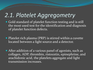
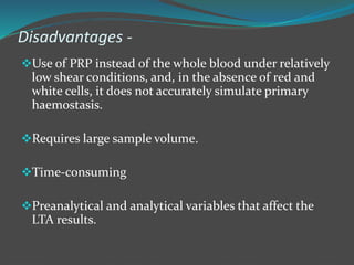
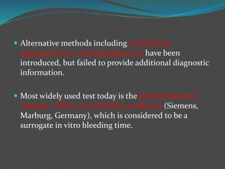
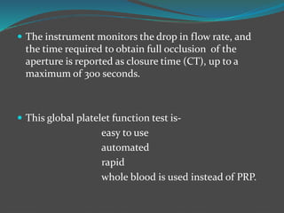
This document summarizes platelet function testing. It discusses how platelets are formed from megakaryocytes in the bone marrow and circulate in the bloodstream. The major platelet function tests are platelet aggregometry, flow cytometry, and point-of-care tests like the impact cone and plate analyzer and thromboelastography. These tests are used to diagnose platelet disorders and monitor antiplatelet therapy. The document also briefly discusses platelet-derived microparticles and microRNAs, which can provide information about platelet activation and signaling.

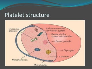
![ Platelets contain three major types of granules:
𝛼- Granules- most abundant granules in platelets and are
rapidly exocytosed upon activation to enhance hemostasis
and inflammation.
Dense bodies- contain adenine nucleotides (ADP and
ATP) and serotonin which induce platelet aggregation,
vasoconstriction, cytokine production, and modulators of
inflammation.
Lysosomes- contain glycohydrolases and proteases that
can aid in pathogen clearance, breakdown of extracellular
matrix, and contribute to the clearance of platelet thrombi
and degradation of heparin [1–3].](https://image.slidesharecdn.com/plateletfunctiontests-160108113642/85/Platelet-function-tests-pptx-2-pptx-final-5-320.jpg)
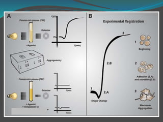
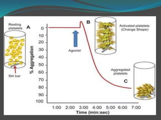
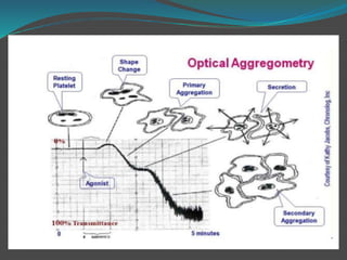
![ The platelet aggregation pattern is thought as a
primary response to an exogenous agonist, followed by
a secondary response to the release of dense granule
contents.
This biphasic response can be masked if high
concentrations of agonists are added.
Parameters measured include the rate or slope of
aggregation (%/min) and the maximal amplitude (%)
or percentage of aggregation after a fixed period of
time, usually 6–10min [7–12].](https://image.slidesharecdn.com/plateletfunctiontests-160108113642/85/Platelet-function-tests-pptx-2-pptx-final-12-320.jpg)
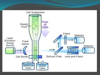
![The most common platelet activation
markers assessed by flow cytometry are --
P-selectin expression on the platelet surface (as a marker of
𝛼-granule secretion),
the conformational change of GP IIb/IIIa into its active
state (measured with monoclonal antibody PAC-1),
platelet-leukocyte conjugates,
microparticle examination,
exposure of anionic, negatively charged phospholipids on
the platelet surface (procoagulant activity),
and phosphorylation of vasodilatorstimulated
phosphoprotein-phosphorylation (VASP-P) (Bio- Cytex,
Marseille, France), as a marker of P2Y12 receptor
activation-dependent signaling [18–20]](https://image.slidesharecdn.com/plateletfunctiontests-160108113642/85/Platelet-function-tests-pptx-2-pptx-final-21-320.jpg)
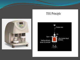
![ This review focuses on the clinical utility of two
representative instruments:
Impact cone and plate analyzer (DiaMed, Cressier,
Switzerland) [2, 18, 19]
Thrombelastography (TEG) (Hemoscope, Niles, IL,
USA) [7–10, 21, 22]](https://image.slidesharecdn.com/plateletfunctiontests-160108113642/85/Platelet-function-tests-pptx-2-pptx-final-24-320.jpg)
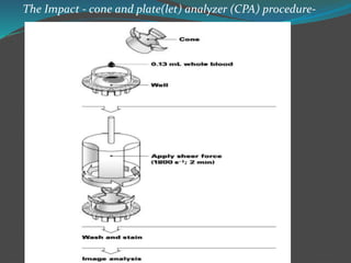
![ The assay is
-Fully automated,
-Simple
-Rapid to use
- Requiring small whole blood.
Emerging data suggest that the impact cone can detect
numerous platelet defects and vWD and could also be
potentially used as a screening method.
But the fully automated version of impact cone has
limited use and therefore further studies are required
[2, 7– 10, 18, 19].](https://image.slidesharecdn.com/plateletfunctiontests-160108113642/85/Platelet-function-tests-pptx-2-pptx-final-26-320.jpg)
![ Therefore, they proposed a therapeutic window
concept for P2Y12 inhibitor therapy [29–31].
The most widely used platelet function assays, Verify
Now P2Y12 assay (Accumetrics, San Diego, CA, USA)
and, at present, Multiplate analyzer (H. Hoffmann-La
Roche Ltd., Basel, Switzerland) and VASP assay,](https://image.slidesharecdn.com/plateletfunctiontests-160108113642/85/Platelet-function-tests-pptx-2-pptx-final-33-320.jpg)
![ Thus, at the present time, HPR and LPR in the setting
of PCI have been defined by the receiver-operator
characteristic (ROC) curve analyses using the
following criteria, respectively:
(1) >208 and <85 P2Y12 reaction units (PRU) by Verify
Now P2Y12 assay,
(2) >50% and <16% platelet reactivity index (PRI) by
VASP-P, and
(3) >46 and <19 arbitrary aggregation units (AU) in
response to ADP by Multiplate analyzer [29–31].](https://image.slidesharecdn.com/plateletfunctiontests-160108113642/85/Platelet-function-tests-pptx-2-pptx-final-34-320.jpg)
![4. Platelet-Derived Microparticles
(PMP)
Defined as small and anucleoid phospholipid vesicles,
approximately 0.1–1.0 𝜇m in diameter, and derived
from different cell types such as platelets, erythrocytes,
leukocytes, endothelial cells, and vascular smooth
muscle cells [35].
Under steady-state conditions, MP originating from
platelet and megakaryocytes are the most abundant
microparticles, constituting up to 70–90% of all MP in
circulation [38].](https://image.slidesharecdn.com/plateletfunctiontests-160108113642/85/Platelet-function-tests-pptx-2-pptx-final-36-320.jpg)