Downloaded 503 times
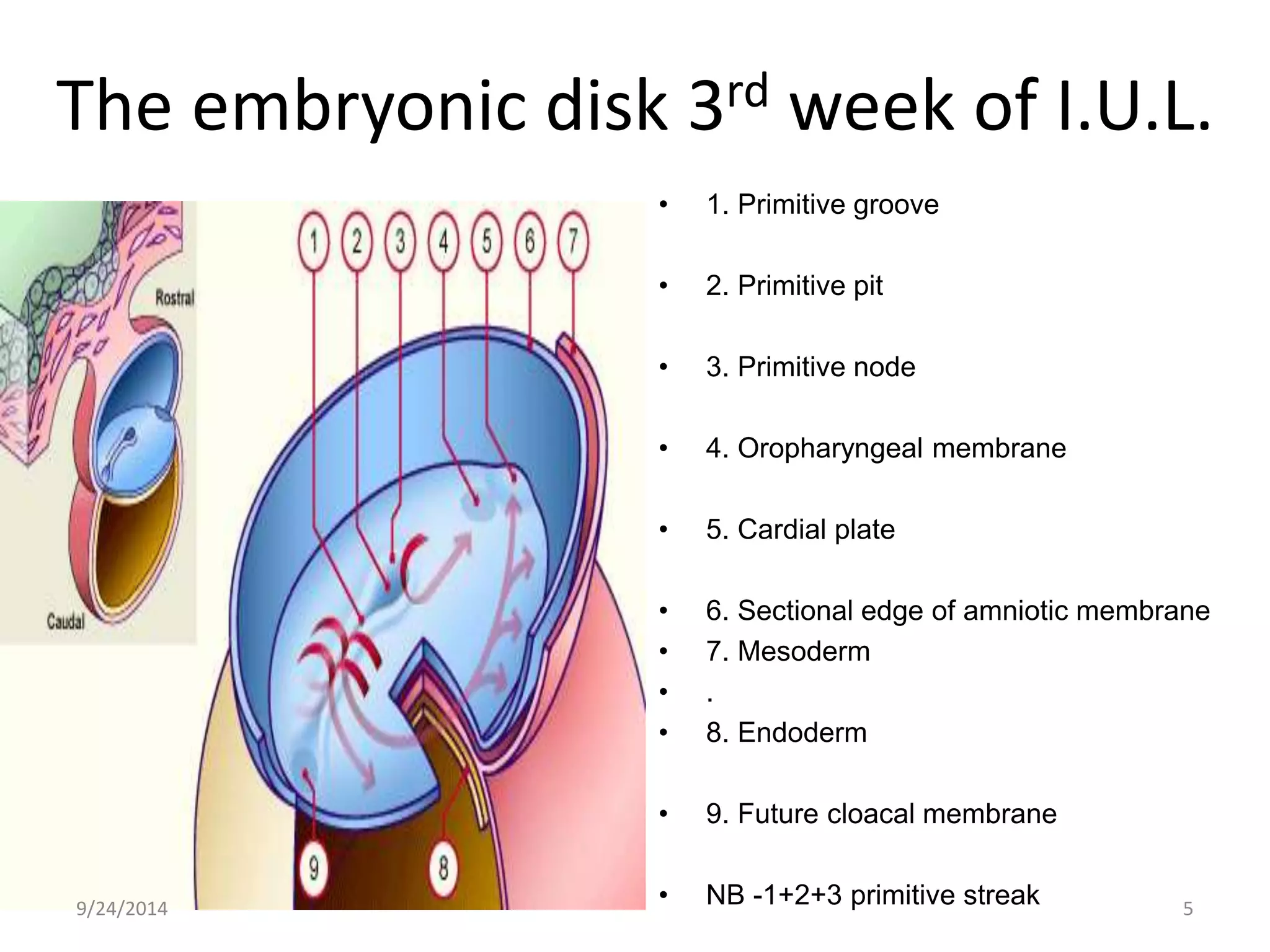
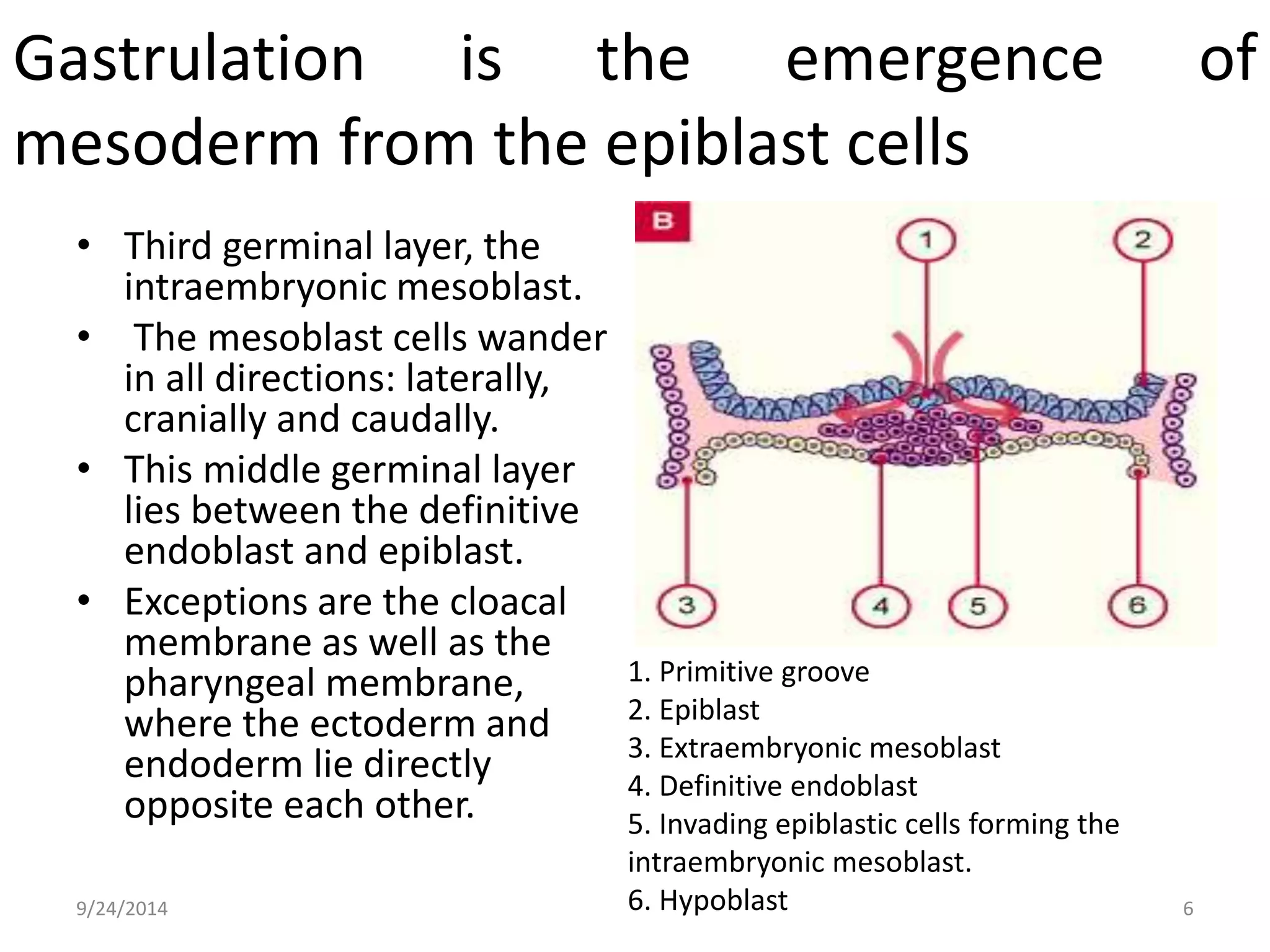
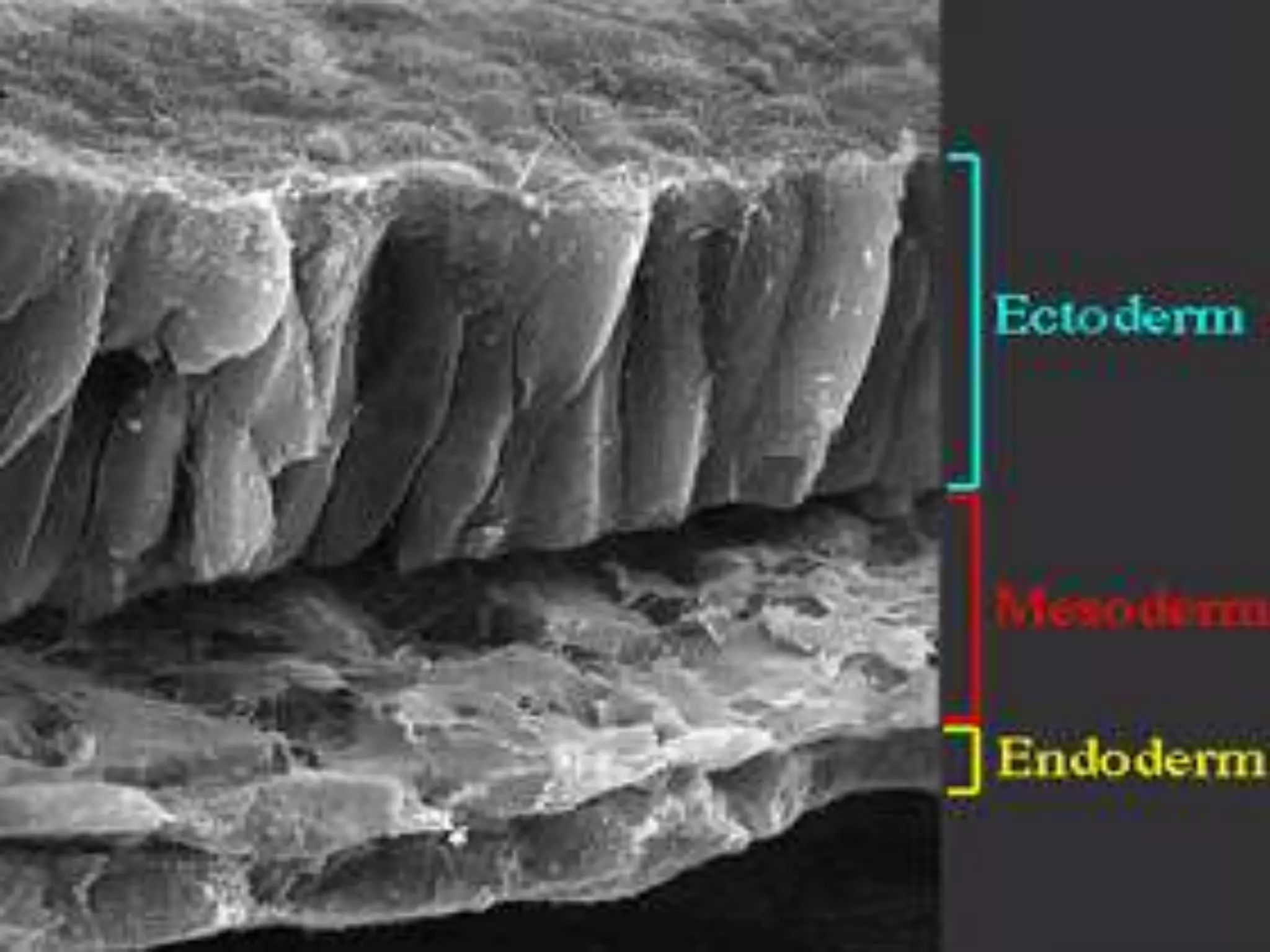
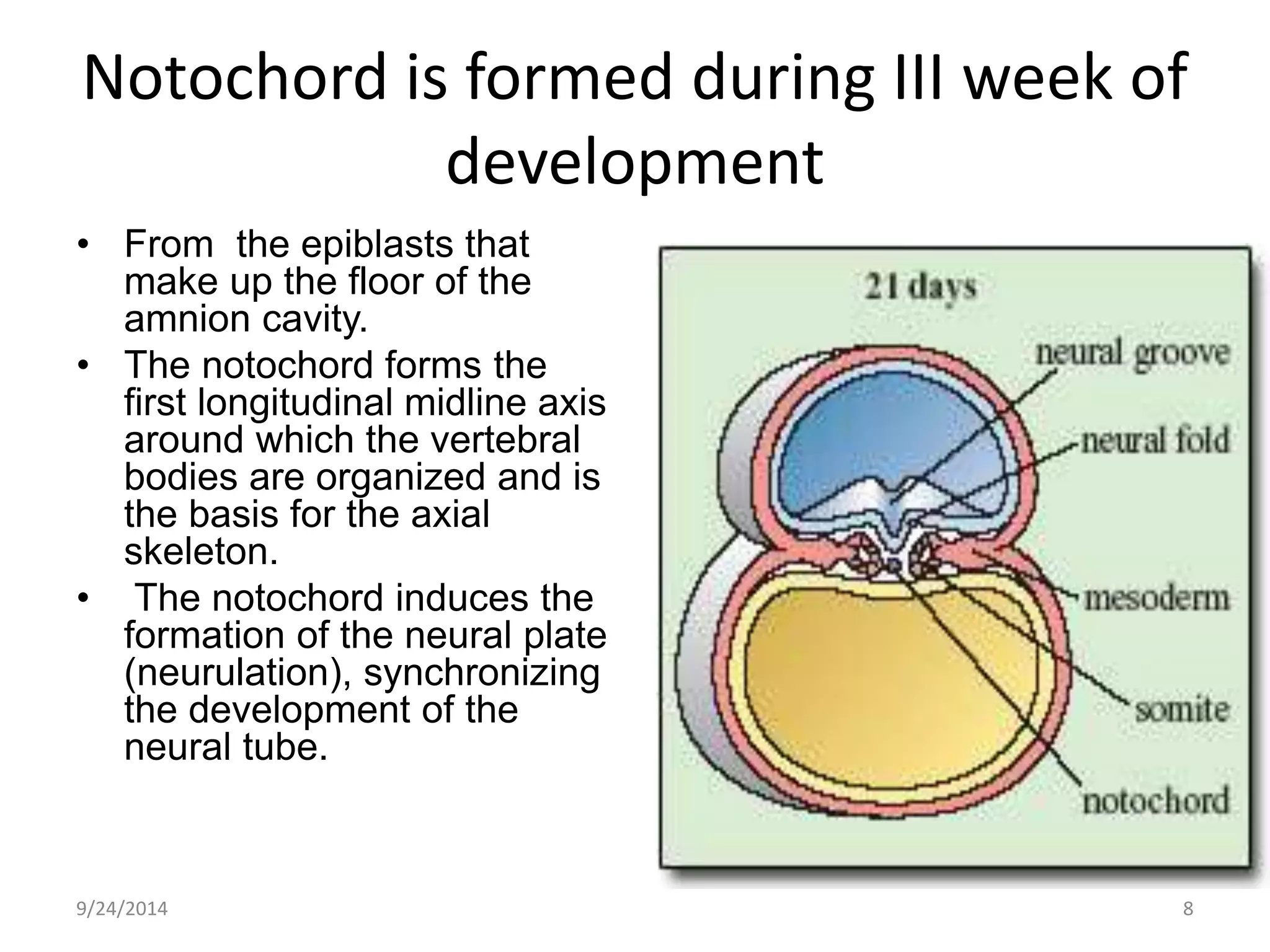
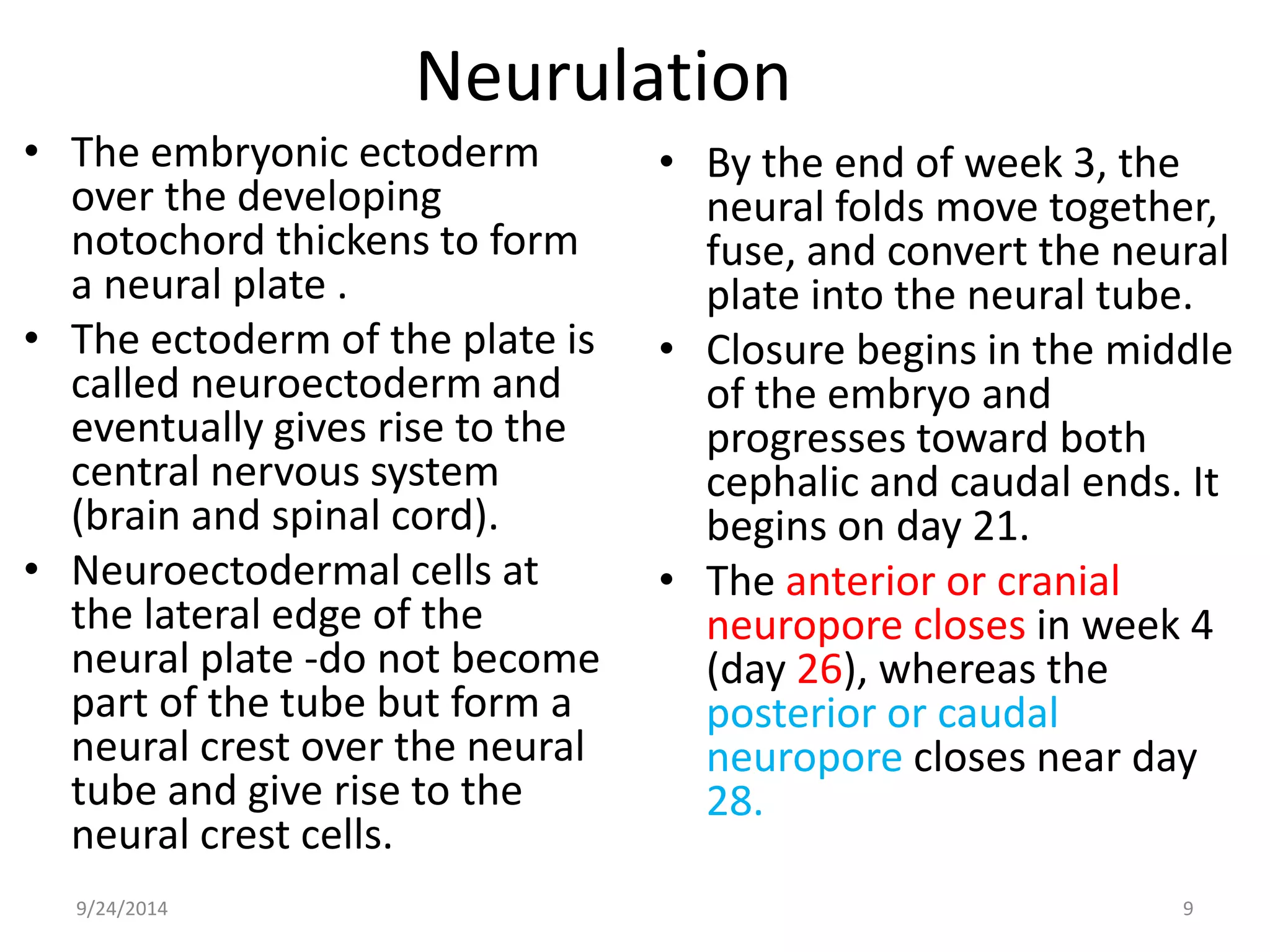
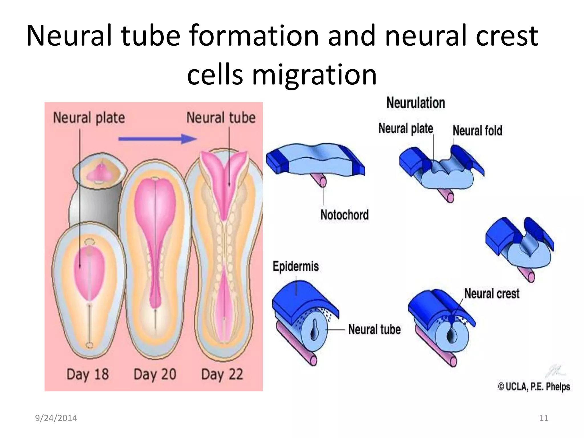
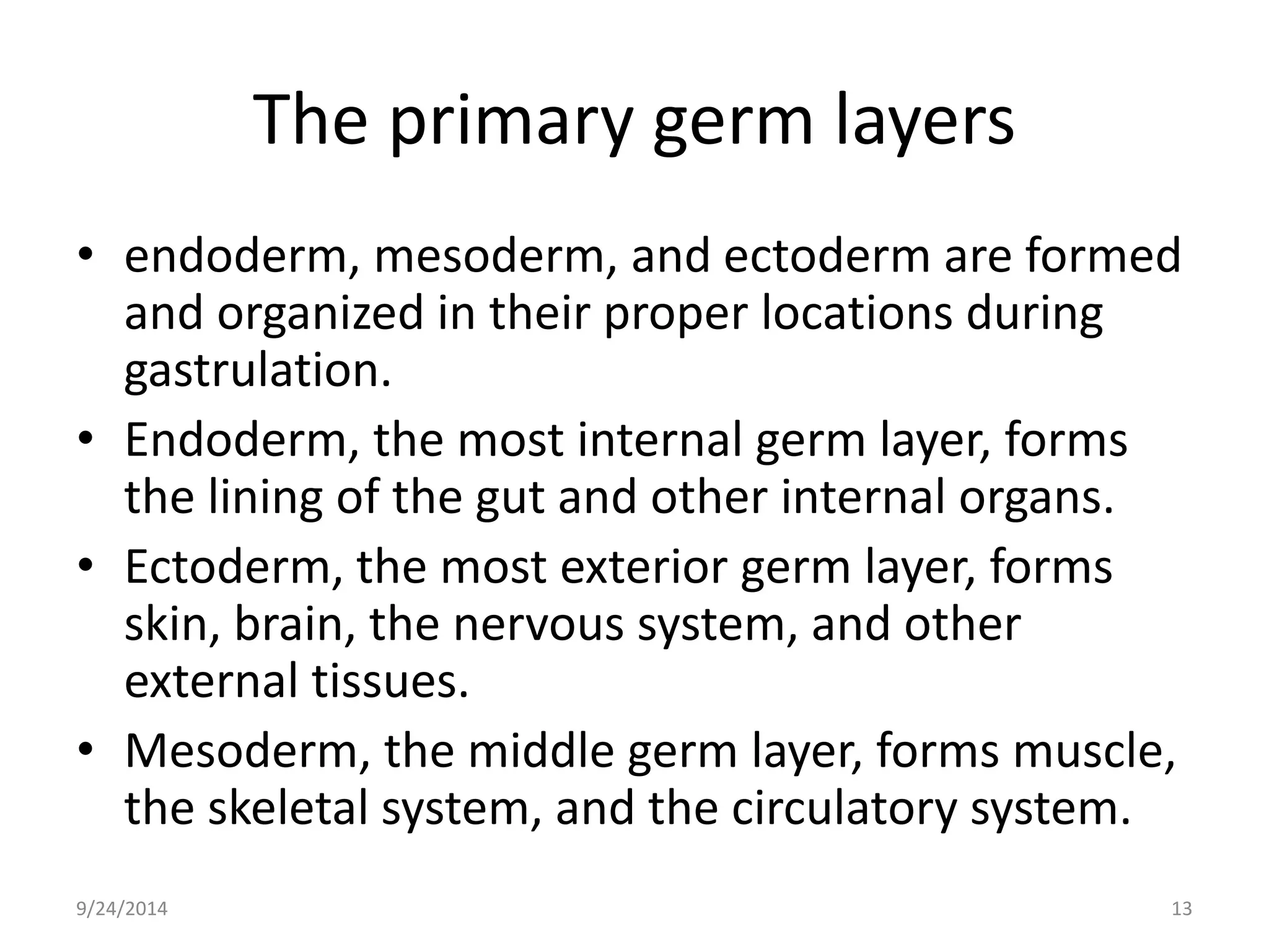
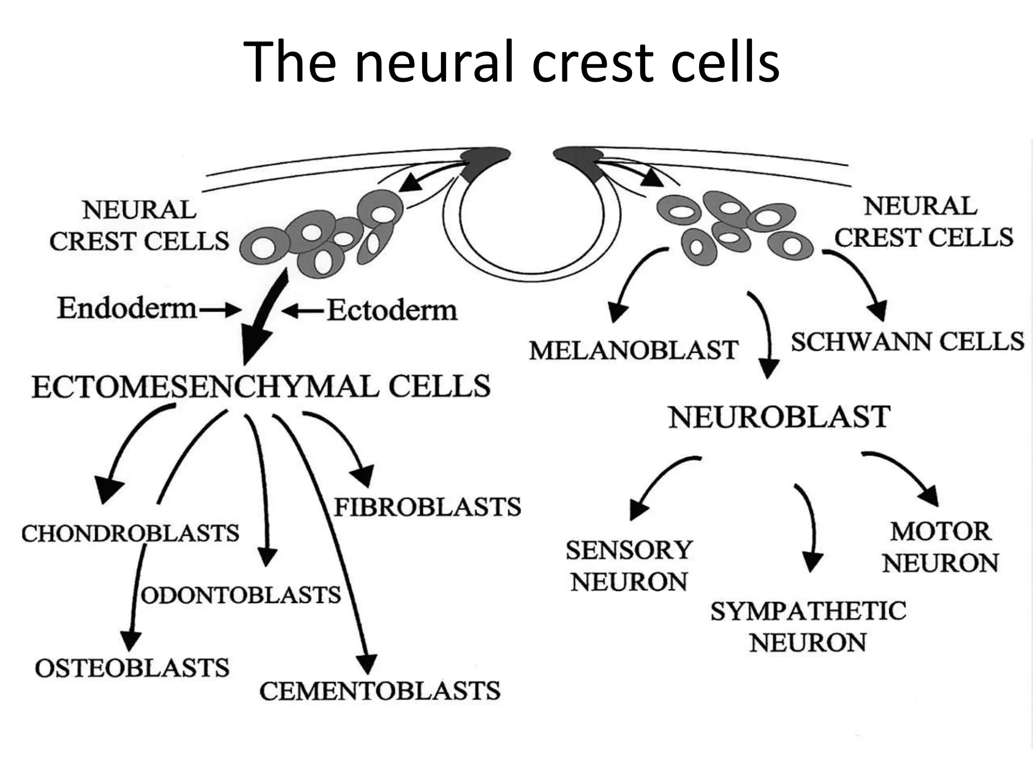
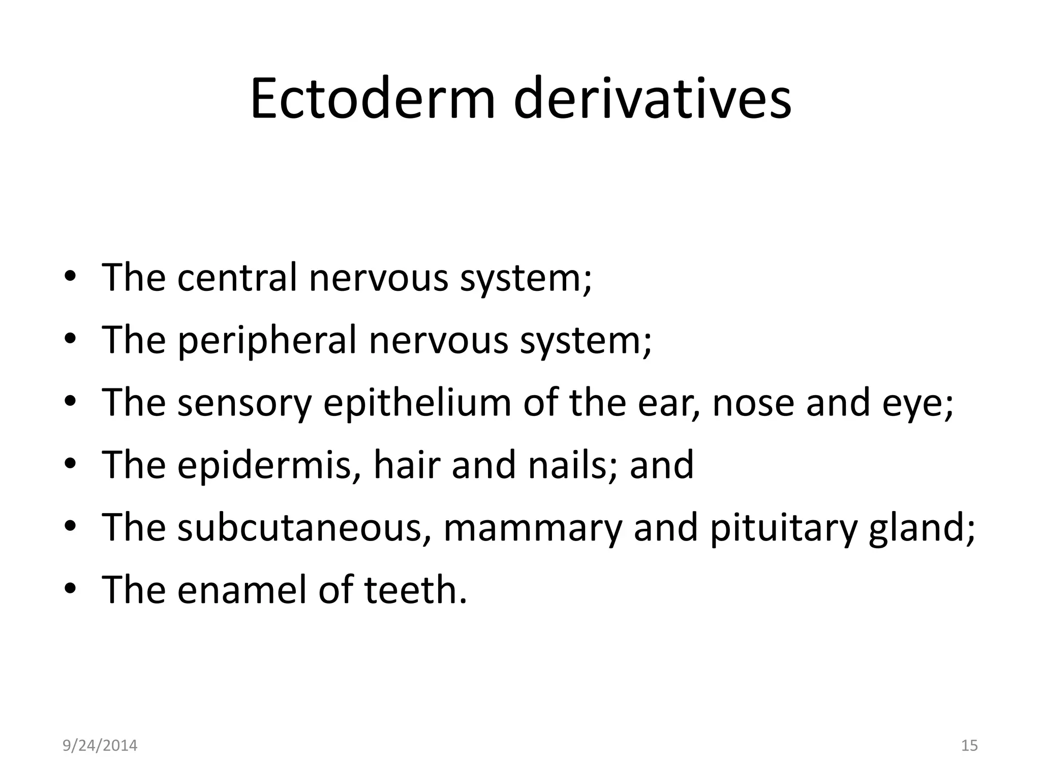
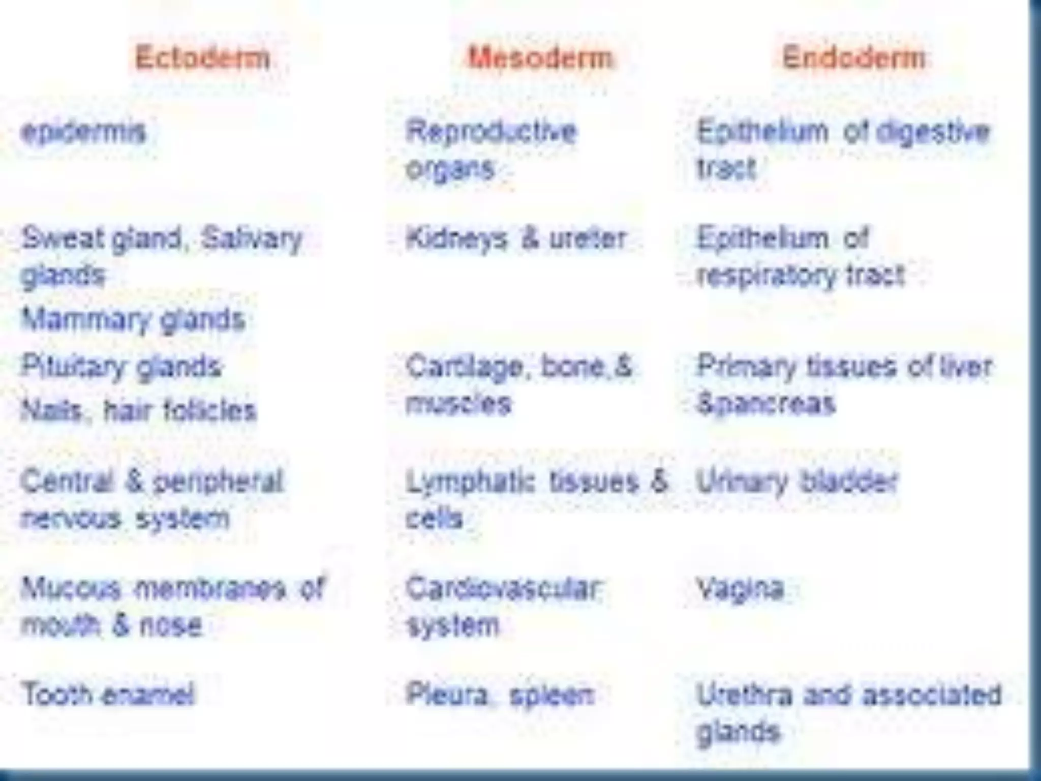

During the third week of development, the bilaminar embryonic disk transforms into a trilaminar structure through the process of gastrulation. The primitive streak forms and epiblast cells migrate through it to form the mesoderm germ layer. Neurulation also occurs as the neural plate forms and folds in on itself to become the neural tube. This establishes the basis for the nervous system. By the end of the third week, the three germ layers—ectoderm, mesoderm, and endoderm—have formed and begun developing into tissues and organs.


![3.1_Third_Week_of_Development[1].pptx](https://cdn.slidesharecdn.com/ss_thumbnails/3-221118124853-25c6f4d7-thumbnail.jpg?width=640&height=640&fit=bounds)


![Apporach to lung biopsy [Auto-saved].pptx latest](https://cdn.slidesharecdn.com/ss_thumbnails/apporachtolungbiopsyauto-saved-251211225655-93258539-thumbnail.jpg?width=640&height=640&fit=bounds)