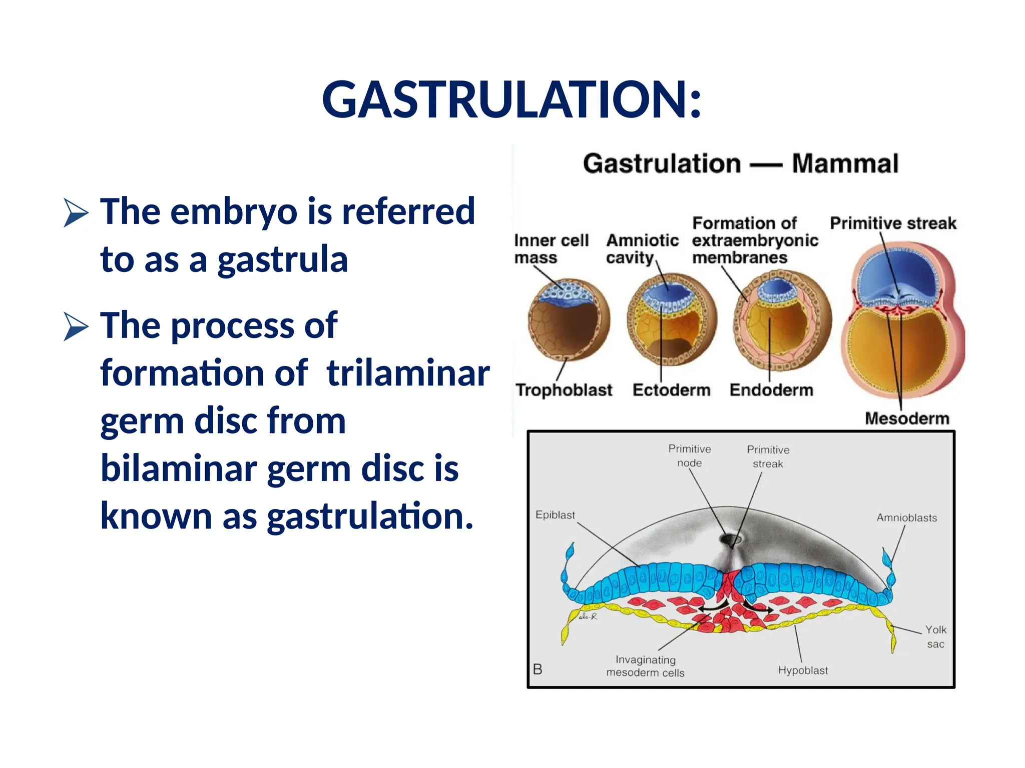
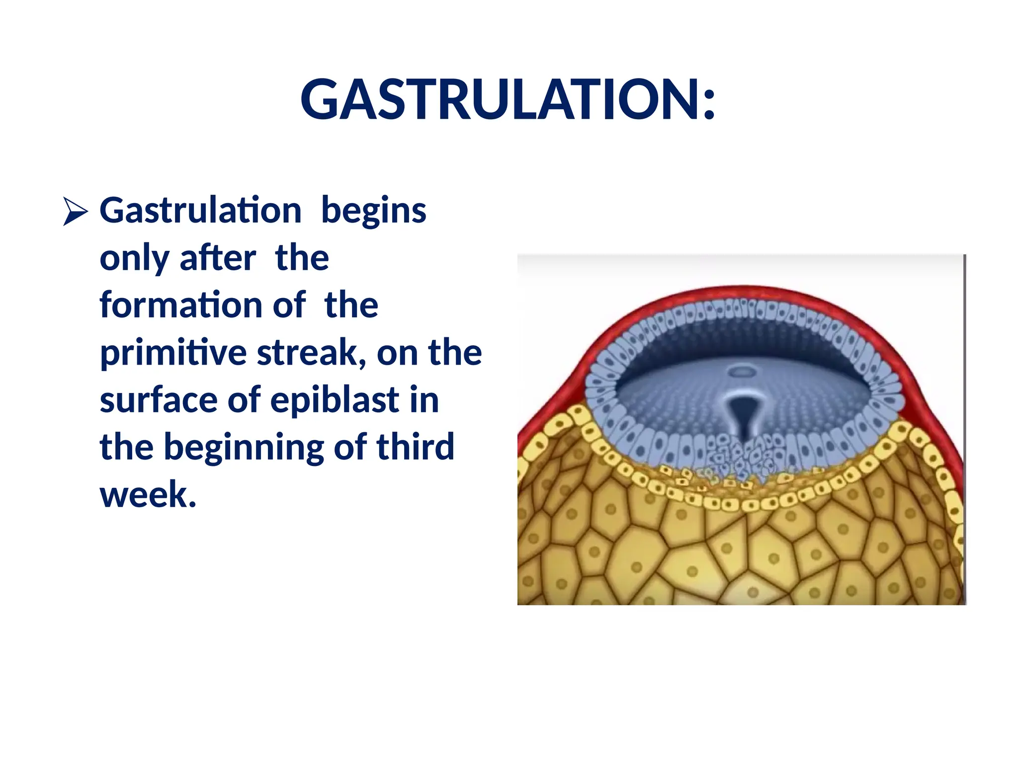
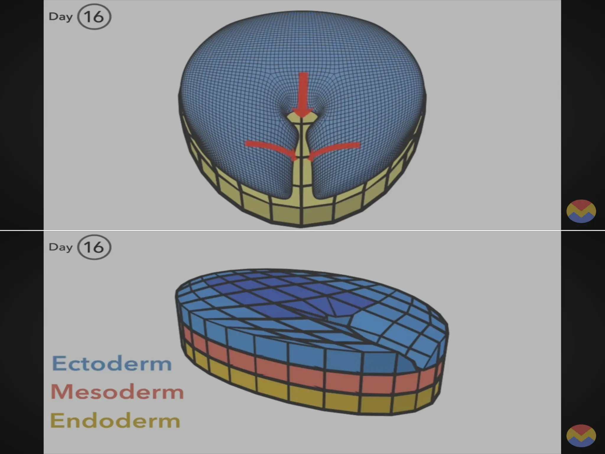
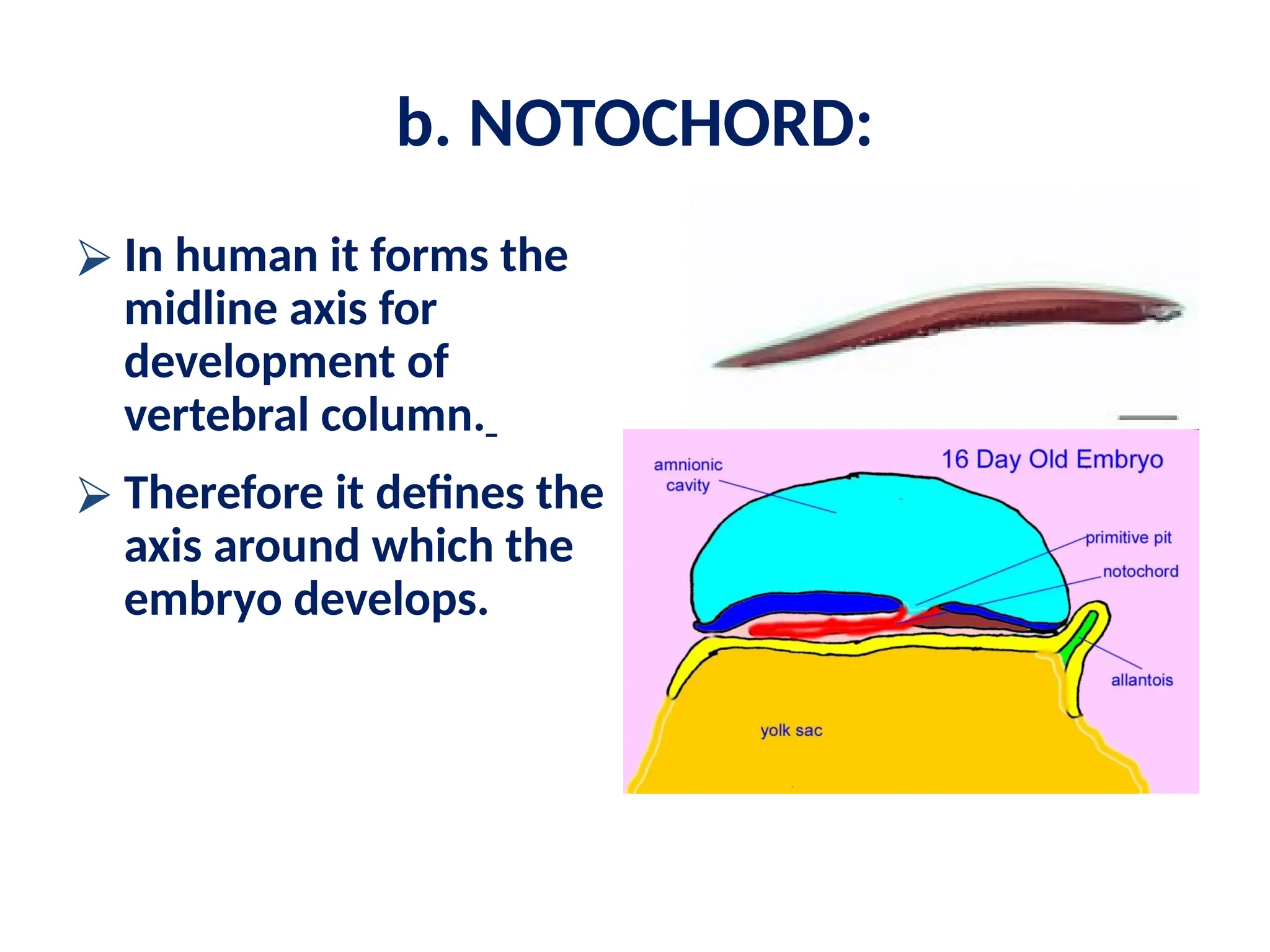
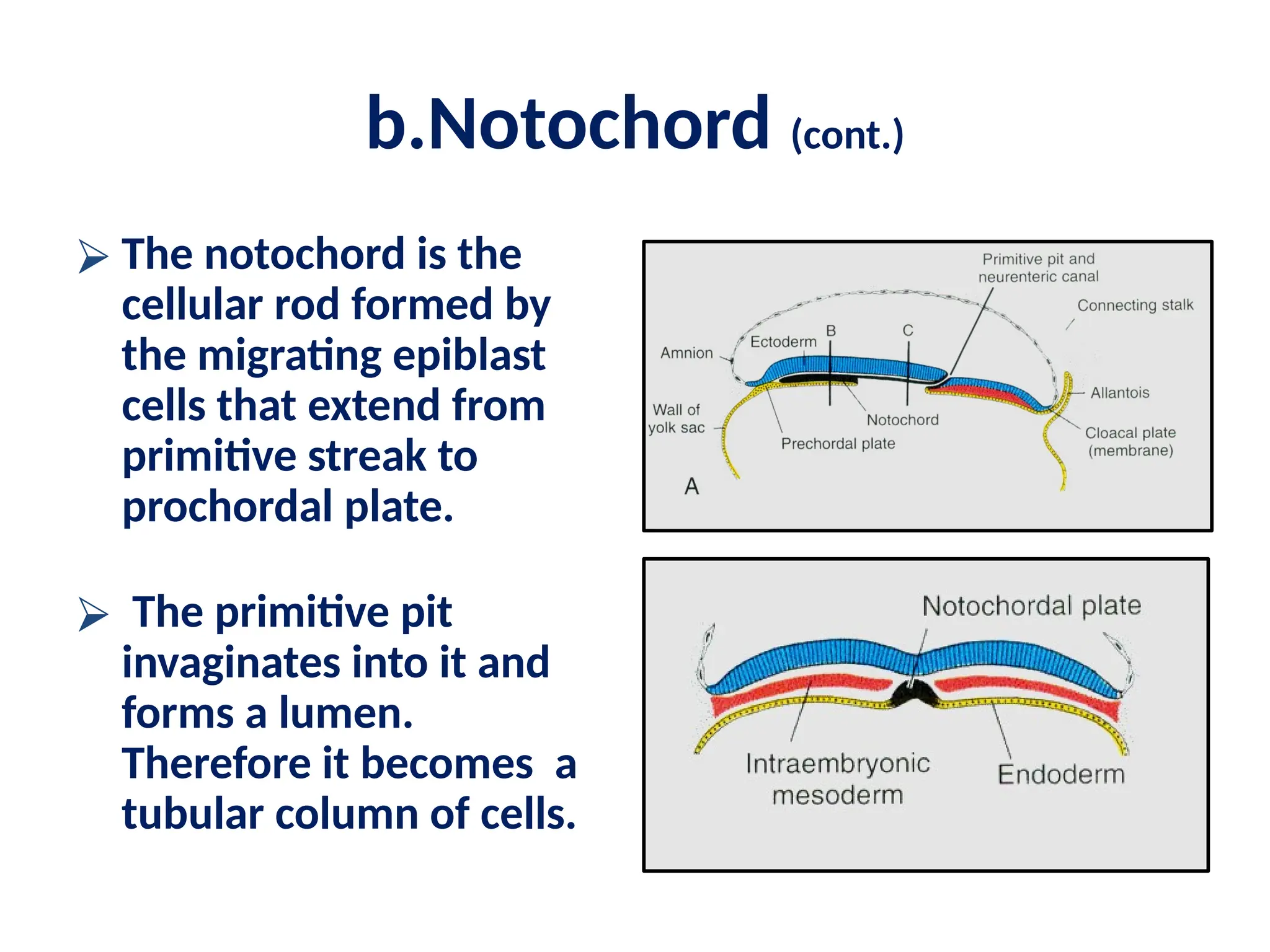
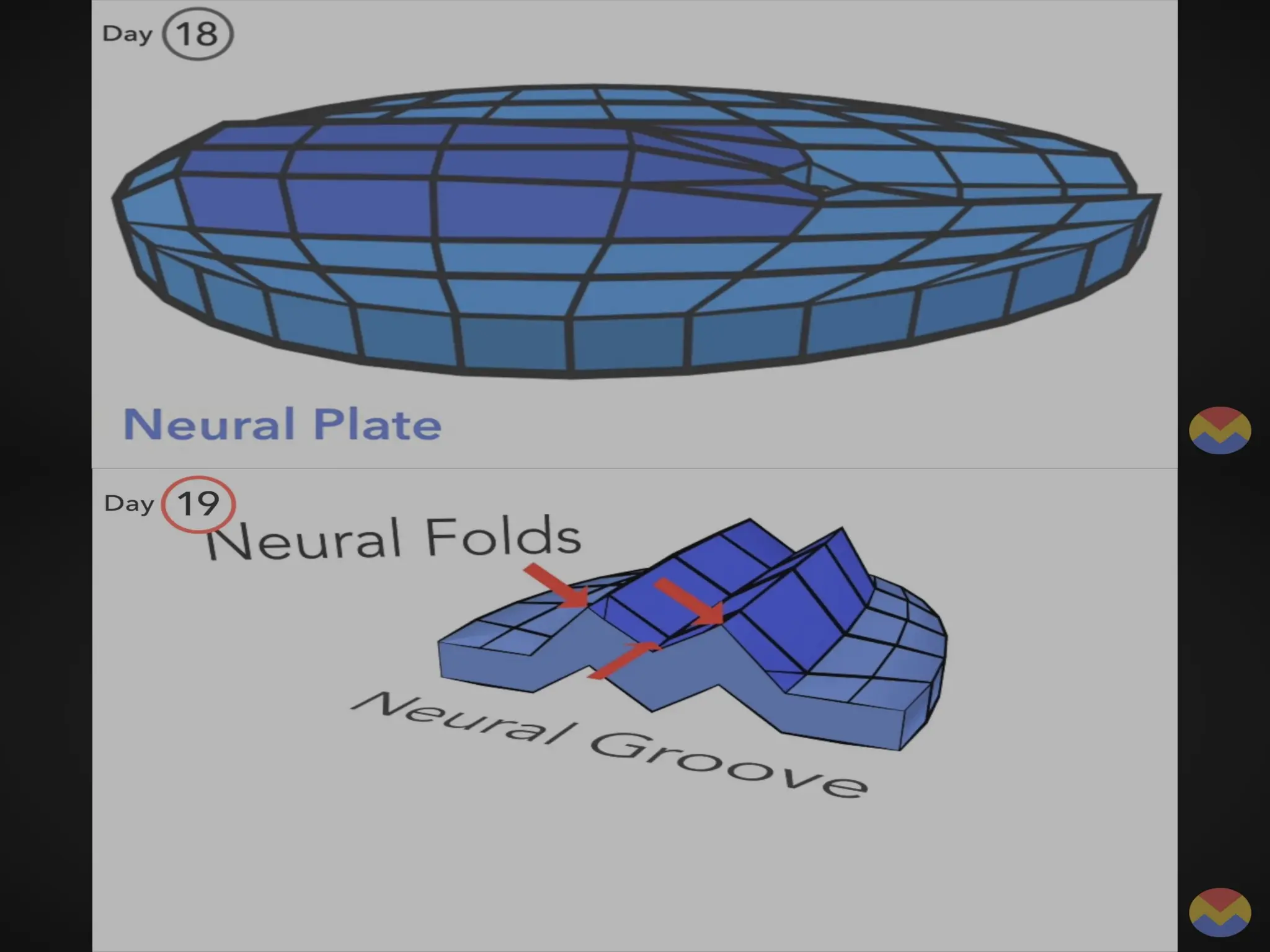
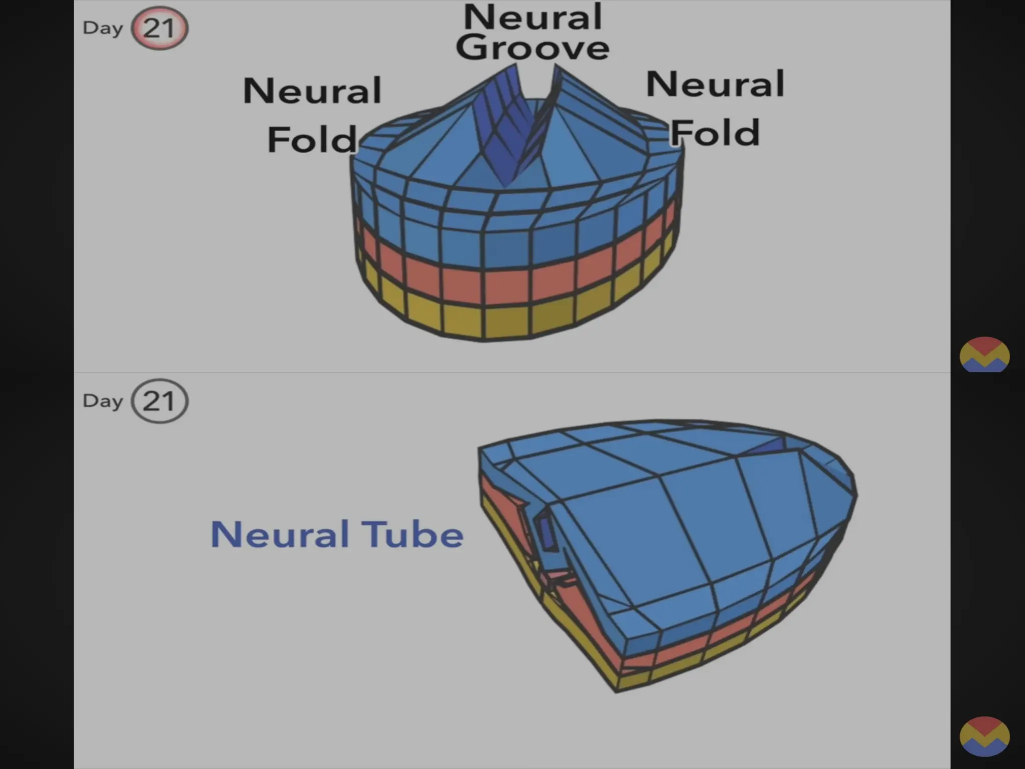
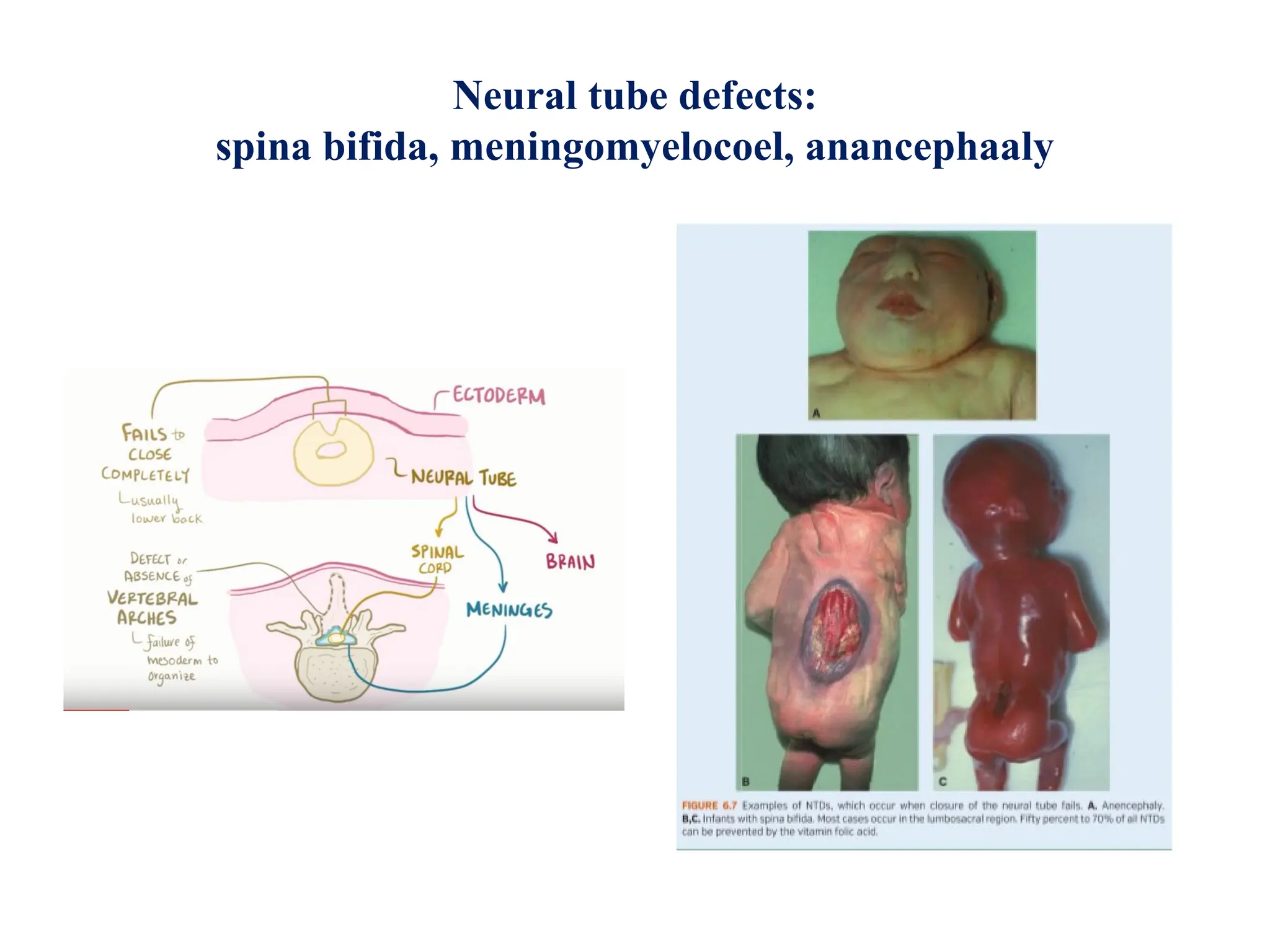
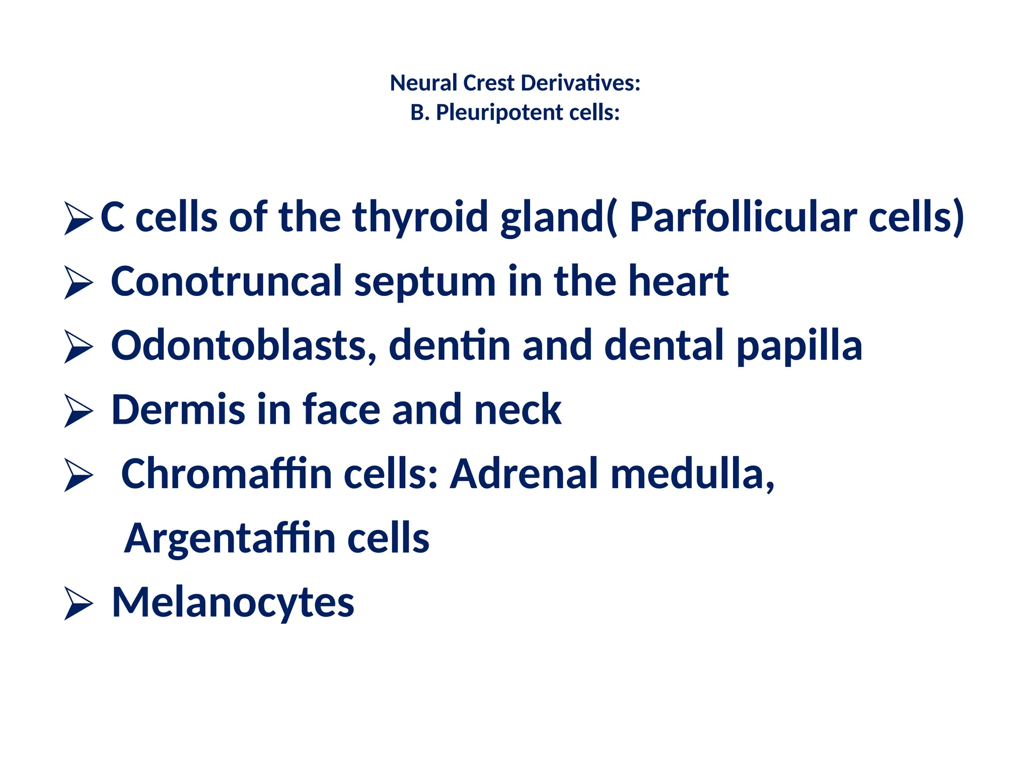
Neurulation
Third week
Events of three:
⮚ Three germ layers comprising
of endoderm, mesoderm &
ectoderm. ( Gastrulation)
⮚ Formation of three new
structures:
a. Primitive streak,
b. Notochord
c. Neural tube.
GASTRULATION:
⮚ The embryo is referred
to as a gastrula
⮚ The process of
formation of trilaminar
germ disc from
bilaminar germ disc is
known as gastrulation.
GASTRULATION:
⮚ Gastrulation begins
only after the
formation of the
primitive streak, on the
surface of epiblast in
the beginning of third
week.
Primitive Streak:
⮚ Therefore before gastrulation
begins, there is formation of
primitive streak on 15th.
day.
⮚ It is a rounded thickening
on the epiblast at the
midline near the caudal
end of the bilaminar germ
disc.
⮚ It becomes linear along
with the elongation of the
germ disc.
Primitive Streak:
⮚ it defines the major body
axis with cranial and
caudal ends and left and
right sides.
⮚ At its anterior end
( cephalic end) - a knob-like
thickening - primitive node
[Hensen’s node] appear
whose centre has a pit
“primitive pit”.
⮚ This continue caudally as
primitive groove.
Primitive Streak (Cont.) :
⮚ Cells from epiblast detached
and slip beneath the primitive
groove and completely replace
the hypoblast cells. Thus giving
rise to definite endoderm by
day 16.
⮚ This process is known as
invagination.
⮚ The remaining cells of epiblast
are known as ectoderm.
⮚ Epiblast also give rise to a new
population of cells-
“gastrulation”.
Fertilization
↓
Cleavage (Repeated mitotic divisions)
↓
Blastula Formation (Hollow ball of cells)
↓
Gastrulation Begins
↓
Formation of Primitive Streak
↓
Invagination (Cells move inward)
↓
Involution & Ingression (Cells migrate to new positions)
↓
Epiboly (Expansion of ectoderm)
↓
Formation of Germ Layers:
- Ectoderm (Outer layer → Skin, Nervous system)
- Mesoderm (Middle layer → Muscles, Bones, Blood)
- Endoderm (Inner layer → Digestive & Respiratory tracts)
b. NOTOCHORD:
⮚ In human it forms the
midline axis for
development of
vertebral column.
⮚ Therefore it defines the
axis around which the
embryo develops.
b.Notochord (cont.)
⮚ The notochord is the
cellular rod formed by
the migrating epiblast
cells that extend from
primitive streak to
prochordal plate.
⮚ The primitive pit
invaginates into it and
forms a lumen.
Therefore it becomes a
tubular column of cells.
b. NOTOCHORD( Cont.):
⮚ By 18th
. Day the floor of
the notochord fuses with
the underlying endoderm
and they disappear. The
remaining cells
proliferate and forms a
solid cellular rod.
⮚ At this stage, a
temporary canal the
neuroenteric canal
connects the yolk sac
with amniotic cavity.
Formation of Primitive Streak
↓
Formation of Hensen’s Node (Primitive Node)
↓
Epiblast Cells Invaginate Through Primitive Streak
↓
Formation of Notochordal Process (Hollow Tube)
↓
Development of Notochordal Canal (Lumen Inside Process)
↓
Fusion of Notochordal Process with Endoderm → Formation of
Notochordal Plate
↓
Detachment & Rolling of Notochordal Plate → Formation of Solid
Notochord
↓
Notochord Induces Neural Plate Formation → Neurulation Begins
↓
Formation of Neural Tube (Future Brain & Spinal Cord)
Notochord( Cont.)
Fate:
With further development
the notochord is replaced
by vertebral column.
Most of it disappear
except in the region of
the intervertebral disc,
where it remain as
nucleus pulposus,
surrounded by annulus
fibrosus.
C. Neural tube:
C. NEURULATION :
⮚ Neurulation is the process
of formation of neural
plate, fold, groove, and it’s
closure, the tube.
⮚ The growing embryo at this
stage is known as neurula.
⮚ By the end of the 19th
.day
the initial thickening of
ectoderm takes place under
the influence of notochord.
Formation of neural Tube:
⮚ Initially the process begins
from cervical regions.
⮚ The thickened part is known
as neural plate.
⮚ By the end of the third week
more and more neural folds
are formed.
⮚ Because of formation of
neural folds laterally the
central part becomes
grooved –neural groove
Formation of neural tube:
⮚ The lateral ends of the
folds fuse in the midline
and neural tube is
formed.
⮚ The two ends of the tube
remain open into
amniotic cavity.
⮚ Closure of the anterior
neuropore takes place on
25th.
, day and posterior
neuropore on 27th.
,day.
Summary of neural tube:
⮚ The ectoderm overlying the
notochord thickens
⮚ Inducer: notochord.
⮚ Extends from buccopharyngeal
membrane to primitive node.
⮚ Formation process: plate, fold,
groove, and tube.
⮚ give rise to neural crest cells
and neural tube.
⮚ Neural tube forms:
▪ Brain.
▪ Spinal cord
Day 6-7: Implantation
↓ (Trophoblast invades endometrium)
Day 8-9: Bilaminar Germ Disc Formation
↓ (Epiblast & Hypoblast form)
Day 12-13: Extraembryonic Structures Develop
↓ (Amniotic cavity, yolk sac, chorionic cavity form)
Day 14-15: Gastrulation Begins
↓ (Primitive streak formation)
Day 16: Trilaminar Germ Disc Formation
↓ (Ectoderm, Mesoderm, Endoderm develop)
Day 17-18: Notochord Formation & Induction
↓ (Signals neural plate development)
Day 18-19: Neural Plate Formation
↓ (Thickening of ectoderm)
Day 19-20: Neural Groove & Neural Fold Formation
↓ (Neural folds start elevating)
Day 21-22: Neural Tube Formation Begins
↓ (Neural folds fuse at the midline)
Day 22-23: Neural Tube Closure Starts (Cervical Region)
Day 25-27: Anterior (Day 25) & Posterior Neuropore (Day 27) Closure
CNS (Brain & Spinal Cord) and PNS Formation
Neural tube defects:
spina bifida, meningomyelocoel, anancephaaly
Neural crest cells:
⮚ arise from the embryonic
ectoderm cell layer,
⮚ give rise to a
diverse cell lineage—
including melanocytes,
craniofacial cartilage and
bone, smooth muscle,
peripheral and enteric
neurons and glia.
Neural groove & neural crest cells :
⮚ Neural crest cells are
first seen in the lateral
margin of the
developing neural
groove.
⮚ They migrate to
dorsolateral aspect of
the tube.
⮚ Give rise to different
structures.
Neural Crest Derivatives:
A) Related to nervous system:
a) Supporting tissue of nervous system :
i) Schwann cells and Glial cells
ii) Arachnoid and pia mater
(leptomeninges)
b) Sensory ganglia : i) dorsal root ganglia
ii) sensory ganglia of V, VII, VIII,
IX & X cr.nvs.
ii) Peripheral autonomic
sensory ganglia (GIT)
c) ANS: i) Sympathetic ganglia
ii) Parasympathetic ganglia.
Neural Crest Derivatives:
B. Pleuripotent cells:
⮚C cells of the thyroid gland( Parfollicular cells)
⮚ Conotruncal septum in the heart
⮚ Odontoblasts, dentin and dental papilla
⮚ Dermis in face and neck
⮚ Chromaffin cells: Adrenal medulla,
Argentaffin cells
⮚ Melanocytes
Neural Crest Derivatives:
⮚ Connective tissue and
bones of the face and
skull
Thank You !

Neurulation.pptxvvvbbbbhbbbbvffghhjjjjjh

  • 1.
  • 2.
    Third week Events ofthree: ⮚ Three germ layers comprising of endoderm, mesoderm & ectoderm. ( Gastrulation) ⮚ Formation of three new structures: a. Primitive streak, b. Notochord c. Neural tube.
  • 3.
    GASTRULATION: ⮚ The embryois referred to as a gastrula ⮚ The process of formation of trilaminar germ disc from bilaminar germ disc is known as gastrulation.
  • 4.
    GASTRULATION: ⮚ Gastrulation begins onlyafter the formation of the primitive streak, on the surface of epiblast in the beginning of third week.
  • 7.
    Primitive Streak: ⮚ Thereforebefore gastrulation begins, there is formation of primitive streak on 15th. day. ⮚ It is a rounded thickening on the epiblast at the midline near the caudal end of the bilaminar germ disc. ⮚ It becomes linear along with the elongation of the germ disc.
  • 8.
    Primitive Streak: ⮚ itdefines the major body axis with cranial and caudal ends and left and right sides. ⮚ At its anterior end ( cephalic end) - a knob-like thickening - primitive node [Hensen’s node] appear whose centre has a pit “primitive pit”. ⮚ This continue caudally as primitive groove.
  • 9.
    Primitive Streak (Cont.): ⮚ Cells from epiblast detached and slip beneath the primitive groove and completely replace the hypoblast cells. Thus giving rise to definite endoderm by day 16. ⮚ This process is known as invagination. ⮚ The remaining cells of epiblast are known as ectoderm. ⮚ Epiblast also give rise to a new population of cells- “gastrulation”.
  • 10.
    Fertilization ↓ Cleavage (Repeated mitoticdivisions) ↓ Blastula Formation (Hollow ball of cells) ↓ Gastrulation Begins ↓ Formation of Primitive Streak ↓ Invagination (Cells move inward) ↓ Involution & Ingression (Cells migrate to new positions) ↓ Epiboly (Expansion of ectoderm) ↓ Formation of Germ Layers: - Ectoderm (Outer layer → Skin, Nervous system) - Mesoderm (Middle layer → Muscles, Bones, Blood) - Endoderm (Inner layer → Digestive & Respiratory tracts)
  • 11.
    b. NOTOCHORD: ⮚ Inhuman it forms the midline axis for development of vertebral column. ⮚ Therefore it defines the axis around which the embryo develops.
  • 12.
    b.Notochord (cont.) ⮚ Thenotochord is the cellular rod formed by the migrating epiblast cells that extend from primitive streak to prochordal plate. ⮚ The primitive pit invaginates into it and forms a lumen. Therefore it becomes a tubular column of cells.
  • 13.
    b. NOTOCHORD( Cont.): ⮚By 18th . Day the floor of the notochord fuses with the underlying endoderm and they disappear. The remaining cells proliferate and forms a solid cellular rod. ⮚ At this stage, a temporary canal the neuroenteric canal connects the yolk sac with amniotic cavity.
  • 15.
    Formation of PrimitiveStreak ↓ Formation of Hensen’s Node (Primitive Node) ↓ Epiblast Cells Invaginate Through Primitive Streak ↓ Formation of Notochordal Process (Hollow Tube) ↓ Development of Notochordal Canal (Lumen Inside Process) ↓ Fusion of Notochordal Process with Endoderm → Formation of Notochordal Plate ↓ Detachment & Rolling of Notochordal Plate → Formation of Solid Notochord ↓ Notochord Induces Neural Plate Formation → Neurulation Begins ↓ Formation of Neural Tube (Future Brain & Spinal Cord)
  • 16.
    Notochord( Cont.) Fate: With furtherdevelopment the notochord is replaced by vertebral column. Most of it disappear except in the region of the intervertebral disc, where it remain as nucleus pulposus, surrounded by annulus fibrosus.
  • 17.
  • 18.
    C. NEURULATION : ⮚Neurulation is the process of formation of neural plate, fold, groove, and it’s closure, the tube. ⮚ The growing embryo at this stage is known as neurula. ⮚ By the end of the 19th .day the initial thickening of ectoderm takes place under the influence of notochord.
  • 19.
    Formation of neuralTube: ⮚ Initially the process begins from cervical regions. ⮚ The thickened part is known as neural plate. ⮚ By the end of the third week more and more neural folds are formed. ⮚ Because of formation of neural folds laterally the central part becomes grooved –neural groove
  • 25.
    Formation of neuraltube: ⮚ The lateral ends of the folds fuse in the midline and neural tube is formed. ⮚ The two ends of the tube remain open into amniotic cavity. ⮚ Closure of the anterior neuropore takes place on 25th. , day and posterior neuropore on 27th. ,day.
  • 26.
    Summary of neuraltube: ⮚ The ectoderm overlying the notochord thickens ⮚ Inducer: notochord. ⮚ Extends from buccopharyngeal membrane to primitive node. ⮚ Formation process: plate, fold, groove, and tube. ⮚ give rise to neural crest cells and neural tube. ⮚ Neural tube forms: ▪ Brain. ▪ Spinal cord
  • 27.
    Day 6-7: Implantation ↓(Trophoblast invades endometrium) Day 8-9: Bilaminar Germ Disc Formation ↓ (Epiblast & Hypoblast form) Day 12-13: Extraembryonic Structures Develop ↓ (Amniotic cavity, yolk sac, chorionic cavity form) Day 14-15: Gastrulation Begins ↓ (Primitive streak formation) Day 16: Trilaminar Germ Disc Formation ↓ (Ectoderm, Mesoderm, Endoderm develop) Day 17-18: Notochord Formation & Induction ↓ (Signals neural plate development) Day 18-19: Neural Plate Formation ↓ (Thickening of ectoderm) Day 19-20: Neural Groove & Neural Fold Formation ↓ (Neural folds start elevating) Day 21-22: Neural Tube Formation Begins ↓ (Neural folds fuse at the midline) Day 22-23: Neural Tube Closure Starts (Cervical Region) Day 25-27: Anterior (Day 25) & Posterior Neuropore (Day 27) Closure CNS (Brain & Spinal Cord) and PNS Formation
  • 28.
    Neural tube defects: spinabifida, meningomyelocoel, anancephaaly
  • 29.
    Neural crest cells: ⮚arise from the embryonic ectoderm cell layer, ⮚ give rise to a diverse cell lineage— including melanocytes, craniofacial cartilage and bone, smooth muscle, peripheral and enteric neurons and glia.
  • 30.
    Neural groove &neural crest cells : ⮚ Neural crest cells are first seen in the lateral margin of the developing neural groove. ⮚ They migrate to dorsolateral aspect of the tube. ⮚ Give rise to different structures.
  • 31.
    Neural Crest Derivatives: A)Related to nervous system: a) Supporting tissue of nervous system : i) Schwann cells and Glial cells ii) Arachnoid and pia mater (leptomeninges) b) Sensory ganglia : i) dorsal root ganglia ii) sensory ganglia of V, VII, VIII, IX & X cr.nvs. ii) Peripheral autonomic sensory ganglia (GIT) c) ANS: i) Sympathetic ganglia ii) Parasympathetic ganglia.
  • 32.
    Neural Crest Derivatives: B.Pleuripotent cells: ⮚C cells of the thyroid gland( Parfollicular cells) ⮚ Conotruncal septum in the heart ⮚ Odontoblasts, dentin and dental papilla ⮚ Dermis in face and neck ⮚ Chromaffin cells: Adrenal medulla, Argentaffin cells ⮚ Melanocytes
  • 33.
    Neural Crest Derivatives: ⮚Connective tissue and bones of the face and skull
  • 34.