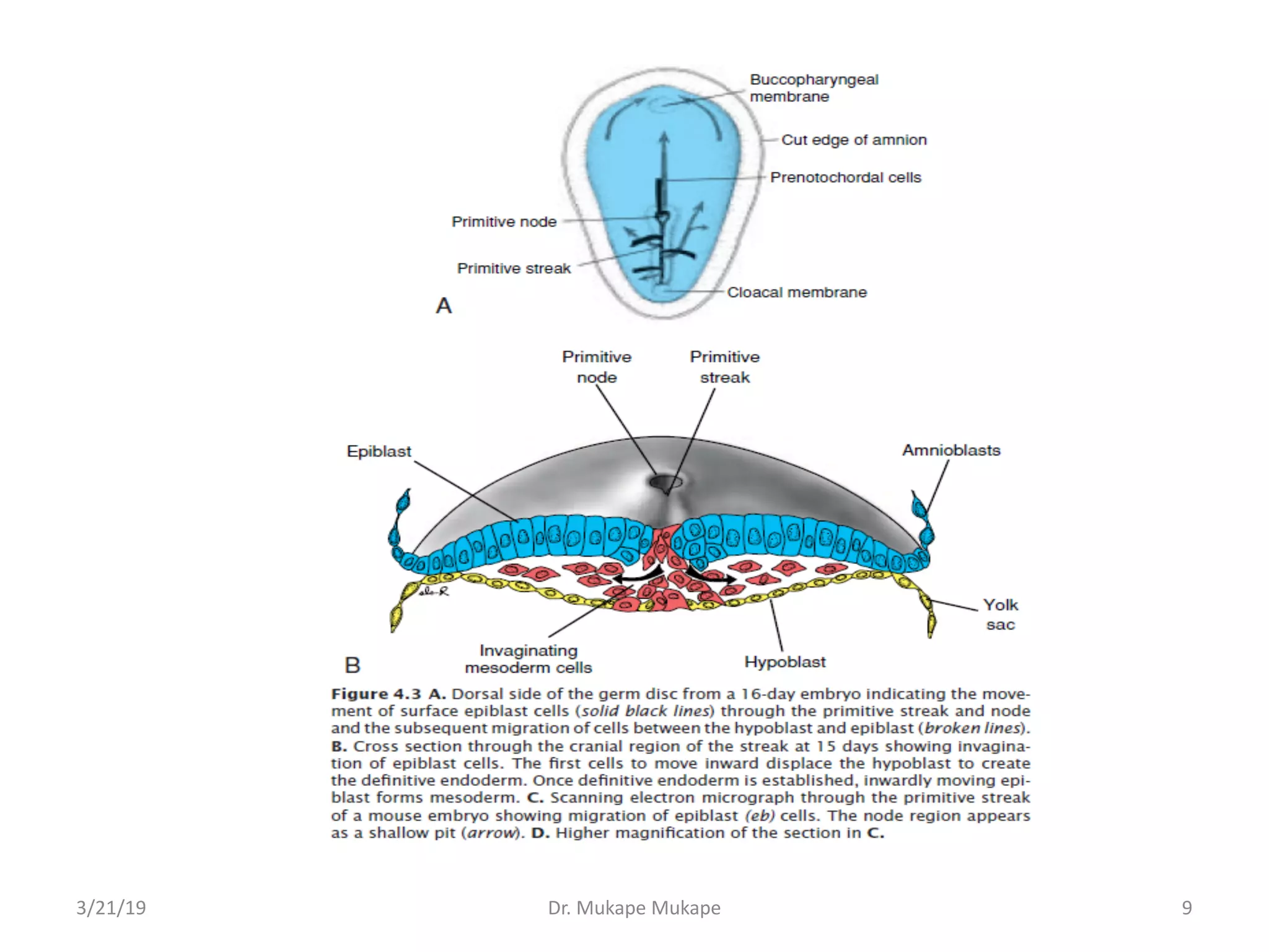
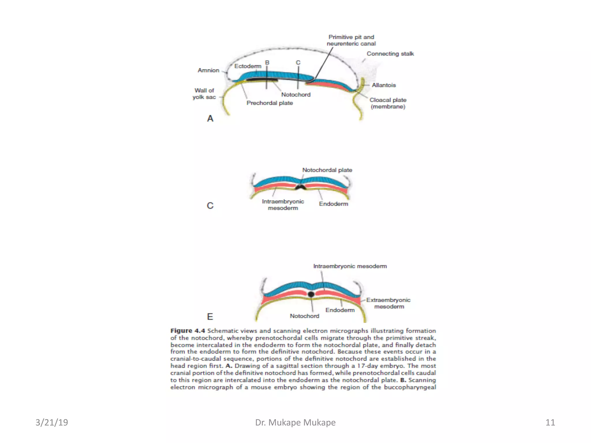
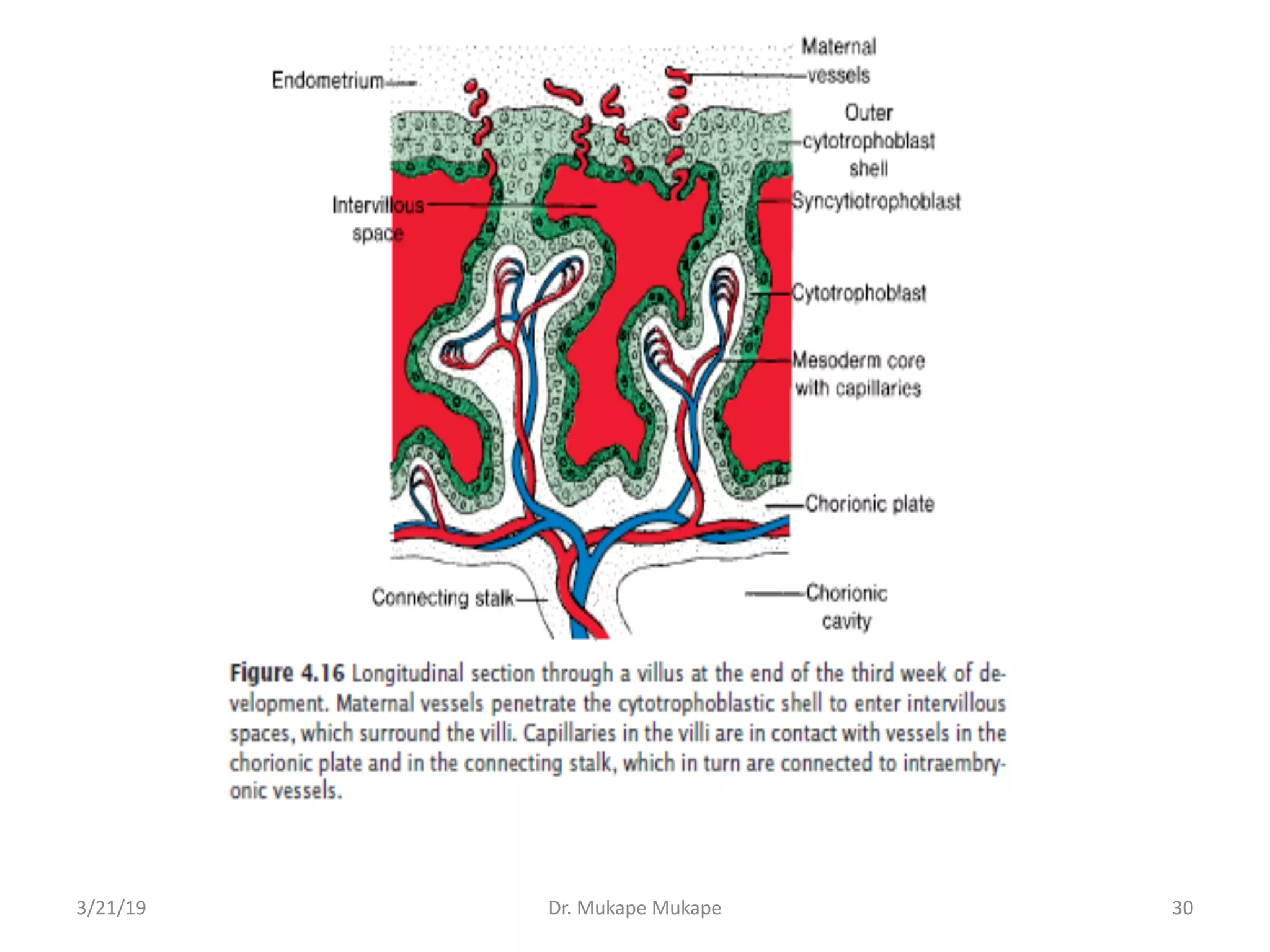
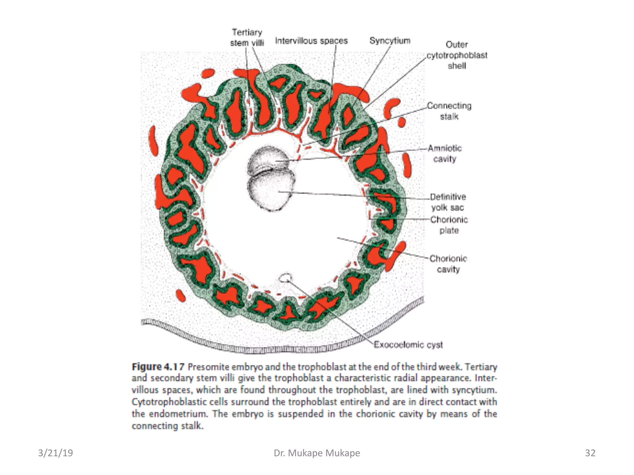
During the third week of development, gastrulation establishes the three germ layers through the formation of the primitive streak on the epiblast surface. Cells migrate through the streak and invaginate to form the endoderm, mesoderm and ectoderm. The notochord also begins forming from prenotochordal cells that migrate through the primitive pit. By the end of the third week, the embryonic disc has elongated and the placenta has developed stem villi that will facilitate nutrient exchange with the maternal blood.
1. UNIVERSITY OF ZAMBIA
SCHOOL OF MEDICINE
HUMAN EMBRYOLOGY
3/21/19 Dr. Mukape Mukape 0
Topic: Third Week of Development
- Trilaminar Germ Disc
Dr. Mukape Mukape
- UNZA (BSc.HB, MBChB, MSc)
- ZIDIS (Diplomatic Practice and Public Relations)
2. Gastrulation: Formation of the 3 Germ Layers
• The most characteristic event occurring during the third
week of gestation is gastrulation, the process that
establishes all the 3 germ layers (ectoderm, mesoderm,
and endoderm) in the embryo
• Gastrulation begins with formation of the primitive
streak on the surface of the epiblast
• Initially, the streak is vaguely defined, but in a 15- to 16-
day embryo, it is clearly visible as a narrow groove with
slightly bulging regions on either side and limiting itself
to the caudal region of the embryo
• The cephalic end of the streak, called the primitive
node, consists of a slightly elevated area surrounding
the small primitive pit
3/21/19 Dr. Mukape Mukape 2
5. • Cells of the epiblast migrate toward the primitive
streak
• Upon arrival in the region of the streak, they become
flask-shaped, detach from the epiblast, and slip
beneath it
• This inward movement is known as invagination
• Hence, the primitive streak region is an ingression
point for the process of gastrulation
• Once the cells have invaginated, some displace the
hypoblast, creating the embryonic endoderm, and
others come to lie between the epiblast and newly
created endoderm to form mesoderm
3/21/19 Dr. Mukape Mukape 5
6. • Cells remaining in the epiblast then form ectoderm
• Thus, the epiblast, through the process of
gastrulation, is the source of all of the germ layers,
and cells in these layers will give rise to all of the
tissues and organs in the embryo
• As more and more cells move between the epiblast
and hypoblast layers, they begin to spread laterally
and cephalad
• Gradually, they migrate beyond the margin of the
disc and establish contact with the extraembryonic
splanchnopleuric mesoderm covering the yolk sac
and amnion
3/21/19 Dr. Mukape Mukape 6
7. • In the cephalic direction, they pass on each side of
the prechordal plate
• The prechordal plate itself forms between the tip of
the notochord and the buccopharyngeal membrane
and is derived from some of the first cells that
migrate through the node in a cephalic direction
• Later, the prechordal plate will be important for
induction of the forebrain
• The buccopharyngeal membrane at the cranial end
of the disc consists of a small region of tightly
adherent ectoderm and endoderm cells with no
intervening mesoderm that represents the future
opening of the oral cavity
3/21/19 Dr. Mukape Mukape 7
8. Formation of the Notochord
• Prenotochordal cells invaginating in the primitive
pit move forward cephalad until they reach the
prechordal plate
• These prenotochordal cells become intercalated in
the hypoblast so that, for a short time, the midline
of the embryo consists of two cell layers that form
the notochordal plate
• As the hypoblast is replaced by endoderm cells
moving in at the streak, cells of the notochordal
plate proliferate and detach from the endoderm
• They then form a solid cord of cells, the definitive
notochord which underlies the neural tube and
serves as the basis for the axial skeleton
3/21/19 Dr. Mukape Mukape 8
10. • Because elongation of the notochord is a
dynamic process, the cranial end forms first, and
caudal regions are added as the primitive streak
assumes a more caudal position
• The notochord and prenotochordal cells extend
cranially to the prechordal plate (an area just
caudal to the buccopharyngeal membrane) and
caudally to the primitive pit
• At the point where the primitive pit forms an
indentation in the epiblast, the neurenteric
canal temporarily connects the amniotic and
yolk sac cavities
3/21/19 Dr. Mukape Mukape 10
12. • The cloacal membrane is formed at the caudal
end of the embryonic disc
• This membrane, which is similar in structure
to the buccopharyngeal membrane, consists
of tightly adherent ectoderm and endoderm
cells with no intervening mesoderm
• When the cloacal membrane appears, the
posterior wall of the yolk sac forms a small
diverticulum that extends into the connecting
stalk
3/21/19 Dr. Mukape Mukape 12
13. • This diverticulum, the allantoenteric
diverticulum, or allantois, appears around the
16th day of development
• Although in some lower vertebrates the
allantois serves as a reservoir for excretion
products of the renal system, in humans it
remains rudimentary but may be involved in
abnormalities of bladder development
3/21/19 Dr. Mukape Mukape 13
14. Fate Map Established During Gastrulation
• Regions of the epiblast that migrate and ingress through
the primitive streak have been mapped and their ultimate
fates determined
- Cells that ingress through the cranial region of the node
become notochord
- Those migrating at the lateral edges of the node and from
the cranial end of the streak become paraxial mesoderm
- Cells migrating through the midstreak region become
intermediate mesoderm
- Those migrating through the more caudal part of the
streak form lateral plate mesoderm
- Cells migrating through the caudal-most part of the streak
contribute to extraembryonic mesoderm (the other
source of this tissue is the primitive yolk sac [hypoblast])
3/21/19 Dr. Mukape Mukape 14
15. Growth of the Embryonic Disc
• The embryonic disc, initially flat and almost
round, gradually becomes elongated, with a
broad cephalic and a narrow caudal end
• Expansion of the embryonic disc occurs mainly in
the cephalic region; the region of the primitive
streak remains more or less the same size
• Growth and elongation of the cephalic part of the
disc are caused by a continuous migration of cells
from the primitive streak region in a cephalic
direction
3/21/19 Dr. Mukape Mukape 15
16. • Invagination of surface cells in the primitive streak
and their subsequent migration forward and
laterally continues until the end of the 4th wk
• At that stage, the primitive streak shows regressive
changes, rapidly shrinks, and soon disappears
• That the primitive streak at the caudal end of the
disc continues to supply new cells until the end of
the 4th wk has an important bearing on
development of the embryo
3/21/19 Dr. Mukape Mukape 16
17. • In the cephalic part, germ layers begin their
specific differentiation by the middle of the 3rd
wk, whereas in the caudal part, differentiation
begins by the end of the 4th wk
• Thus gastrulation, or formation of the germ
layers, continues in caudal segments while
cranial structures are differentiating, causing
the embryo to develop cephalocaudally
3/21/19 Dr. Mukape Mukape 17
18. Clinical correlates
Teratogenesis Associated With Gastrulation
• The beginning of the third week of development,
when gastrulation is initiated, is a highly sensitive
stage for teratogenic insult
• At this time, fate maps can be made for various
organ systems, such as the eyes and brain anlage,
and these cell populations may be damaged by
teratogens
• For example, high doses of alcohol at this stage kill
cells in the anterior midline of the germ disc,
producing a deficiency of the midline in craniofacial
structures and resulting in holoprosencephaly
3/21/19 Dr. Mukape Mukape 18
19. • In such a child, the forebrain is small, the two
lateral ventricles often merge into a single
ventricle, and the eyes are close together
(hypotelorism)
• Gastrulation itself may be disrupted by genetic
abnormalities and toxic insults
• In caudal dysgenesis (sirenomelia), insufficient
mesoderm is formed in the caudal-most region
of the embryo
3/21/19 Dr. Mukape Mukape 19
20. • Because this mesoderm contributes to
formation of the lower limbs, urogenital system
(intermediate mesoderm), and lumbosacral
vertebrae, abnormalities in these structures
ensue
• Affected individuals exhibit a variable range of
defects, including hypoplasia and fusion of the
lower limbs, vertebral abnormalities, renal
agenesis, imperforate anus, and anomalies of the
genital organs
• In humans, the condition is associated with
maternal diabetes mellitus and other causes
3/21/19 Dr. Mukape Mukape 20
22. • Situs inversus is a condition in which transposition of
the viscera in the thorax and abdomen occurs
• This includes dextrocardia, when the heart is located
on the right hemithorax instead of the usual left side
• Despite this organ reversal, other structural
abnormalities occur only slightly more frequently in
these individuals
• Approximately 20% of patients with complete situs
inversus also have bronchiectasis and chronic
sinusitis, infertility if female because of abnormal
cilia (Kartagener syndrome)
3/21/19 Dr. Mukape Mukape 22
23. • Other conditions of abnormal sidedness are
known as laterality sequences
• Patients with these conditions do not have
complete situs inversus but appear to be
predominantly bilaterally left sided or right sided
• The spleen reflects the differences; those with
left-sided bilaterality have polysplenia, and
those with right-sided bilaterality have asplenia
or hypoplastic spleen
• Patients with laterality sequences also are likely
to have other malformations, especially heart
defects
3/21/19 Dr. Mukape Mukape 23
24. Tumours Associated With Gastrulation
• Sometimes, remnants of the primitive streak persist
in the sacrococcygeal region
• These clusters of pluripotent cells proliferate and
form tumours, known as sacrococcygeal teratomas,
that commonly contain tissues derived from all 3
germ layers
• This is the most common tumour in newborns,
occurring with a frequency of one in 37,000
• These tumors may also arise from primordial germ
cells (PGCs) that fail to migrate to the gonadal ridge
3/21/19 Dr. Mukape Mukape 24
26. Further Development of the Trophoblast
• By the beginning of the 3rd wk, the trophoblast is
characterized by primary villi that consist of a
cytotrophoblastic core covered by a syncytial layer
• During further development, mesodermal cells
penetrate the core of primary villi and grow toward
the decidua
• The newly formed structure is known as a
secondary villus
3/21/19 Dr. Mukape Mukape 26
27. • By the end of the 3rd wk, mesodermal cells in the
core of the villus begin to differentiate into blood
cells and small blood vessels, forming the villous
capillary system
• The villus is now known as a tertiary villus or
definitive placental villus
• Capillaries in tertiary villi make contact with
capillaries developing in mesoderm of the
chorionic plate and in the connecting stalk
• These vessels, in turn, establish contact with the
intraembryonic circulatory system, connecting the
placenta and the embryo
3/21/19 Dr. Mukape Mukape 27
29. • Hence, when the heart begins to beat in the 4th wk
of development, the villous system is ready to
supply the embryo proper with essential nutrients
and oxygen
• Meanwhile, cytotrophoblastic cells in the villi
penetrate progressively into the overlying syncytium
until they reach the maternal endometrium
• Here they establish contact with similar extensions
of neighboring villous stems, forming a thin outer
cytotrophoblast shell
• This shell gradually surrounds the trophoblast
entirely and attaches the chorionic sac firmly to the
maternal endometrial tissue
3/21/19 Dr. Mukape Mukape 29
31. • Villi that extend from the chorionic plate to the
decidua basalis (decidual plate: the part of the
endometrium where the placenta will form;) are
called stem or anchoring villi
• Those that branch from the sides of stem villi are
free (terminal) villi, through which exchange of
nutrients and other factors will occur
• The chorionic cavity, meanwhile, becomes larger, and
by the 19th or 20th day, the embryo is attached to its
trophoblastic shell by a narrow connecting stalk
• The connecting stalk later develops into the
umbilical cord, which forms the connection between
placenta and embryo
3/21/19 Dr. Mukape Mukape 31