The document summarizes key topics in general embryology, including the subdivision of prenatal and postnatal periods, development of male and female reproductive systems, and placental structures. It then describes important events that occur during the third and fourth weeks of human development, such as formation of the primitive streak and trilaminar disc, development of the notochord, neurulation, chorionic villi development, and early cardiovascular development. Teratology, which is the study of birth defects, is also mentioned.
The third week of embryonic development is characterized by gastrulation and the formation of the three germ layers. The most important event is the appearance of the primitive streak, which marks the beginning of gastrulation. During gastrulation, cells from the epiblast migrate through the primitive streak to form the endoderm and mesoderm, while those remaining form the ectoderm. This conversion of the bilaminar disc into a trilaminar disc containing three germ layers by the end of the third week is a significant developmental milestone.
The document discusses prenatal development in humans, which begins with fertilization and is divided into three main periods - the proliferative period, embryonic period, and fetal period. It describes the formation of the germ layers and early embryonic structures in the first few weeks after conception. Key events include formation of the bilaminar disc, separation of the neural crest cells, and development of the pharyngeal arches which contribute to facial structures.
Gastrulation occurs during weeks 2-4 of gestation and involves the inward migration of cells from the blastula to form the three germ layers - ectoderm, mesoderm, and endoderm. Each germ layer then gives rise to different tissues and organs. Neurulation follows gastrulation and occurs during weeks 3-4, forming the neural tube from the ectoderm. Somites form from the paraxial mesoderm on either side of the neural tube and differentiate into structures like bone, cartilage, muscle and tendons. Neural crest cells form at the border of the neural plate and migrate to form parts of the nervous system, face and pigmentation.
The document summarizes the development of the face, paranasal sinuses, and associated structures from early embryonic development through the fetal period. It describes how the germ layers form and give rise to the ectoderm, endoderm, and mesoderm. It then explains how the pharyngeal arches develop and contribute to structures of the face, nose, mouth, and neck. It provides details on the development of specific structures including the lips, cheeks, nose, eyes, ears, palate, and paranasal sinuses. It also briefly mentions anomalies that can arise from abnormalities during development of each structure.
During the 3rd week of development, gastrulation occurs which involves the formation of the three germ layers - ectoderm, mesoderm, and endoderm. This transforms the bilaminar embryo into a trilaminar embryo with distinct layers. Neurulation also occurs, forming the neural tube which will later become the central nervous system. By the end of the 3rd week, the foundation is laid for all major organ systems as each germ layer gives rise to specific tissues and organs.
Basics of Human Embryology including dental applicationssadafcmw1
The document provides an overview of basic human embryology, including:
1. The formation and development of the three germ layers (ectoderm, endoderm, mesoderm) from the zygote to blastocyst stage.
2. The development of the neural crest cells, which give rise to many structures including ganglia and connective tissues.
3. The formation of the pharyngeal arches and their derivatives, including muscles, arteries, nerves and cartilages that give rise to parts of the skull, face and neck.
This document summarizes human embryonic and fetal development from the first week through birth. It describes the major developmental milestones that occur each week, including formation of the germ layers and organ systems. The embryonic period lasts from weeks 3-8, characterized by organogenesis. The fetal period lasts from month 3 until birth, marked by rapid growth and tissue maturation. Key events include closure of the neural tube, development of limbs, and the shift of erythropoiesis from the liver to the spleen.
The third week of development is characterized by major events including the formation of the primitive streak, three germ layers, notochord, allantois, neural plate and tube, somites, and intra-embryonic coelom. Gastrulation occurs as epiblast cells migrate through the primitive streak, forming the definitive endoderm, mesoderm and ectoderm. The notochord develops from the primitive node and defines the embryonic axis. Chorionic villi also develop further with mesodermal cores and capillaries. Neurulation involves the formation and closure of the neural tube from the neural plate.
Growth and development of cranium and faceRajesh Bariker
The document discusses prenatal human growth and development, beginning with definitions of growth and development and covering topics such as critical periods, signaling growth factors, prenatal development including pre-implantation, embryonic, and fetal periods, postnatal development, osteogenesis, basic growth movements, theories of growth, and normal and abnormal development. It provides details on the derivation and development of structures from the germ layers and pharyngeal arches during important periods such as pre-somite, somite, and post-somite.
This document provides an outline for a lecture on general embryology. It covers topics like erythropoiesis, gastrulation, formation of the three germ layers, neurulation, somite formation, and differentiation of the mesoderm and endoderm. During gastrulation, cells migrate through the primitive streak to form the ectoderm, mesoderm and endoderm layers. The notochord forms in the midline and guides development. Neurulation involves the formation of the neural plate and tube which will become the central nervous system. Somites form from the paraxial mesoderm and differentiate into sclerotome, myotome and dermatome. The intermediate and lateral plate mes
During the third week of development, gastrulation occurs where the three germ layers (ectoderm, mesoderm, endoderm) are formed. The notochord also begins developing from epiblast cells that ingress through the primitive streak and primitive node. These cells form the notochordal process which then fuses with endoderm and detaches to form the definitive notochord cord between the ectoderm and endoderm. The mesoderm organizes into three segments - paraxial, intermediate, and lateral plate mesoderm - which will give rise to muscles, skeleton, urinary/genital systems, and other tissues.
prenatal growth - Dr.Hiba Abdullah MurshidHibaAbdullah8
The document discusses prenatal growth and development from weeks 1 through 4. In week 1 and 2, the zygote undergoes cleavage and implants in the uterus as a blastocyst. In week 3, known as the trilaminar disk stage, gastrulation occurs as the primitive streak forms and the three germ layers - ectoderm, endoderm, and mesoderm - develop. In week 4, organ systems begin to form rapidly from the germ layers and the neural tube develops as neurulation occurs.
During the third week of development, gastrulation occurs where the three germ layers - ectoderm, mesoderm and endoderm - are formed. The notochord also begins developing from epiblast cells that ingress through the primitive streak and primitive node. These cells form the notochordal process which then fuses with endoderm to form the definitive notochord, a cellular cord that defines the embryonic axes. The mesoderm organizes into three segments - paraxial, intermediate and lateral plate mesoderm - which will give rise to muscles, skeleton, urinary and other organ systems.
During the third week of development, implantation of the conceptus in the uterus is complete. Gastrulation occurs, converting the bilaminar embryo into a trilaminar embryo with three germ layers. Trophoblast cells continue to invade the uterine wall and develop the placenta, while folding of the embryonic disc transforms its shape from two-dimensional to three-dimensional.
3rd week BDS development in Embryology (Anatomy)kimt9613
During the 3rd week of development, the formation of the three germ layers (ectoderm, mesoderm, and endoderm) occurs through gastrulation. The primitive streak forms and cells migrate through to form the germ layers. The notochord develops from the primitive node and induces the overlying ectoderm to form the neural plate. Somites form from the paraxial mesoderm and will later develop into muscles, bone, and dermis. Neurulation results in the formation of the neural tube from the neural plate and folds. The chorionic villi further develop to aid in nutrient exchange and implantation of the embryo.
Embryology is necessary to understand the growth of various anatomical structures pertinent to orthodontics and will help understand the anomalies associated with its maldevelopment.
The document discusses prenatal development of the face, beginning with formation of the pharyngeal arches and facial prominences in the early embryo. It describes how the maxilla and mandible develop from the first pharyngeal arch. Ossification begins slightly earlier in the mandible. Prenatal growth involves remodeling and reshaping of structures. Postnatally, the mandible grows primarily through deposition at the condyle and ramus. The maxilla is attached to the cranial base and its position depends on cranial growth.
Neural crest cells in 2 parts / dental implant courses by Indian dental academy Indian dental academy
This document provides information about neural crest cells from an educational seminar on the topic. It begins with learning objectives about understanding the history, formation, migration and derivatives of neural crest cells. The contents section outlines topics that will be covered, including introduction, history, evolution, formation of neural crest, migration of neural crest, derivatives of neural crest and applications. Several sections provide details on the history and evolution of neural crest cells. The formation of neural crest cells is described, beginning with the formation of the three germ layers during embryogenesis. The migration and induction of migration of neural crest cells is also summarized. Finally, the document outlines the main cell lineages derived from neural crest cells in the cranial, trunk, vagal/
Neural crest cells / dental implant courses by Indian dental academy Indian dental academy
The Indian Dental Academy is the Leader in continuing dental education , training dentists in all aspects of dentistry and
offering a wide range of dental certified courses in different formats.for more details please visit
www.indiandentalacademy.com
The Indian Dental Academy is the Leader in continuing dental education , training dentists in all aspects of dentistry and
offering a wide range of dental certified courses in different formats.for more details please visit
www.indiandentalacademy.com
The document discusses human craniofacial development from conception through fetal stages. It covers the origin of the human embryo from fertilization, the formation of germ layers, development of branchial arches and clefts, and the differentiation of tissues and structures from the germ layers and arches in the lower, middle, and upper thirds of the face. Key topics include mesenchymal condensations that form the mandibular arch and maxillary processes, ossification centers of the maxilla, and cartilage contributions to mandibular growth.
The document discusses the embryology of the face, beginning with formation of the three germ layers - ectoderm, endoderm, and mesoderm. It describes how the pharyngeal arches and pouches form and their derivatives. Facial development involves outgrowths from the frontonasal process and mandibular arches. Nasal placodes form and sink to become nasal pits. The mandibular processes fuse to form the lower lip and jaw, while maxillary processes fuse with other structures to form the upper lip.
This document provides an overview of basic human embryology. It discusses that embryology deals with prenatal development from the period of fertilization to 8 weeks when the embryo becomes a fetus. During this time critical processes like cleavage, differentiation of the germ layers, implantation, formation of the placenta and organs occur. It then outlines the key developmental events and changes that occur in each of the three trimesters.
Bilaminar and trilaminar embryonic discs form during the third week of gestation through the process of gastrulation. In a bilaminar disc, the inner cell mass differentiates into two germ layers - an outer ectoderm layer and inner endoderm layer. As development continues, the disc becomes pear-shaped and the primitive streak and notochord form along the central axis to establish the embryo's orientation. Mesoderm cells then migrate between the ectoderm and endoderm to form the trilaminar disc consisting of all three germ layers.
The document summarizes key events during the third week of human embryonic development. It describes how the bilaminar embryonic disc undergoes gastrulation to form the trilaminar embryo, with the three germ layers. It also discusses neurulation, somite formation, and the folding of the embryo. The main developments are the formation of the primitive streak, notochord, and somites, which establish the body plan and organize the developing systems and structures.
Similar to 6. Third week of development review (1 part).pptx (20)
Macroeconomics- Movie Location
This will be used as part of your Personal Professional Portfolio once graded.
Objective:
Prepare a presentation or a paper using research, basic comparative analysis, data organization and application of economic information. You will make an informed assessment of an economic climate outside of the United States to accomplish an entertainment industry objective.
Assessment and Planning in Educational technology.pptxKavitha Krishnan
In an education system, it is understood that assessment is only for the students, but on the other hand, the Assessment of teachers is also an important aspect of the education system that ensures teachers are providing high-quality instruction to students. The assessment process can be used to provide feedback and support for professional development, to inform decisions about teacher retention or promotion, or to evaluate teacher effectiveness for accountability purposes.
Physiology and chemistry of skin and pigmentation, hairs, scalp, lips and nail, Cleansing cream, Lotions, Face powders, Face packs, Lipsticks, Bath products, soaps and baby product,
Preparation and standardization of the following : Tonic, Bleaches, Dentifrices and Mouth washes & Tooth Pastes, Cosmetics for Nails.
Strategies for Effective Upskilling is a presentation by Chinwendu Peace in a Your Skill Boost Masterclass organisation by the Excellence Foundation for South Sudan on 08th and 09th June 2024 from 1 PM to 3 PM on each day.
How to Manage Your Lost Opportunities in Odoo 17 CRMCeline George
Odoo 17 CRM allows us to track why we lose sales opportunities with "Lost Reasons." This helps analyze our sales process and identify areas for improvement. Here's how to configure lost reasons in Odoo 17 CRM
A review of the growth of the Israel Genealogy Research Association Database Collection for the last 12 months. Our collection is now passed the 3 million mark and still growing. See which archives have contributed the most. See the different types of records we have, and which years have had records added. You can also see what we have for the future.
This presentation includes basic of PCOS their pathology and treatment and also Ayurveda correlation of PCOS and Ayurvedic line of treatment mentioned in classics.
ISO/IEC 27001, ISO/IEC 42001, and GDPR: Best Practices for Implementation and...PECB
Denis is a dynamic and results-driven Chief Information Officer (CIO) with a distinguished career spanning information systems analysis and technical project management. With a proven track record of spearheading the design and delivery of cutting-edge Information Management solutions, he has consistently elevated business operations, streamlined reporting functions, and maximized process efficiency.
Certified as an ISO/IEC 27001: Information Security Management Systems (ISMS) Lead Implementer, Data Protection Officer, and Cyber Risks Analyst, Denis brings a heightened focus on data security, privacy, and cyber resilience to every endeavor.
His expertise extends across a diverse spectrum of reporting, database, and web development applications, underpinned by an exceptional grasp of data storage and virtualization technologies. His proficiency in application testing, database administration, and data cleansing ensures seamless execution of complex projects.
What sets Denis apart is his comprehensive understanding of Business and Systems Analysis technologies, honed through involvement in all phases of the Software Development Lifecycle (SDLC). From meticulous requirements gathering to precise analysis, innovative design, rigorous development, thorough testing, and successful implementation, he has consistently delivered exceptional results.
Throughout his career, he has taken on multifaceted roles, from leading technical project management teams to owning solutions that drive operational excellence. His conscientious and proactive approach is unwavering, whether he is working independently or collaboratively within a team. His ability to connect with colleagues on a personal level underscores his commitment to fostering a harmonious and productive workplace environment.
Date: May 29, 2024
Tags: Information Security, ISO/IEC 27001, ISO/IEC 42001, Artificial Intelligence, GDPR
-------------------------------------------------------------------------------
Find out more about ISO training and certification services
Training: ISO/IEC 27001 Information Security Management System - EN | PECB
ISO/IEC 42001 Artificial Intelligence Management System - EN | PECB
General Data Protection Regulation (GDPR) - Training Courses - EN | PECB
Webinars: https://pecb.com/webinars
Article: https://pecb.com/article
-------------------------------------------------------------------------------
For more information about PECB:
Website: https://pecb.com/
LinkedIn: https://www.linkedin.com/company/pecb/
Facebook: https://www.facebook.com/PECBInternational/
Slideshare: http://www.slideshare.net/PECBCERTIFICATION
How to Add Chatter in the odoo 17 ERP ModuleCeline George
In Odoo, the chatter is like a chat tool that helps you work together on records. You can leave notes and track things, making it easier to talk with your team and partners. Inside chatter, all communication history, activity, and changes will be displayed.
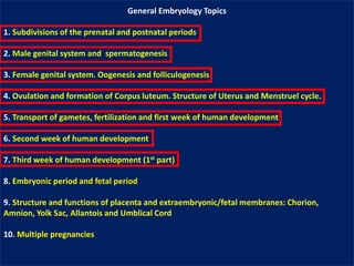
1. General Embryology Topics
1. Subdivisions of the prenatal and postnatal periods
2. Male genital system and spermatogenesis
3. Female genital system. Oogenesis and folliculogenesis
4. Ovulation and formation of Corpus luteum. Structure of Uterus and Menstruel cycle.
5. Transport of gametes, fertilization and first week of human development
6. Second week of human development
7. Third week of human development (1st part)
8. Embryonic period and fetal period
9. Structure and functions of placenta and extraembryonic/fetal membranes: Chorion,
Amnion, Yolk Sac, Allantois and Umblical Cord
10. Multiple pregnancies
2. 2
Embryonic period: This period extends from beginning of the third week to the end of the eight week.
This period is also known as organogenetic period (All organs and systems are formed in this period).
The cells of each germ layer (Ectoderm, Endoderm and Mesoderm) divide, migrate, aggregate, and
differentiate in rather precise patterns as they form the various organ systems.
During this period, embryo has taken on recognizable human shape.
Embryonic period is also most risky and dangerous period of most affected by teratogens (Alcohol,
cigarette, some drugs, radiation and infections-TORCH).
Teratology is a branch of embryology that is concerned with the congenital anomalies or birth defects.
It deals with abnormal embryonic and fetal development.
3. Important Events of Third and Fourth Week of Human Development
I. Formation of Primitive Streak and then development of Trilaminar
Embryonic Disc (Ectoderm, mesoderm and endoderm)
II. Development of Notochord and function and importance of Notochord
III. Neurulation: Formation of Neural Tube (Neural tube will become brain and
spinal cord)
IV. Beginning of the development of chorionic villi of placenta
V. Early development of Cardiyovascular System
VI. Embryonic Folding
VII. Further development of trilaminar embryonic disc (Ectoderm, mesoderm
and endoderm)
4. The most characteristic event occurring during the third week of gestation is gastrulation.
Gastrulation is a process by which the bilaminar embryonic disc is converted into a
trilaminar embryonic disc.
Gastrulation is the beginning of morphogenesis (development of body form).
Epiblast
Hypoblast
Ectoderm
Mesoderm
Endoderm
5. Formation of primitive
streak/primitive groove and
primitive node/primitive pit:
At the beginning of the third week
(day 15), a longitudinal ridge
appears in the midline at the caudal
end of the dorsal aspect of the
bilaminar embryonic disc.
This longitudinal cellular ridge is
called primitive streak.
It is formed due to proliferation of
the Epiplast cells.
It becomes visible on the dorsal
surface of embryonic disc as a
narrow groove (Primitive groove)
flanked by a slight bulge on either
side.
At the cranial end of the primitive
streak, the cells proliferate and
form a rounded elevation called
primitive node (Hensen’s node)
surrounding a small primitive pit.
6. Primitive streak and Primitive
groove
Primitive Node
and Primitive Pit
Caudal Region of
bilaminar disc
7. PA 2010 7
The epiblast cells on both sides of the primitive streak are detached from each other and migrate to the primitive
groove.
Upon arrival in the region of the streak, they become flask-shaped, detach from the epiblast, and slip beneath it.
This inward and downward movement of epiblastic cells is known as invagination.
The epiblast cells enter from the primitive groove between the epiblast and the hypoblast layers.
First targets of invaginated epiblast cells are hypoblastic cells.
Hypoblast cells that see the epiblast cells begin to die with apoptosis.
Hypoblast is removed from here and replaced with new epiblast cells.
This new cell layer is now called the “Intraembryonic Endoderm Layer”.
* Other epiblast cells come to lie between the epiblast and the newly formed endoderm to form the ‘’intraembryonic
mesoderm layer’’.
* The remaining cells of the epiblast now form the ‘’intraembryonic ectoderm layer’’.
* all the three primary germ layers are derived from epiblast cells
9. PA 2010
Mode of spread of intraembryonic
mesoderm:
The intraembryonic mesoderm
spreads in cranial, caudal, and lateral
directions into all parts of the
embryonic disc, except in the
following regions:
* Region of prechordal plate where
the ectoderm and endoderm are in
firm contact with each other and it
will become the buccopharyngeal
membrane and mouth.
* Region of cloacal membrane
(circular area at the caudal end of the
disc): Here also the ectoderm and
endoderm are in an intimate contact
with each other.
It will become anus.
10. Important Events of Third and Fourth Week of Human Development
I. Formation of Primitive Streak and then development of Trilaminar
Embryonic Disc (Ectoderm, mesoderm and endoderm)
II. Development of Notochord and function and importance of Notochord
III. Neurulation: Formation of Neural Tube (Neural tube will become brain and
spinal cord)
IV. Beginning of the development of chorionic villi of placenta
V. Early development of Cardiyovascular System
VI. Embryonic Folding
VII. Further development of trilaminar embryonic disc (Ectoderm, mesoderm
and endoderm)
11. 11
Cranial end
Oropharyngeal
membrane (Mouth)
Caudal end
Cloacal membrane (Anus)
Formation of Notochord:
The notochord is a midline
epiblastic cellular cordon that
develops in the region
between the primitive node
and the prechordal plate
(buccopharyngeal membrane).
Primitive Node and pit
12. PA 2010 12
Formation of Notochord:
The notochord is a midline epiblastic cellular
structure that develops in the region between the
primitive node and the prochordal plate
(buccopharyngeal membrane).
Understanding of development of notochord is
essential because;
* it forms the central axis of the embryonic disc and
gives it some rigidity.
* It induces the ectodermal cells and then formation
neural tube and nervous system.
* It induces the further development of
intraembryonic mesodermal layer cells.
Fate of Notochord:
In human beings, it appears only in embryo.
In later life, it disappears but its remnants are seen
in the form of nucleus pulposus of the intervertebral
discs.
Remnants of notocord can lead to Chordoma
tumors.
15. Important Events of Third and Fourth Week of Human Development
I. Formation of Primitive Streak and then development of Trilaminar
Embryonic Disc (Ectoderm, mesoderm and endoderm)
II. Development of Notochord and function and importance of Notochord
III. Neurulation: Formation of Neural Tube (Neural tube will become brain and
spinal cord)
IV. Beginning of the development of chorionic villi of placenta
V. Early development of Cardiyovascular System
VI. Embryonic Folding
VII. Further development of trilaminar embryonic disc (Ectoderm, mesoderm
and endoderm)
16. PA 2010 16
Notochord cells
induces and
promotes
ectodermal cells to
development of
nervous system
and also induces
further
development of
mesodermal layer.
17. Neurulation (16-22 days): The process of formation of neural tube
1. The notochord cells induce the ectoderm cells
on it and thicken the edges of this layer to form
the neural plate
2. In the middle of the neural plate, a trough
called neural groove and neural folds occurs
3. During the process of neural plate formation, a
group of cells on the most surface of ectoderm
layer are differentiated into neural crest cells
4. Neural folds converge by approaching midline,
and form neural tube surrounded by
neuroepithelial cells around the neural cavity
5. After the neural tube has been formed, the
ectoderm layer is identified by 3 different names:
I. Outermost: Surface Ectoderm (Outer ectoderm)
II. In the innermost: the neuroepithelial layer of
the neural tube (Neuroectoderm)
III. In the middle: Neural Crest cell layer which
moved to migrate to different regions of body
20. Important Events of Third and Fourth Week of Human Development
I. Formation of Primitive Streak and then development of Trilaminar
Embryonic Disc (Ectoderm, mesoderm and endoderm)
II. Development of Notochord and function and importance of Notochord
III. Neurulation: Formation of Neural Tube (Neural tube will become brain and
spinal cord)
IV. Beginning of the development of chorionic villi of placenta
V. Early development of Cardiyovascular System
VI. Embryonic Folding
VII. Further development of trilaminar embryonic disc (Ectoderm, mesoderm
and endoderm)
21. Development of Chorionic Villi of Placenta
1. Primary villus, 12-13 days:
The cytotrophoblast forms fingerlike projections that invade the
syncytiotrophoblast.
This finger-like projection of cytotrophoblast surrounded by a layer of
syncytiotrophoblast is called the primary villus.
23. Development of Chorionic Villi of Placenta
2. Secondary villus, 15-16 days:
The extraembryonic somatopleuric mesoderm lying deep to the cytotrophoblast
now invades the center of each villus.
As a result, now each villus consists of three layers.
From inside to outside these are mesoderm, cytotrophoblast, and
syncytiotrophoblast.
This villus is now termed secondary villus.
25. Development of Chorionic Villi of Placenta
3. Tertiary villus, 20-21 days:
The blood vessels develop in the mesoderm of the secondary villus.
The secondary villus with blood vessels in its mesoderm is called tertiary villus.