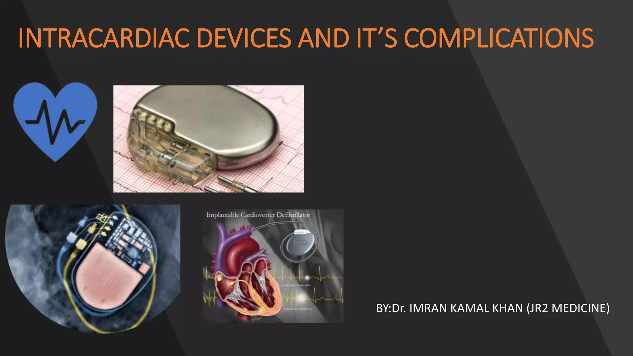
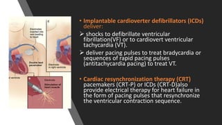
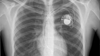
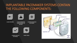
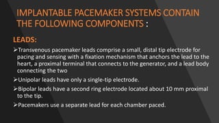
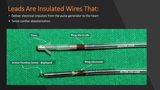
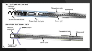
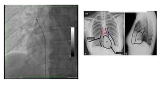
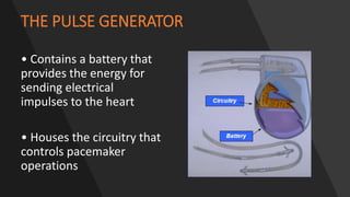
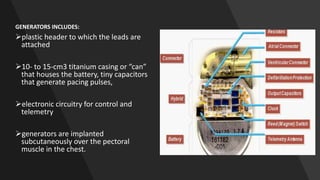
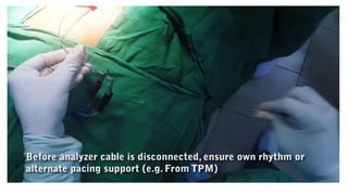
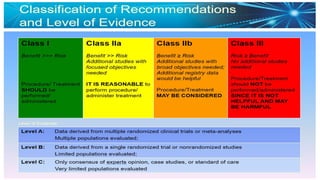
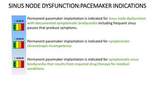
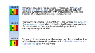
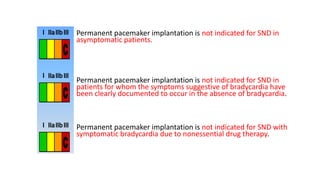
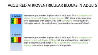
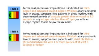
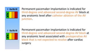
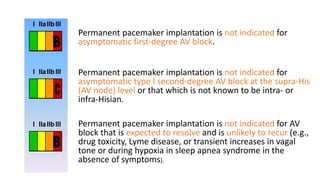
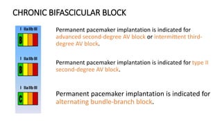
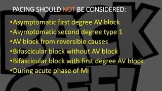
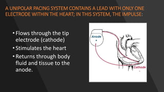
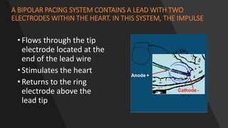
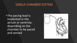
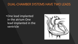
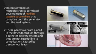
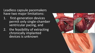
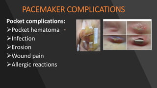
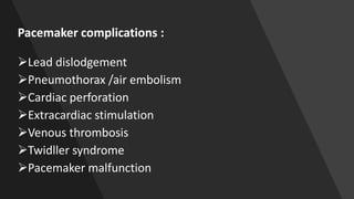
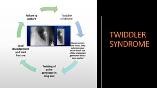
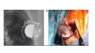
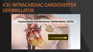
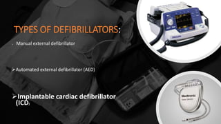
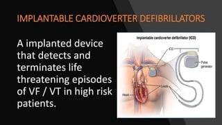
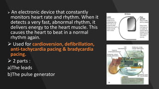
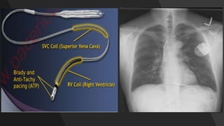
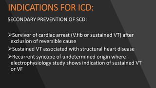
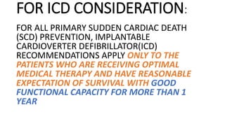
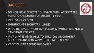
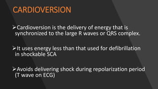
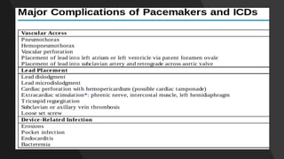
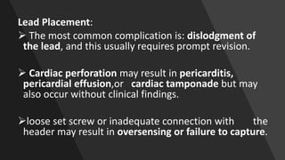
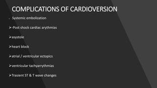
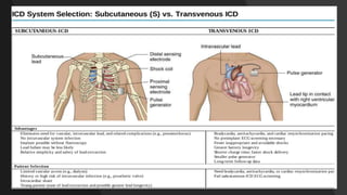
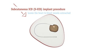
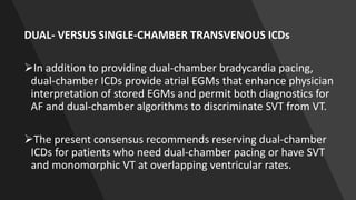
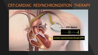
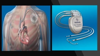
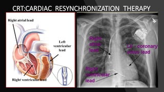
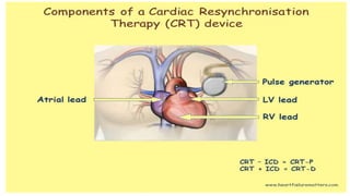
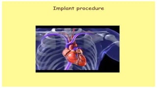
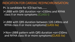
2. • Implantable cardioverter defibrillators (ICDs)
deliver:
shocks to defibrillate ventricular
fibrillation(VF) or to cardiovert ventricular
tachycardia (VT).
deliver pacing pulses to treat bradycardia or
sequences of rapid pacing pulses
(antitachycardia pacing) to treat VT.
• Cardiac resynchronization therapy (CRT)
pacemakers (CRT-P) or ICDs (CRT-D)also
provide electrical therapy for heart failure in
the form of pacing pulses that resynchronize
the ventricular contraction sequence.
3. PACEMAKER
Electrical therapy for cardiac arrhythmias includes:
low-voltage (1 to 5 V) :pacing stimuli (pulses) and
high-voltage (500 to 1400 V) stimuli (shocks).
Pacemaker delivers pacing pulses to treat bradycardia
4.
5.
6. IMPLANTABLE PACEMAKER SYSTEMS CONTAIN
THE FOLLOWING COMPONENTS:
• LEADS OR WIRES • PULSE GENERATOR:
POWER SOURCE OR
BATTERY
• CATHODE (NEGATIVE
ELECTRODE)
• ANODE (POSITIVE
ELECTRODE)
• BODY TISSUE
7. IMPLANTABLE PACEMAKER SYSTEMS CONTAIN
THE FOLLOWING COMPONENTS :
LEADS:
Transvenous pacemaker leads comprise a small, distal tip electrode for
pacing and sensing with a fixation mechanism that anchors the lead to the
heart, a proximal terminal that connects to the generator, and a lead body
connecting the two
Unipolar leads have only a single-tip electrode.
Bipolar leads have a second ring electrode located about 10 mm proximal
to the tip.
Pacemakers use a separate lead for each chamber paced.
8. Leads Are Insulated Wires That:
• Deliver electrical impulses from the pulse generator to the heart
• Sense cardiac depolarisation
9.
10.
11.
12. THE PULSE GENERATOR
• Contains a battery that
provides the energy for
sending electrical
impulses to the heart
• Houses the circuitry that
controls pacemaker
operations
13. GENERATORS INCLUDES:
plastic header to which the leads are
attached
10- to 15-cm3 titanium casing or “can”
that houses the battery, tiny capacitors
that generate pacing pulses,
electronic circuitry for control and
telemetry
generators are implanted
subcutaneously over the pectoral
muscle in the chest.
14.
15.
16.
17. SINUS NODE DYSFUNCTION:PACEMAKER INDICATIONS
Permanent pacemaker implantation is indicated for sinus node dysfunction
with documented symptomatic bradycardia including frequent sinus
pauses that produce symptoms.
Permanent pacemaker implantation is indicated for symptomatic
chronotropic incompetence
Permanent pacemaker implantation is indicated for symptomatic sinus
bradycardia that results from required drug therapy for medical
conditions
18. Permanent pacemaker implantation is reasonable for SND with
heart rate less than 40 bpm when a clear association between
significant symptoms consistent with bradycardia and the actual
presence of bradycardia has not been documented.
Permanent pacemaker implantation is reasonable for syncope
of unexplained origin when clinically significant abnormalities
of sinus node function are discovered or provoked in
electrophysiological studies.
Permanent pacemaker implantation may be considered in
minimally symptomatic patients with chronic heart rate
less than 40 bpm while awake.
19. Permanent pacemaker implantation is not indicated for SND in
asymptomatic patients.
Permanent pacemaker implantation is not indicated for SND in
patients for whom the symptoms suggestive of bradycardia have
been clearly documented to occur in the absence of bradycardia.
Permanent pacemaker implantation is not indicated for SND with
symptomatic bradycardia due to nonessential drug therapy.
20. ACQUIRED ATRIOVENTRICULAR BLOCKS IN ADULTS
Permanent pacemaker implantation is indicated for third-degree and
advanced second-degree atrioventricular (AV) block at any anatomic
level associated with bradycardia with symptoms (including heart
failure) or ventricular arrhythmias presumed to be due to AV block.
Permanent pacemaker implantation is indicated for third-degree and
advanced second-degree AV block at any anatomic level associated
with arrhythmias and other medical conditions that require drug
therapy that results in symptomatic bradycardia.
21. Permanent pacemaker implantation is indicated for third-
degree and advanced second-degree AV block at any anatomic
level in awake, symptom-free patients in sinus rhythm, with
documented periods of asystole greater than or equal to 3.0
seconds or any escape rate less than 40 bpm, or with an
escape rhythm that is below the AV node.
Permanent pacemaker implantation is indicated for third-
degree and advanced second-degree AV block at any anatomic
level in awake, symptom-free patients with atrial fibrillation
(AF) and bradycardia with 1 or more pauses of at least 5
seconds or longer.
22. Permanent pacemaker implantation is indicated for
third-degree and advanced second-degree AV block at
any anatomic level after catheter ablation of the AV
junction.
Permanent pacemaker implantation is indicated for
third-degree and advanced second-degree AV block at
any anatomic level associated with postoperative AV
block that is not expected to resolve after cardiac
surgery.
23.
24. Permanent pacemaker implantation is not indicated for
asymptomatic first-degree AV block.
Permanent pacemaker implantation is not indicated for
asymptomatic type I second-degree AV block at the supra-His
(AV node) level or that which is not known to be intra- or
infra-Hisian.
Permanent pacemaker implantation is not indicated for AV
block that is expected to resolve and is unlikely to recur (e.g.,
drug toxicity, Lyme disease, or transient increases in vagal
tone or during hypoxia in sleep apnea syndrome in the
absence of symptoms).
25. CHRONIC BIFASCICULAR BLOCK
Permanent pacemaker implantation is indicated for
advanced second-degree AV block or intermittent third-
degree AV block.
Permanent pacemaker implantation is indicated for type II
second-degree AV block.
Permanent pacemaker implantation is indicated for
alternating bundle-branch block.
26. PACING SHOULD NOT BE CONSIDERED:
•Asymptomatic first degree AV block
•Asymptomatic second degree type 1
•AV block from reversible causes
•Bifascicular block without AV block
•Bifascicular block with first degree AV block
•During acute phase of MI
27. A UNIPOLAR PACING SYSTEM CONTAINS A LEAD WITH ONLY ONE
ELECTRODE WITHIN THE HEART; IN THIS SYSTEM, THE IMPULSE:
• Flows through the tip
electrode (cathode)
• Stimulates the heart
• Returns through body
fluid and tissue to the
anode.
28. A BIPOLAR PACING SYSTEM CONTAINS A LEAD WITH TWO
ELECTRODES WITHIN THE HEART. IN THIS SYSTEM, THE IMPULSE
• Flows through the tip
electrode located at the
end of the lead wire
• Stimulates the heart
• Returns to the ring
electrode above the
lead tip
30. DUAL-CHAMBER SYSTEMS HAVE TWO LEADS
•One lead implanted
in the atrium One
lead implanted in the
ventricle
31. BENEFITS OF DUAL CHAMBER PACING
Provides AV
synchrony
Lower incidence
of atrial
fibrillation
Lower risk of
systemic
embolism and
stroke
Lower incidence
of new
congestive heart
failure
Lower mortality
and higher
survival rates
32. CAPTURE AND SENSING
Cardiac pacing requires a local stimulus that creates a
field sufficient to depolarize (reduce the membrane
potential of) local myocardium during diastole to
threshold voltage and thus initiate a self propagating
wavefront of depolarization.
A stimulus that brings local myocardium to threshold is
said to “CAPTURE” it.
33. SENSING
When a depolarization
wavefront passes the tip
electrode, a deflection in the
EGM signal travels
instantaneously through the
electrode to the generator.
There, the signal is amplified,
filtered, processed by the
sensing electronics, and
compared to a threshold
voltage (sensing threshold).
A sensed event occurs when
the processed signal exceeds
this sensing threshold, and
the device determines that
an atrial or ventricular
depolarization has occurred.
34. • Most pacemaker sensing thresholds are programmed to
fixed values, typically about 2.0 mV for ventricular
channels and 0.3 to 0.6 mV for atrial channels, with
bipolar sensing to allow sensing of lower-amplitude P
waves and atrial EGMs during atrial fibrillation (AF).
• Highly sensitive programmed values can result in
oversensing, sensing of unintended signals not originating
in the cardiac chamber of interest.
35. Recent advances in
microelectronics permitted
development of leadless
capsule pacemakers that
comprise both the generator
and the lead system.
These pacemakers are placed
in the RV endocardium through
a catheter delivery system and
thus are not susceptible to
complications caused by
transvenous leads.
36. Leadless capsule pacemakers
have two major limitations:
1. first-generation devices
permit only single-chamber
ventricular pacing, and
2. the feasibility of extracting
chronically implanted
devices is unknown
39. TWIDDLER
SYNDROME
Twiddler
syndrome:
Obese women
with loose, fatty
subcutaneous
tissue Small size
of the implanted
generator with a
large pocket
Twisting of
pulse
generator in
long axis
Lead
dislodgement
and lead
fracture
Failure to
capture
42. INTRACARDIAC CARDIOVERTER DEFIBRILLATOR
•™Defibrillator is a device that delivers a
therapeutic dose of electrical energy
(electric shock) to the affected heart
(fibrillated heart or other shockable rhythm)
to force the heart to produce more normal
cardiac rhythm.
45. An electronic device that constantly
monitors heart rate and rhythm. When it
detects a very fast, abnormal rhythm, it
delivers energy to the heart muscle. This
causes the heart to beat in a normal
rhythm again.
Used for cardioversion, defibrillation,
anti-tachycardia pacing & bradycardia
pacing.
2 parts :
a)The leads
b)The pulse generator
46.
47. INDICATIONS FOR ICD:
SECONDARY PREVENTION OF SCD:
Survivor of cardiac arrest (V.fib or sustained VT) after
exclusion of reversible cause
Sustained VT associated with structural heart disease
Recurrent syncope of undetermined origin where
electrophysiology study shows indication of sustained VT
or VF
49. FOR ICD CONSIDERATION:
FOR ALL PRIMARY SUDDEN CARDIAC DEATH
(SCD) PREVENTION, IMPLANTABLE
CARDIOVERTER DEFIBRILLATOR(ICD)
RECOMMENDATIONS APPLY ONLY TO THE
PATIENTS WHO ARE RECEIVING OPTIMAL
MEDICAL THERAPY AND HAVE REASONABLE
EXPECTATION OF SURVIVAL WITH GOOD
FUNCTIONAL CAPACITY FOR MORE THAN 1
YEAR
50. BACK OFF!
• DO NOT HAVE EXPECTED SURVIVAL WITH ACCEPTABLE
FUNCTIONAL STATUS FOR ATLEAST 1 YEAR
• INCESSANT VT or VF
• SIGNIFICANT PSYCHIATRY ILLNESS
• DRUG REFRACTORY CHF (NYHA class IV )WHICH ARE NOT A
CANDIDATE FOR CRT
• IF VF or VT IS AMENABLE TO SURGICAL OR CATHETER
ABLATION (Afib with WPW;OUTFLOW TRACT VTs)
• VF ,VT DUE TO REVERSIBLE CAUSE
51. CARDIOVERSION
Cardioversion is the delivery of energy that is
synchronized to the large R waves or QRS complex.
It uses energy less than that used for defibrillation
in shockable SCA
Avoids delivering shock during repolarization period
(T wave on ECG)
53. CONTRAINDICATIONS:
Digitalis induced dysrhythmias:
refractory to cardioversion
may precipitate to more serious ventricular dysarrythmias
Junctional tachycardia or ectopics / multifocal atrial tachycardia
automatic foci not reverted by cardioversion
54. CARDIOVERSION VS DEFIBRILLATOR
Elective planned procedure emergency life saving procedure
Synchronized shock un synchronized shock
Low energy shock high energy shock
There can be some delay no delay,immediate
Anticoagulation needed no anticoagulation needed
Less damage to myocardium more damage to myocardium
Used in most of the arrhythmias used in VT/VF
except VT /VF
55.
56. Lead Placement:
The most common complication is: dislodgment of
the lead, and this usually requires prompt revision.
Cardiac perforation may result in pericarditis,
pericardial effusion,or cardiac tamponade but may
also occur without clinical findings.
loose set screw or inadequate connection with the
header may result in oversensing or failure to capture.
57. COMPLICATIONS OF CARDIOVERSION
-Systemic embolization
-Post shock cardiac arythmias
asystole
heart block
atrial / ventricular ectopics
ventricular tachyarrythmias
Trasient ST & T wave changes
58.
59.
60. DUAL- VERSUS SINGLE-CHAMBER TRANSVENOUS ICDs
In addition to providing dual-chamber bradycardia pacing,
dual-chamber ICDs provide atrial EGMs that enhance physician
interpretation of stored EGMs and permit both diagnostics for
AF and dual-chamber algorithms to discriminate SVT from VT.
The present consensus recommends reserving dual-chamber
ICDs for patients who need dual-chamber pacing or have SVT
and monomorphic VT at overlapping ventricular rates.
65. CARDIAC RESYNCHRONIZATION THERAPY
• Several conduction abnormalities are commonly seen in association with
chronic heart failure.
• Among these are abnormalities of ventricular conduction,such as bundle
branch blocks, that alter the timing and pattern of ventricular contraction so as
to place the already failing heart at a further mechanical disadvantage.
• These ventricular conduction delays produce :
suboptimal ventricular filling,
a reduction in left ventricular (LV) contractility,
prolonged duration of mitral regurgitation, and
paradoxical septal wall motion.
• These mechanical manifestations of altered ventricular conduction have been
termed ventricular dyssynchrony.
66.
67. • Ventricular dyssynchrony has been defined by :
a prolonged QRS duration, generally greater than 120
milliseconds, on the surface electrocardiogram (ECG).
• By this definition,about one third of patients with systolic HF
have ventricular dyssynchrony.
• In addition to reducing the ability of the failing heart to eject
blood, ventricular dyssynchrony has also been associated with
increased mortality in HF patients.
68. •Ventricular dyssynchrony is now addressed with
pacing therapy, through the implantation of
pacing leads to both right and left ventricles.
•This form of pacing therapy is now known as
CARDIAC RESYNCHRONIZATION THERAPY.OR
BI-VENTRICULAR PACING
69.
70. INDICATION FOR CARDIAC RESYNCHRONISATION:
Pt. is candidate for ICD but has…….
LBBB with QRS duration =or >150ms and NYHA
class II or more symptoms.(CLASS I)
LBBB with QRS duration between 120-149ms and
NYHA class II or more symptoms(CLASS IIa)
Non-LBBB pattern with QRS duration =or>150ms
and NYHA class III or more symptoms(CLASS IIa)
71.
72.
73. BACK OFF!
•Non-LBBB with QRS
<150 ms and NYHA class
I or II symptoms
•Do not have expected
survival with acceptable
functional status for at
least 1 year.
74. •Implant-related complications are
similar to those seen with standard
pacemakers and defibrillators, with
the additional risk of dissectionor
perforation of the coronary sinus.