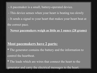
CARDIAC PACEMAKERS
N. LEMUEL MATHEW
MSc. Cardiac Catheterization
& Interventional Technology
- A pacemaker is a small, battery-operated device.
- This device senses when your heart is beating too slowly
- It sends a signal to your heart that makes your heart beat at
the correct pace.
Newer pacemakers weigh as little as 1 ounce (28 grams)
Most pacemakers have 2 parts:
 The generator contains the battery and the information to
control the heartbeat.
 The leads which are wires that connect the heart to the
generator and carry the electrical messages to the heart.
Procedure:
- A pacemaker is implanted under the skin.
- This procedure takes about 1 hour in most cases.
- You will be given a sedative to help you relax and you will be
awake during the procedure.
- A small incision (cut) is made. Most often, the cut is on the
left side (if you are right handed) of the chest below your
collarbone.
- The pacemaker generator is then placed under the skin at this
location.
-The generator may also be placed in the abdomen, but this is
less common.
- A new "leadless" pacemaker is a self-contained unit that is
implanted in the right ventricle of the heart.
- Using live x-rays to see the area, the doctor (most often a
heart specialist called a cardiologist) puts the leads through the
cut, into a vein, and then into the heart.
- The leads are connected to the generator.
- The skin is closed with stitches.
- Most people go home within 1 day of the procedure.
There are 2 kinds of pacemakers used only in medical
emergencies. They are:
- Transcutaneous pacemakers
- Transvenous pacemakers
They are not permanent pacemakers.
Risks:
Possible complications of pacemaker surgery are:
- Abnormal heart rhythms -Bleeding
- Punctured lung. This is rare. - Infection
- Puncture of the heart, which can lead to bleeding around
the heart. This is rare.
Signs you need a pacemaker:
- Chest pain (also known as angina).
- Tachycardia: Unusually fast heartbeat (more than 100 beats /min)
- Bradycardia: Unusually slow heartbeat (fewer than 60 beats/min).
- Irregular heartbeat (arrhythmia): Heartbeat that skips beats or adds in
extra beats.
- Heart palpitations: Happens when you can feel your heartbeat
- Shortness of breath: Especially when you’re more active.
- Unexplained dizziness or lightheadedness, nausea or fainting.
- Unexplained confusion.
- Swelling in your ankles, legs and abdomen.
- Needing to urinate multiple times at night.
Approach:
Catheter-based approach: This method is used with leadless pacemakers,
which only treat conditions involving a single chamber of your heart.
Transvenous (through a vein) approach :This method is frequently used
in adults.
Surgical-based (epicardial) approach :A surgeon creates a small incision
in your chest and inserts the lead(s) through that incision, and then attaches
the lead(s) to your heart
Indications:
Common Indications for Pacemaker Placement
- Sinus node dysfunction (Class I indication)
- Acquired AV block
- Post myocardial infarction
Less Common Indications
- Congenital complete heart block
- Long QT syndrome
- Hypertrophic Cardiomyopathy
- Heart failure
Pacemaker Related Complications
Pacemaker Syndrome: is a disease that represents the clinical consequences of
suboptimal atrioventricular (AV) synchrony or AV dyssynchrony
Symptoms of Pacemaker Syndrome are:
- Cannon A-waves - Chest pain
- Confusion - Dizziness
- Fatigue - Palpitations
- Shortness of breath - Syncope
Other complications associated with Pacemakers include:
- Pneumothorax - Cardiac perforation
- Significant pocket hematoma - Lead dislodgement
- Venous thrombosis and obstruction
- Mechanical lead complications
Pacemaker Types and System:
Transvenous Systems: Most cardiac pacing systems use transvenous
electrodes to transmit electrical impulses from the pulse generator to the
heart musculature.
Epicardial systems work by direct stimulation through the pulse generator
by attaching directly to the heart's surface.
Leadless systems: There have been some innovations in developing
leadless systems
Types of Pacemakers:
There are 3 basic kinds of pacemakers:
Single chamber. One lead attaches to the upper or lower heart chamber.
Dual-chamber. Uses 2 leads, 1 for the upper and 1 for the lower chamber
Biventricular pacemakers (used in cardiac resynchronization therapy).
Single chamber cardiac pacemakers are cardiac conduction devices with
one lead terminating in (most commonly) the right ventricular apex or the
right atrium.
Components:
Includes one of the following:
- Lead in the right atrium
- Lead in the right ventricle
Function:
-Single chamber Atrial pacemakers are uncommonly placed
- Indications include severely symptomatic sinus Bradycardia and
the sick sinus syndrome (with the tachycardia- Bradycardia syndrome)
- Lead placement is typically in the right Atrial appendage
ECG:
- Atrial pacing will result in a sharp, vertical pacemaker spike
followed by ectopic P wave, upright polarity in the limb leads, with the
exception of aVR.
- Ventricular pacing from right ventricular apex result in abnormal
right-to-left depolarization of the ventricular myocardium, shows LBBB like
morphology of the QRS complex.
Indications for permanent single-chamber ventricular
pacing include :
- Sinus node dysfunction
- Advanced atrioventricular (AV) block
- Neurocardiogenic syncope
- Cardiac transplant
- Hypertrophic Cardiomyopathy
with LVOT obstruction cardiac sarcoidosis
with atrioventricular block
Indications for Temporary single-chamber ventricular
pacing include :
-Bradycardia
- When associated with myocardial infarction (MI)
- Not associated with MI
- Ventricular dysrhythmias
- High grade or complete AV block
- Risk of post-procedural bradycardia
- Tachycardia (overdrive suppression)
- Supraventricular tachycardia (SVT)
- Ventricular tachycardia (VT)
Dual-chamber pacemakers: Two leads are placed, one in the right atrium
and one in the right ventricle.
The right atrial lead is typically placed in the
right atrial appendage,
The right ventricular lead is placed in the apex
of the right ventricle
Function:
By pacing both the atria and ventricles, dual-chamber
pacemakers help to coordinate the heart's electrical activity, improving the
efficiency and overall function of the heart.
Modes of Cardiac Pacing:
Typically consist of 5 letters.
Letter 1. In the area being paced, A stands for atria, V stands
for Ventricle, D stands for Dual, O stands for none
Letter 2. For the area that is sensed, A stands for atria, V
stands for Ventricle, D stands for Dual, O stands for none
Letter 3. The response of the pacemaker to sensing: O stands
for none, I stands for inhibiting, T stands for triggering, D
stands for dual
Letter 4. Rate adaptiveness. O stands for none, and R stands
for rate adaptiveness.
Single Chamber Modes:
VOO
V- Pacing in the ventricle
O- Sensing is OFF
O- Response to sensing is OFF
VVI
V- Pacing in the ventricle
V- Sensing in the ventricle
I-Inhibit
In this mode, the pacemaker can sense the electrical activity and withhold
pacing when not required.
AOO
A- Pacing in the atrium
O- Sensing is OFF
O- Response to sensing is OFF
In this mode, the pacemaker paces at a programmed rate regardless of the
heart's intrinsic electrical activity.
AAI
A- Pacing in the atrium
A- Sensing in the atrium
I- Inhibit
In this mode, the pacemaker can adapt to the intrinsic atrial rate and should
be able to pace when needed and inhibit when not required.
Dual Chamber Modes:
Dual Chamber Modes can further subdivide into
Tracking Modes and Non-Tracking modes.
Tracking Modes:
DDD
D- Pacing in the atrium and ventricle
D- Sensing in the atrium and ventricle
D- Inhibit and or trigger
VDD
V- Pacing in the ventricle
D- Sensing in the atrium and ventricle
D- Inhibit and or trigger
Non-Tracking Modes:
DDI
D - Pacing in the atrium and ventricle
D - Sensing in the atrium and ventricle
I - Response to that sensing be to either pace or inhibit
DOO
D- Pacing in the atrium and ventricle
O- Sensing is OFF
O- Response to that sensing is OFF
Choosing a Pacing Mode:
- Wants to ensure that the patient has a viable Atrial activity.
- Whether the sinus node function is intact.
If sinus node function is not intact and the patient has Atrial
arrhythmias:
Chronic Atrial Arrhythmias, e.g., Atrial Fibrillation or Atrial Flutter:
If the patient is chronotropically incompetent, the mode of choice is VVIR.
If the patient is chronotropically competent, the mode of choice is VVI.
Paroxysmal Atrial Arrhythmias:
If the patient is chronotropically incompetent, the mode of choice is
DDDR.
If the patient is chronotropically competent, the mode of choice is DDD.
If the sinus node is intact and the patient has normal sinus rhythm or
sinus Bradycardia:
Intact AV Node conduction:
If the patient is chronotropically incompetent, the mode of choice is AAIR.
If the patient is chronotropically competent, the mode of choice is AAI.
AV Node conduction not Intact:
If the patient is chrontropically incompetent, the mode of choice is DDDR.
If the patient is chronotropically competent, the mode of choice is DDD.
THANK YOU

Cardiac Pacemakers and its Modes.pptx

  • 1.
    CARDIAC PACEMAKERS N. LEMUELMATHEW MSc. Cardiac Catheterization & Interventional Technology
  • 2.
    - A pacemakeris a small, battery-operated device. - This device senses when your heart is beating too slowly - It sends a signal to your heart that makes your heart beat at the correct pace. Newer pacemakers weigh as little as 1 ounce (28 grams) Most pacemakers have 2 parts:  The generator contains the battery and the information to control the heartbeat.  The leads which are wires that connect the heart to the generator and carry the electrical messages to the heart.
  • 3.
    Procedure: - A pacemakeris implanted under the skin. - This procedure takes about 1 hour in most cases. - You will be given a sedative to help you relax and you will be awake during the procedure. - A small incision (cut) is made. Most often, the cut is on the left side (if you are right handed) of the chest below your collarbone. - The pacemaker generator is then placed under the skin at this location.
  • 4.
    -The generator mayalso be placed in the abdomen, but this is less common. - A new "leadless" pacemaker is a self-contained unit that is implanted in the right ventricle of the heart. - Using live x-rays to see the area, the doctor (most often a heart specialist called a cardiologist) puts the leads through the cut, into a vein, and then into the heart. - The leads are connected to the generator. - The skin is closed with stitches. - Most people go home within 1 day of the procedure.
  • 5.
    There are 2kinds of pacemakers used only in medical emergencies. They are: - Transcutaneous pacemakers - Transvenous pacemakers They are not permanent pacemakers. Risks: Possible complications of pacemaker surgery are: - Abnormal heart rhythms -Bleeding - Punctured lung. This is rare. - Infection - Puncture of the heart, which can lead to bleeding around the heart. This is rare.
  • 6.
    Signs you needa pacemaker: - Chest pain (also known as angina). - Tachycardia: Unusually fast heartbeat (more than 100 beats /min) - Bradycardia: Unusually slow heartbeat (fewer than 60 beats/min). - Irregular heartbeat (arrhythmia): Heartbeat that skips beats or adds in extra beats. - Heart palpitations: Happens when you can feel your heartbeat - Shortness of breath: Especially when you’re more active. - Unexplained dizziness or lightheadedness, nausea or fainting. - Unexplained confusion. - Swelling in your ankles, legs and abdomen. - Needing to urinate multiple times at night.
  • 7.
    Approach: Catheter-based approach: Thismethod is used with leadless pacemakers, which only treat conditions involving a single chamber of your heart. Transvenous (through a vein) approach :This method is frequently used in adults. Surgical-based (epicardial) approach :A surgeon creates a small incision in your chest and inserts the lead(s) through that incision, and then attaches the lead(s) to your heart
  • 8.
    Indications: Common Indications forPacemaker Placement - Sinus node dysfunction (Class I indication) - Acquired AV block - Post myocardial infarction Less Common Indications - Congenital complete heart block - Long QT syndrome - Hypertrophic Cardiomyopathy - Heart failure
  • 9.
    Pacemaker Related Complications PacemakerSyndrome: is a disease that represents the clinical consequences of suboptimal atrioventricular (AV) synchrony or AV dyssynchrony Symptoms of Pacemaker Syndrome are: - Cannon A-waves - Chest pain - Confusion - Dizziness - Fatigue - Palpitations - Shortness of breath - Syncope Other complications associated with Pacemakers include: - Pneumothorax - Cardiac perforation - Significant pocket hematoma - Lead dislodgement - Venous thrombosis and obstruction - Mechanical lead complications
  • 10.
    Pacemaker Types andSystem: Transvenous Systems: Most cardiac pacing systems use transvenous electrodes to transmit electrical impulses from the pulse generator to the heart musculature. Epicardial systems work by direct stimulation through the pulse generator by attaching directly to the heart's surface. Leadless systems: There have been some innovations in developing leadless systems
  • 11.
    Types of Pacemakers: Thereare 3 basic kinds of pacemakers: Single chamber. One lead attaches to the upper or lower heart chamber. Dual-chamber. Uses 2 leads, 1 for the upper and 1 for the lower chamber Biventricular pacemakers (used in cardiac resynchronization therapy). Single chamber cardiac pacemakers are cardiac conduction devices with one lead terminating in (most commonly) the right ventricular apex or the right atrium. Components: Includes one of the following: - Lead in the right atrium - Lead in the right ventricle
  • 12.
    Function: -Single chamber Atrialpacemakers are uncommonly placed - Indications include severely symptomatic sinus Bradycardia and the sick sinus syndrome (with the tachycardia- Bradycardia syndrome) - Lead placement is typically in the right Atrial appendage ECG: - Atrial pacing will result in a sharp, vertical pacemaker spike followed by ectopic P wave, upright polarity in the limb leads, with the exception of aVR. - Ventricular pacing from right ventricular apex result in abnormal right-to-left depolarization of the ventricular myocardium, shows LBBB like morphology of the QRS complex.
  • 13.
    Indications for permanentsingle-chamber ventricular pacing include : - Sinus node dysfunction - Advanced atrioventricular (AV) block - Neurocardiogenic syncope - Cardiac transplant - Hypertrophic Cardiomyopathy with LVOT obstruction cardiac sarcoidosis with atrioventricular block
  • 14.
    Indications for Temporarysingle-chamber ventricular pacing include : -Bradycardia - When associated with myocardial infarction (MI) - Not associated with MI - Ventricular dysrhythmias - High grade or complete AV block - Risk of post-procedural bradycardia - Tachycardia (overdrive suppression) - Supraventricular tachycardia (SVT) - Ventricular tachycardia (VT)
  • 15.
    Dual-chamber pacemakers: Twoleads are placed, one in the right atrium and one in the right ventricle. The right atrial lead is typically placed in the right atrial appendage, The right ventricular lead is placed in the apex of the right ventricle Function: By pacing both the atria and ventricles, dual-chamber pacemakers help to coordinate the heart's electrical activity, improving the efficiency and overall function of the heart.
  • 16.
    Modes of CardiacPacing: Typically consist of 5 letters. Letter 1. In the area being paced, A stands for atria, V stands for Ventricle, D stands for Dual, O stands for none Letter 2. For the area that is sensed, A stands for atria, V stands for Ventricle, D stands for Dual, O stands for none Letter 3. The response of the pacemaker to sensing: O stands for none, I stands for inhibiting, T stands for triggering, D stands for dual Letter 4. Rate adaptiveness. O stands for none, and R stands for rate adaptiveness.
  • 17.
    Single Chamber Modes: VOO V-Pacing in the ventricle O- Sensing is OFF O- Response to sensing is OFF VVI V- Pacing in the ventricle V- Sensing in the ventricle I-Inhibit In this mode, the pacemaker can sense the electrical activity and withhold pacing when not required.
  • 18.
    AOO A- Pacing inthe atrium O- Sensing is OFF O- Response to sensing is OFF In this mode, the pacemaker paces at a programmed rate regardless of the heart's intrinsic electrical activity. AAI A- Pacing in the atrium A- Sensing in the atrium I- Inhibit In this mode, the pacemaker can adapt to the intrinsic atrial rate and should be able to pace when needed and inhibit when not required.
  • 19.
    Dual Chamber Modes: DualChamber Modes can further subdivide into Tracking Modes and Non-Tracking modes. Tracking Modes: DDD D- Pacing in the atrium and ventricle D- Sensing in the atrium and ventricle D- Inhibit and or trigger
  • 20.
    VDD V- Pacing inthe ventricle D- Sensing in the atrium and ventricle D- Inhibit and or trigger Non-Tracking Modes: DDI D - Pacing in the atrium and ventricle D - Sensing in the atrium and ventricle I - Response to that sensing be to either pace or inhibit DOO D- Pacing in the atrium and ventricle O- Sensing is OFF O- Response to that sensing is OFF
  • 21.
    Choosing a PacingMode: - Wants to ensure that the patient has a viable Atrial activity. - Whether the sinus node function is intact. If sinus node function is not intact and the patient has Atrial arrhythmias: Chronic Atrial Arrhythmias, e.g., Atrial Fibrillation or Atrial Flutter: If the patient is chronotropically incompetent, the mode of choice is VVIR. If the patient is chronotropically competent, the mode of choice is VVI. Paroxysmal Atrial Arrhythmias: If the patient is chronotropically incompetent, the mode of choice is DDDR. If the patient is chronotropically competent, the mode of choice is DDD.
  • 22.
    If the sinusnode is intact and the patient has normal sinus rhythm or sinus Bradycardia: Intact AV Node conduction: If the patient is chronotropically incompetent, the mode of choice is AAIR. If the patient is chronotropically competent, the mode of choice is AAI. AV Node conduction not Intact: If the patient is chrontropically incompetent, the mode of choice is DDDR. If the patient is chronotropically competent, the mode of choice is DDD.
  • 23.