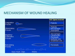
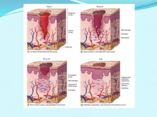
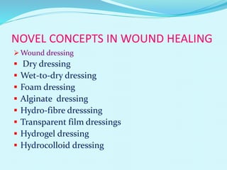
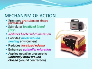
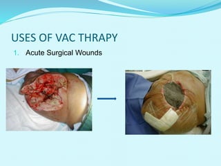
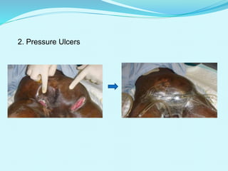
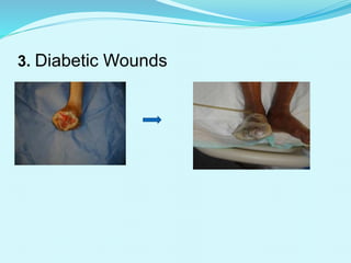
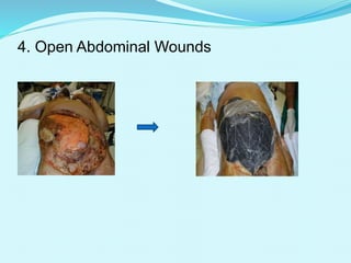
Presented by Guide:
BINUJA S.S. PRASANTH M.S
CONTENTS
 INTRODUCTION
 MECHANISM OF WOUND HEALING
 NOVEL CONCEPTS IN WOUND HEALING
 VACUUM ASSISSTED CLOSURE WOUND THERAPY
 MECHANISM OF ACTION OF VAC
 METHODOLOGY
 USES OF VAC
 ADVANTAGES AND DISADVANTAGES
 APPLICATIONS
 FUTURE DEVELPOMENT
 CONCLUSION
 REFERENCES
INTRODUCTION
 Wounds may result from trauma or from a surgical incision. In
addition, pressure ulcers (also known as decubitus ulcers or bed
sores), a type of skin ulcer, might also be considered wounds.
 Wound healing is the process of repair that follows injury to the
skin and other soft tissues.
 The capacity of a wound to heal depends in part on its depth,
as well as on the overall health and nutritional status of the
individual.
 Following injury, an inflammatory response occurs and the
cells below the dermis (the deepest skin layer) begin to increase
collagen (connective tissue) production. Later, the epithelial
tissue (the outer skin layer) is regenerated.
Standard wound management consists of:
 Initial surgical debridement (a rapid and effective
technique to remove devitalised tissue).
 Wet-to moist (WM) gauze dressings , which need to
be changed at least twice daily.
 These dressings are relatively inexpensive, readily
available.
Disadvantages: non-selective debridement with
dressing removal, possible wound desiccation, and the
need for frequent dressing changes.
The vacuum-assisted closure (VAC) device was
pioneered by Dr Louis Argenta and Dr Michael
Morykwas in 1993.
Vacuum-assisted closure (VAC) therapy-
 Alternative to the standard forms of wound
management, which incorporates the use of negative
pressure to optimise conditions for wound healing and
requires fewer painful dressing changes.
MECHANISM OF WOUND HEALING
NOVEL CONCEPTS IN WOUND HEALING
 Wound dressing
 Dry dressing
 Wet-to-dry dressing
 Foam dressing
 Alginate dressing
 Hydro-fibre dresssing
 Transparent film dressings
 Hydrogel dressing
 Hydrocolloid dressing
VACUUM ASSISSTED CLOSURE WOUND
THERAPY
 The application of controlled levels of negative
pressure accelerates debridement and promote healing
in many different types of wounds.
 The optimum level of negative pressure appears to be
around 125 mmHg.
 Negative pressure assists;
Removal of interstitial fluid.
Decreases localised oedema.
Increases blood flow.
MECHANISM OF ACTION
 Promotes granulation tissue
formation .
 Stimulates localized blood
flow .
 Reduces bacterial colonization
 Provides moist wound
healing environment
 Reduces localized edema
 Enhances epithelial migration
 Applies negative pressure to
uniformly draw wound
closed (wound contraction)
VAC SYSTEM
VAC PUMP
SPONGE
Polyurethane Foam Poly-vinyl-alcohol
Pore size:400-600 microns Pore size: 0.2 -1mm
Tube
For fluid For measurement
Multi-lumen
METHODOLOGY
Materials needed:
 Scissors (sterile or clean)
 Gloves (sterile or clean)
 Dressing kit
 Canister
 V.A.C. Unit
Optional:
 Skin prep Tincture
Benzoin
 Non-adherent dressing,
such as Mepitel
Aggressively clean wound
Debride necrotic tissue or eschar if possible
Achieve hemostasis
Shave hair around border if needed
Irrigate wound with normal saline
Dry and prep skin as appropriate
Cut foam to size of wound
Gently lay foam in wound, including tunnels,
undermining, and all surfaces
1.Clean wound thoroughly
Aggressive cleaning of the wound at each dressing change is
imperative to decrease bacterial load and minimize odor
Cut foam
Cut the foam to fit the size and shape of the wound, including tunnels and
undermined areas
Lay foam in wound
Gently place the foam into the wound cavity, covering the entire
wound base and sides, tunneling and undermining
Cut the drape
Cut the drape large enough to cover the foam and 3-5 cm of surrounding
healthy tissue with drape.
Applying the drape
Apply the drape beginning on one side of the foam, toward the tubing. Do not
stretch the drape and do not compress the foam into the wound with drape.
This helps minimize tension or shearing forces on periwound tissue
Applying the suction tubing
Cut hole in drape about 1.5
cm and apply tubing
Connect to canister
Connect dressing tubing to canister tubing, making sure clamps are open
Y - connecting
A Y-connector is available to connect 2 or more wounds to one V.A.C. pump
Canister CANISTER WITH ISOLYSER
Canister comes with Isolyser gel that gels fluid on contact and helps
eliminate odor
USES OF VAC THRAPY
1. Acute Surgical Wounds
2. Pressure Ulcers
3. Diabetic Wounds
4. Open Abdominal Wounds
ADVANTAGES
 Provides more effective therapy because target sub
atmospheric pressure is monitored and maintained at
Maximizes accuracy and effectiveness of V.A.C.® Therapy.
Reduced frequency of dressing changes.
Reduced bacterial cell count.
Enhanced dermal perfusion.
Provision of closed, moist wound healing
environment.control of odour and exudate.
Reduction in complexity and number of surgical
procedure.
DISADVANTAGES
 Pain and discomfort when suction is applied initially.
 Allergies to adhesive drape.
 Noise of vac therapy unit.
 If the wound deteriorates after the first dressing
change discontinue vac therapy.
 Fulminant or incipient skin necrosis.
 Excoriation of the skin if foam is not correctly cut to
use.
 Drain require fixation.
APPLICATIONS
Treatment of early hip joint infections.
Post operative ascetic fluid leaks in cirrhotic patients.
Wound temporation in composite scalp and calvarial
defects.
Sea water-immersed wound treatment under different
negative pressure.
Treatment of perineal war wound related to rectum.
In patients with wound dehiscence after abdominal
open surgery.
Management of Postpneumonectomy Empyema.
Management of lung abscess.
Treatment of mastitis assossiated chronic breast
wounds.
FUTURE DEVELOPMENT
 Emerging use of VAC therapy in the paediatric
population. Clarification is needed on the type of foam
dressing and pressure settings to be used in these
patients.
 Research is needed to establish the relationship
between negative pressure and blood flow and the
optimal pressure for wound healing.
 As new negative pressure devices are developed, there
will be a need to compare the effectiveness of the
V.A.C. Therapy system with these emerging systems.
CONCLUSION
 New tool.
 Convert complicated wound into simpler wound.
 Improved efficacy
 Safety outcomes
 Limited cost effectiveness
 Fewer painful dressing changes
 Smoother transition from hospital to community
REFERENCE
 Sziklavari Z, Grosser C, Neu R, Schemm R, Kortner A,(
2011) “Complex pleural empyema can be safely treated
with vacuum-assisted closure." Cardiothorac Surgery, 6-
130.
 Labler L, Keel M, Trentz O. (2004) Vacuum-assisted
closure (V.A.C.) for temporary coverage of soft-tissue
injury in type III open fracture of lower extremities.
European Journal of Trauma ; 30(5):305-12.
 Hunter JE, Teot L, Horch R, Banwell PE (2007).
Evidence based medicine: vacuum assisted closure in
wound care management. Wound J ; 4(3): 256-69.
 M. J. Morykwas, J. Simpson, K. Punger, A. Argenta, L.
Kremers, and J. Argenta,(2006) “Vacuum-assisted
closure: state of basic research and physiologic
foundation,” Plastic and Reconstructive Surgery, vol.
117, no. 7, pp. 121S–126S.
Palmen M, van Breugel HN, Geskes GG, (1997). Open
window thoracostomy treatment of empyema is
accelerated by vacuum-assisted closure. Ann Thorac
Surg;88:1131-6.
VACUUM ASSISTED WOUND THERAPY

VACUUM ASSISTED WOUND THERAPY

  • 2.
    Presented by Guide: BINUJAS.S. PRASANTH M.S
  • 3.
    CONTENTS  INTRODUCTION  MECHANISMOF WOUND HEALING  NOVEL CONCEPTS IN WOUND HEALING  VACUUM ASSISSTED CLOSURE WOUND THERAPY  MECHANISM OF ACTION OF VAC  METHODOLOGY  USES OF VAC  ADVANTAGES AND DISADVANTAGES  APPLICATIONS  FUTURE DEVELPOMENT  CONCLUSION  REFERENCES
  • 4.
    INTRODUCTION  Wounds mayresult from trauma or from a surgical incision. In addition, pressure ulcers (also known as decubitus ulcers or bed sores), a type of skin ulcer, might also be considered wounds.  Wound healing is the process of repair that follows injury to the skin and other soft tissues.  The capacity of a wound to heal depends in part on its depth, as well as on the overall health and nutritional status of the individual.  Following injury, an inflammatory response occurs and the cells below the dermis (the deepest skin layer) begin to increase collagen (connective tissue) production. Later, the epithelial tissue (the outer skin layer) is regenerated.
  • 5.
    Standard wound managementconsists of:  Initial surgical debridement (a rapid and effective technique to remove devitalised tissue).  Wet-to moist (WM) gauze dressings , which need to be changed at least twice daily.  These dressings are relatively inexpensive, readily available. Disadvantages: non-selective debridement with dressing removal, possible wound desiccation, and the need for frequent dressing changes.
  • 6.
    The vacuum-assisted closure(VAC) device was pioneered by Dr Louis Argenta and Dr Michael Morykwas in 1993. Vacuum-assisted closure (VAC) therapy-  Alternative to the standard forms of wound management, which incorporates the use of negative pressure to optimise conditions for wound healing and requires fewer painful dressing changes.
  • 7.
  • 9.
    NOVEL CONCEPTS INWOUND HEALING  Wound dressing  Dry dressing  Wet-to-dry dressing  Foam dressing  Alginate dressing  Hydro-fibre dresssing  Transparent film dressings  Hydrogel dressing  Hydrocolloid dressing
  • 10.
    VACUUM ASSISSTED CLOSUREWOUND THERAPY  The application of controlled levels of negative pressure accelerates debridement and promote healing in many different types of wounds.  The optimum level of negative pressure appears to be around 125 mmHg.  Negative pressure assists; Removal of interstitial fluid. Decreases localised oedema. Increases blood flow.
  • 11.
    MECHANISM OF ACTION Promotes granulation tissue formation .  Stimulates localized blood flow .  Reduces bacterial colonization  Provides moist wound healing environment  Reduces localized edema  Enhances epithelial migration  Applies negative pressure to uniformly draw wound closed (wound contraction)
  • 12.
  • 13.
  • 14.
    SPONGE Polyurethane Foam Poly-vinyl-alcohol Poresize:400-600 microns Pore size: 0.2 -1mm
  • 15.
    Tube For fluid Formeasurement Multi-lumen
  • 16.
    METHODOLOGY Materials needed:  Scissors(sterile or clean)  Gloves (sterile or clean)  Dressing kit  Canister  V.A.C. Unit Optional:  Skin prep Tincture Benzoin  Non-adherent dressing, such as Mepitel
  • 17.
    Aggressively clean wound Debridenecrotic tissue or eschar if possible Achieve hemostasis Shave hair around border if needed Irrigate wound with normal saline Dry and prep skin as appropriate Cut foam to size of wound Gently lay foam in wound, including tunnels, undermining, and all surfaces
  • 18.
    1.Clean wound thoroughly Aggressivecleaning of the wound at each dressing change is imperative to decrease bacterial load and minimize odor
  • 19.
    Cut foam Cut thefoam to fit the size and shape of the wound, including tunnels and undermined areas
  • 20.
    Lay foam inwound Gently place the foam into the wound cavity, covering the entire wound base and sides, tunneling and undermining
  • 21.
    Cut the drape Cutthe drape large enough to cover the foam and 3-5 cm of surrounding healthy tissue with drape.
  • 22.
    Applying the drape Applythe drape beginning on one side of the foam, toward the tubing. Do not stretch the drape and do not compress the foam into the wound with drape. This helps minimize tension or shearing forces on periwound tissue
  • 23.
    Applying the suctiontubing Cut hole in drape about 1.5 cm and apply tubing
  • 24.
    Connect to canister Connectdressing tubing to canister tubing, making sure clamps are open
  • 25.
    Y - connecting AY-connector is available to connect 2 or more wounds to one V.A.C. pump
  • 26.
    Canister CANISTER WITHISOLYSER Canister comes with Isolyser gel that gels fluid on contact and helps eliminate odor
  • 27.
    USES OF VACTHRAPY 1. Acute Surgical Wounds
  • 28.
  • 29.
  • 30.
  • 31.
    ADVANTAGES  Provides moreeffective therapy because target sub atmospheric pressure is monitored and maintained at Maximizes accuracy and effectiveness of V.A.C.® Therapy. Reduced frequency of dressing changes. Reduced bacterial cell count. Enhanced dermal perfusion. Provision of closed, moist wound healing environment.control of odour and exudate. Reduction in complexity and number of surgical procedure.
  • 32.
    DISADVANTAGES  Pain anddiscomfort when suction is applied initially.  Allergies to adhesive drape.  Noise of vac therapy unit.  If the wound deteriorates after the first dressing change discontinue vac therapy.  Fulminant or incipient skin necrosis.  Excoriation of the skin if foam is not correctly cut to use.  Drain require fixation.
  • 33.
    APPLICATIONS Treatment of earlyhip joint infections. Post operative ascetic fluid leaks in cirrhotic patients. Wound temporation in composite scalp and calvarial defects. Sea water-immersed wound treatment under different negative pressure. Treatment of perineal war wound related to rectum.
  • 34.
    In patients withwound dehiscence after abdominal open surgery. Management of Postpneumonectomy Empyema. Management of lung abscess. Treatment of mastitis assossiated chronic breast wounds.
  • 35.
    FUTURE DEVELOPMENT  Emerginguse of VAC therapy in the paediatric population. Clarification is needed on the type of foam dressing and pressure settings to be used in these patients.  Research is needed to establish the relationship between negative pressure and blood flow and the optimal pressure for wound healing.  As new negative pressure devices are developed, there will be a need to compare the effectiveness of the V.A.C. Therapy system with these emerging systems.
  • 36.
    CONCLUSION  New tool. Convert complicated wound into simpler wound.  Improved efficacy  Safety outcomes  Limited cost effectiveness  Fewer painful dressing changes  Smoother transition from hospital to community
  • 37.
    REFERENCE  Sziklavari Z,Grosser C, Neu R, Schemm R, Kortner A,( 2011) “Complex pleural empyema can be safely treated with vacuum-assisted closure." Cardiothorac Surgery, 6- 130.  Labler L, Keel M, Trentz O. (2004) Vacuum-assisted closure (V.A.C.) for temporary coverage of soft-tissue injury in type III open fracture of lower extremities. European Journal of Trauma ; 30(5):305-12.  Hunter JE, Teot L, Horch R, Banwell PE (2007). Evidence based medicine: vacuum assisted closure in wound care management. Wound J ; 4(3): 256-69.
  • 38.
     M. J.Morykwas, J. Simpson, K. Punger, A. Argenta, L. Kremers, and J. Argenta,(2006) “Vacuum-assisted closure: state of basic research and physiologic foundation,” Plastic and Reconstructive Surgery, vol. 117, no. 7, pp. 121S–126S. Palmen M, van Breugel HN, Geskes GG, (1997). Open window thoracostomy treatment of empyema is accelerated by vacuum-assisted closure. Ann Thorac Surg;88:1131-6.