SlideShare a Scribd company logo
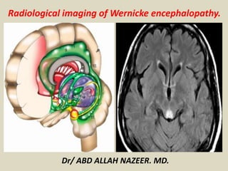
Radiological imaging of Wernicke encephalopathy.
Dr/ ABD ALLAH NAZEER. MD.
Wernicke encephalopathy, also referred as Wernicke-Korsakoff
syndrome, is due to thiamine (vitamin B1) deficiency, and is typically
seen in alcoholics.
On imaging, it is commonly seen on MRI as areas of symmetrical
increased T2/FLAIR signal involving the mammillary bodies,
dorsomedial thalami, tectal plate, periaqueductal area, and around
the third ventricle.
Clinical presentation
It was originally described as characterised by the triad of:
Acute confusion.
Ataxia.
Ophthalmoplegia.
Wernicke encephalopathy can evolve into the chronic form of thiamine
deficiency known as Korsakoff psychosis, characterised by:
Memory loss (global amnesia).
Confabulation.
The two terms are often concatenated to form Wernicke-Korsakoff
syndrome.
Aetiology
Thiamine deficiency results from malnutrition or
malabsorption, which can occur for a number of
reasons:
Alcohol abuse (up to 90% in industrialized countries)
Starvation/fasting.
Prolonged total parental nutrition without
supplementation.
Post bariatric surgery.
Hyperemesis gravidum.
Gastrointestinal malignancy.
Chronic dialysis.
Radiographic features
CT
usually normal
MRI
T2/FLAIR: symmetrically increased signal intensity in the:
mammillary bodies
dorsomedial thalami
tectal plate
periaqueductal area
around the third ventricle
T1 C+ (Gd): contrast enhancement can also be seen in the same
regions, most commonly of the mamillary bodies
DWI/ADC: restricted diffusion can also be seen in the same
regions
MR spectroscopy: may show decreased or normal NAA with the
notable presence of lactate
Midsagittal T2-weighted MR image with gray-scale inversion in healthy 37-
year-old man shows schematic representation of anatomic regions typically
(circles) and infrequently (asterisks) affected by Wernicke's encephalopathy.
Note that caudate capita and dentate nuclei are not seen in this view.
33-year-old man affected by acute Wernicke's encephalopathy caused by severe malnutrition.
A 54-year-old woman with leukemia, changes in consciousness, and ataxia. FLAIR axial images (11,000/140/2
[TR/TE/NEX]). A, The prepositus hypoglossal nuclei show symmetric high-signal-intensity alterations (arrows). B,
The medial vestibular nuclei show symmetric hyperintense lesions (arrows). C, Symmetric high-signal-intensity
alterations in the facial nuclei (arrows) are detected. Subtle signal-intensity alterations in the abducens nuclei are
seen (arrowheads). D, The tectum of the midbrain and the periaqueductal gray matter shows signal-intensity
alterations (arrow). E, The mamillary bodies (arrows) show signal-intensity alterations. F, Note signal-intensity
alterations (arrows) of the medial thalami and periventricular region of the third ventricle.
A 54-year-old woman with a history of food refusal had changes in consciousness. FLAIR coronal
images (11,000/140/2 [TR/TE/NEX]). A, Signal-intensity alterations with different intensity
patterns are seen in the thalami (arrows). Diffuse signal-intensity alterations of the frontal cortex
(arrowheads) are present. B, Note signal-intensity alterations in the mamillary bodies (arrows),
periventricular region of the third ventricle (empty arrows), and brain cortex (arrowheads).
Wernicke encephalopathy
With bilateral thalami
abnormal signal and
restricted diffusion.
CT and MRI images of
35 year-old man with
schizophrenia and
acute nutritional
deficiency-induced
WE.
CT.
T2- weighted late-
echo fast spine echo
image.
Fluid-attenuated
inversion recovery
image.
Diffusion-weighted
image.
Typical MR findings of acute Wernicke Encephalopathy Fluid-attenuated inversion recovery
images presenting symmetric high-signal intensity alterations in the mammillary bodies
and colliculi (a), periventricular gray matter (b), and fornix and thalamus (c).
Wernicke Encephalopathy: MRI in a sixteen weeks pregnant
woman with recent mental status change, repeated vomiting, and
weight loss. Axial FLAIR shows abnormal bright signal.
Hemorrhagic Wernicke encephalopathy
In a patient with liver transplantation.
A, The prepositus hypoglossi nuclei regions show symmetric high signal-intensity alterations (arrows). B, The
abducens nuclei regions show symmetric hyperintense alterations (black arrows). Symmetric high signal-intensity
alterations in the facial nuclei (white arrows) are seen. C, The tectum of the midbrain and the periaqueductal gray
matter demonstrates signal-intensity alterations (black arrows). Focal lesions in the medial lemniscus are seen
bilaterally (white arrows). D, The inferior quadrigeminal plate, the periaqueductal gray matter (black arrows), and
the corticospinal tracts (white arrows) show contrast enhancement. E, Signal-intensity alterations of the mamillary
bodies are detected (white arrows). F and G, Symmetric alterations of the posterior putamen (white arrows) and
periventricular region of the third ventricle (black arrows) are seen on fluid-attenuated inversion recovery and
diffusion-weighted images, respectively. H, Signal-intensity alterations are seen in the motor strip (arrows).
T2-weighted images show
the high signal intensities in
the bilateral paramedian
thalami with mamillary
bodies (A) and in the
periaqueductal gray matter
(B). Diffusion-weighted
images show the bright high
signal intensities in the
corresponding lesions (C and
D). The corresponding
apparent diffusion
coefficient values of the
lesions range from 512 to
545 × 10−6mm2/s.
Changes on MRI with Wernicke's encephalopathy.
Magnetic resonance images in a patient affected by alcoholic Wernicke’s encephalopathy. A-D: MR images
before intravenous administration of thiamine therapy; E-H: MR images after intravenous administration of
thiamine therapy. Axial FLAIR images (A,B) and DWI images (C,D) show signal abnormalities in the
periaqueductal area and in the medial thalami. Axial FLAIR (E,F) and DWI (G,H) follow-up MR images, after
intravenous administration of thiamine therapy, show resolution of the signal abnormalities previously
observed. MR: Magnetic resonance; FLAIR: Fluid-attenuated inversion recovery; DWI: Diffusion weighted image.
Wernicke-Korsakoff syndrome in a nonalcoholic patient: (A) Gadolinium-enhanced T1-weighted axial
MRI shows symmetric enhancement of the mamillary bodies (paired arrowheads). (B) Enlarged axial
view of the region of the hypothalamus showing mamillary body enhancement (paired arrowheads).
(C) Enlarged coronal view of the mamillary body enhancement (paired arrowheads). (D) FLAIR
hyperintensity of the hypothalamus is seen in an axial view (arrows). (E) FLAIR hyperintensity of the
periaqueductal gray (arrows). (F) FLAIR hyperintensity of the dorsomedian thalamus (arrows). (G)
FLAIR hyperintensity of the floor of the fourth ventricle (arrows). (H) FLAIR hyperintensity is seen
throughout the low medulla (arrows).
Alcoholic 65 years-old patient with Wernicke’s encephalopathy. There are symmetrical
hyperintense lesions on these axial T2-WIs in the paraventricular regions of the thalamus
and hypothalamus, periaqueductal regions of the midbrain, and floor of the fourth ventricle
Symmetrical
hyperintense lesions
on these axial FLAIR
images in the
paraventricular
regions of the
thalamus and
hypothalamus and
periaqueductal
regions of the
midbrain.
33-year-old woman, primipara, in her 16th week of gestation: coronal FLAIR (a) and
sagittal T2-weighted (b) MR images showing bilateral and symmetric hyperintense signal
alteration at the level of the medial portion of the thalami and of the tectal plate.
Wernicke encephalopathy in a 29-year-old woman who had severe hyperemesis
gravidarum throughout pregnancy. Axial fluid-attenuated inversion-recovery and Diffusion
weighted MR image shows bilateral symmetrical FLAIR hyperintensity with some diffusion
restriction in dorsomedial thalami, mammillary bodies and periaqueductal grey (arrows)
CONCLUSION.
Wernicke's encephalopathy is characterized
by a quite distinct pattern of MR alterations,
which include symmetrical alterations in the
thalami, mamillary bodies, tectal plate, and
periaqueductal area, but atypical alterations
may also been seen. A thorough knowledge
of the neuroimaging findings of Wernicke's
encephalopathy will assist in arriving at an
early diagnosis, thus reducing the morbidity
and mortality associated with this disease.
Thank You.

More Related Content

What's hot

Presentation1.pptx white matter disorder in pediatric
Presentation1.pptx white matter disorder in pediatricPresentation1.pptx white matter disorder in pediatric
Presentation1.pptx white matter disorder in pediatricAbdellah Nazeer
 
Presentation1.pptx, radiological imaging of cerebral venous thrombosis.
Presentation1.pptx, radiological imaging of cerebral venous thrombosis.Presentation1.pptx, radiological imaging of cerebral venous thrombosis.
Presentation1.pptx, radiological imaging of cerebral venous thrombosis.
Abdellah Nazeer
 
Cp angle tumours
Cp angle tumoursCp angle tumours
Cp angle tumours
Khalidmsayed
 
Cisterns of brain
Cisterns of brainCisterns of brain
Cisterns of brain
suresh Bishokarma
 
imaging in neurology - demyelinating diseases
 imaging in neurology - demyelinating diseases imaging in neurology - demyelinating diseases
imaging in neurology - demyelinating diseases
NeurologyKota
 
Presentation1, radiological imaging of wilson disease.
Presentation1, radiological imaging of wilson disease.Presentation1, radiological imaging of wilson disease.
Presentation1, radiological imaging of wilson disease.
Abdellah Nazeer
 
Dandy-Walker Malformation: Classification and Management
Dandy-Walker Malformation: Classification and ManagementDandy-Walker Malformation: Classification and Management
Dandy-Walker Malformation: Classification and Management
Dr. Shahnawaz Alam
 
Congenital brain anomalies
Congenital brain anomaliesCongenital brain anomalies
Congenital brain anomalies
Thorsang Chayovan
 
Imaging of cns viral infection
Imaging of cns viral infectionImaging of cns viral infection
Imaging of cns viral infection
NeurologyKota
 
Bilateral basal ganglia abnormalities - MRI
Bilateral basal ganglia abnormalities - MRIBilateral basal ganglia abnormalities - MRI
Bilateral basal ganglia abnormalities - MRI
Roshan Valentine
 
Sulcal and gyral neuroanatomy
Sulcal and gyral neuroanatomySulcal and gyral neuroanatomy
Sulcal and gyral neuroanatomy
Navni Garg
 
Imaging in stroke
Imaging in strokeImaging in stroke
Imaging in stroke
ikramdr01
 
Imaging in multiple ring enhancing brain lesions
Imaging in multiple ring enhancing brain lesionsImaging in multiple ring enhancing brain lesions
Imaging in multiple ring enhancing brain lesions
Sumiya Arshad
 
Presentation1.pptx, radiological imaging of intra cranial calcification.
Presentation1.pptx, radiological imaging of intra cranial calcification.Presentation1.pptx, radiological imaging of intra cranial calcification.
Presentation1.pptx, radiological imaging of intra cranial calcification.
Abdellah Nazeer
 
Presentation1, radiological imaging of pediatric leukodystrophy.
Presentation1, radiological imaging of pediatric leukodystrophy.Presentation1, radiological imaging of pediatric leukodystrophy.
Presentation1, radiological imaging of pediatric leukodystrophy.
Abdellah Nazeer
 
Neuroradiology in dementia
Neuroradiology in dementiaNeuroradiology in dementia
Neuroradiology in dementia
NeurologyKota
 
Presentation2.pptx. posterior fossa tumour
Presentation2.pptx. posterior fossa tumourPresentation2.pptx. posterior fossa tumour
Presentation2.pptx. posterior fossa tumourAbdellah Nazeer
 
Presentation2, radiological imaging of phakomatosis.
Presentation2, radiological imaging of phakomatosis.Presentation2, radiological imaging of phakomatosis.
Presentation2, radiological imaging of phakomatosis.
Abdellah Nazeer
 
MRI imaging hypothalamus Dr Ahmed Esawy
MRI imaging hypothalamus Dr Ahmed Esawy MRI imaging hypothalamus Dr Ahmed Esawy
MRI imaging hypothalamus Dr Ahmed Esawy
AHMED ESAWY
 

What's hot (20)

Presentation1.pptx white matter disorder in pediatric
Presentation1.pptx white matter disorder in pediatricPresentation1.pptx white matter disorder in pediatric
Presentation1.pptx white matter disorder in pediatric
 
Presentation1.pptx, radiological imaging of cerebral venous thrombosis.
Presentation1.pptx, radiological imaging of cerebral venous thrombosis.Presentation1.pptx, radiological imaging of cerebral venous thrombosis.
Presentation1.pptx, radiological imaging of cerebral venous thrombosis.
 
Cp angle tumours
Cp angle tumoursCp angle tumours
Cp angle tumours
 
Cisterns of brain
Cisterns of brainCisterns of brain
Cisterns of brain
 
imaging in neurology - demyelinating diseases
 imaging in neurology - demyelinating diseases imaging in neurology - demyelinating diseases
imaging in neurology - demyelinating diseases
 
Presentation1, radiological imaging of wilson disease.
Presentation1, radiological imaging of wilson disease.Presentation1, radiological imaging of wilson disease.
Presentation1, radiological imaging of wilson disease.
 
Dandy-Walker Malformation: Classification and Management
Dandy-Walker Malformation: Classification and ManagementDandy-Walker Malformation: Classification and Management
Dandy-Walker Malformation: Classification and Management
 
Congenital brain anomalies
Congenital brain anomaliesCongenital brain anomalies
Congenital brain anomalies
 
Ring enhancing lesions
Ring enhancing lesionsRing enhancing lesions
Ring enhancing lesions
 
Imaging of cns viral infection
Imaging of cns viral infectionImaging of cns viral infection
Imaging of cns viral infection
 
Bilateral basal ganglia abnormalities - MRI
Bilateral basal ganglia abnormalities - MRIBilateral basal ganglia abnormalities - MRI
Bilateral basal ganglia abnormalities - MRI
 
Sulcal and gyral neuroanatomy
Sulcal and gyral neuroanatomySulcal and gyral neuroanatomy
Sulcal and gyral neuroanatomy
 
Imaging in stroke
Imaging in strokeImaging in stroke
Imaging in stroke
 
Imaging in multiple ring enhancing brain lesions
Imaging in multiple ring enhancing brain lesionsImaging in multiple ring enhancing brain lesions
Imaging in multiple ring enhancing brain lesions
 
Presentation1.pptx, radiological imaging of intra cranial calcification.
Presentation1.pptx, radiological imaging of intra cranial calcification.Presentation1.pptx, radiological imaging of intra cranial calcification.
Presentation1.pptx, radiological imaging of intra cranial calcification.
 
Presentation1, radiological imaging of pediatric leukodystrophy.
Presentation1, radiological imaging of pediatric leukodystrophy.Presentation1, radiological imaging of pediatric leukodystrophy.
Presentation1, radiological imaging of pediatric leukodystrophy.
 
Neuroradiology in dementia
Neuroradiology in dementiaNeuroradiology in dementia
Neuroradiology in dementia
 
Presentation2.pptx. posterior fossa tumour
Presentation2.pptx. posterior fossa tumourPresentation2.pptx. posterior fossa tumour
Presentation2.pptx. posterior fossa tumour
 
Presentation2, radiological imaging of phakomatosis.
Presentation2, radiological imaging of phakomatosis.Presentation2, radiological imaging of phakomatosis.
Presentation2, radiological imaging of phakomatosis.
 
MRI imaging hypothalamus Dr Ahmed Esawy
MRI imaging hypothalamus Dr Ahmed Esawy MRI imaging hypothalamus Dr Ahmed Esawy
MRI imaging hypothalamus Dr Ahmed Esawy
 

Similar to Presentation1, radiological imaging of wernicke encephalopathy.

Marchiafava–Bignami disease (MBD)
Marchiafava–Bignami disease (MBD) Marchiafava–Bignami disease (MBD)
Marchiafava–Bignami disease (MBD) Dr Surendra Khosya
 
Imaging in MESIAL TEMPORAL EPILESPY
Imaging in MESIAL TEMPORAL EPILESPYImaging in MESIAL TEMPORAL EPILESPY
Imaging in MESIAL TEMPORAL EPILESPY
Sarath Menon
 
57 DAVID SUTTON PICTURES INTRACRANIAL LESIONS I
57 DAVID SUTTON PICTURES INTRACRANIAL LESIONS I57 DAVID SUTTON PICTURES INTRACRANIAL LESIONS I
57 DAVID SUTTON PICTURES INTRACRANIAL LESIONS I
Dr. Muhammad Bin Zulfiqar
 
Presentation1, ultrasound examination of the chest.
Presentation1, ultrasound examination of the chest.Presentation1, ultrasound examination of the chest.
Presentation1, ultrasound examination of the chest.
Abdellah Nazeer
 
Case record...Friedreich ataxia
Case record...Friedreich ataxiaCase record...Friedreich ataxia
Case record...Friedreich ataxia
Professor Yasser Metwally
 
Presentation1, radiological film reading of the hip joint.
Presentation1, radiological film reading of the hip joint.Presentation1, radiological film reading of the hip joint.
Presentation1, radiological film reading of the hip joint.
Abdellah Nazeer
 
23205062
2320506223205062
23205062
radgirl
 
Ecr2017 c 0909
Ecr2017 c 0909Ecr2017 c 0909
Ecr2017 c 0909
MarintamaIndra
 
Paraplegia and spinal cord syndromes
Paraplegia and spinal cord syndromesParaplegia and spinal cord syndromes
Paraplegia and spinal cord syndromes
ramtinyoung
 
THE SPINE IN THE COURSE OF SPONDYLOARTHROPATHIES.pptx
THE SPINE IN THE COURSE OF SPONDYLOARTHROPATHIES.pptxTHE SPINE IN THE COURSE OF SPONDYLOARTHROPATHIES.pptx
THE SPINE IN THE COURSE OF SPONDYLOARTHROPATHIES.pptx
kavitjetly
 
Presentation1, radiological imaging of corpus callosum lesios.
Presentation1, radiological imaging of corpus callosum lesios.Presentation1, radiological imaging of corpus callosum lesios.
Presentation1, radiological imaging of corpus callosum lesios.
Abdellah Nazeer
 
Head & neck anatomy and neuroanatomy multiple choice questions
Head & neck anatomy and neuroanatomy multiple choice questionsHead & neck anatomy and neuroanatomy multiple choice questions
Head & neck anatomy and neuroanatomy multiple choice questions
Shubhamm Baldha
 
Figure 3 sign fleischner sign-fissure sign-headcheese sign-hilar overlay sign...
Figure 3 sign fleischner sign-fissure sign-headcheese sign-hilar overlay sign...Figure 3 sign fleischner sign-fissure sign-headcheese sign-hilar overlay sign...
Figure 3 sign fleischner sign-fissure sign-headcheese sign-hilar overlay sign...
Minstry of health ,Ibn alnafis hoapital, Damascus
 
Presentation1, radiological imaging of leigh disease.
Presentation1, radiological imaging of leigh disease.Presentation1, radiological imaging of leigh disease.
Presentation1, radiological imaging of leigh disease.
Abdellah Nazeer
 
Imaging modalities of the petrous bone
Imaging modalities of the petrous boneImaging modalities of the petrous bone
Imaging modalities of the petrous boneAbdellah Nazeer
 
Presentation1 radiological film reading of wrist joint.
Presentation1 radiological film reading of wrist joint.Presentation1 radiological film reading of wrist joint.
Presentation1 radiological film reading of wrist joint.
Abdellah Nazeer
 
Presentation1, radiological imaging of aicardi syndrome.
Presentation1, radiological imaging of aicardi syndrome.Presentation1, radiological imaging of aicardi syndrome.
Presentation1, radiological imaging of aicardi syndrome.
Abdellah Nazeer
 
Congenital lung diseases
Congenital lung diseasesCongenital lung diseases
Congenital lung diseases
Milan Silwal
 

Similar to Presentation1, radiological imaging of wernicke encephalopathy. (20)

Marchiafava–Bignami disease (MBD)
Marchiafava–Bignami disease (MBD) Marchiafava–Bignami disease (MBD)
Marchiafava–Bignami disease (MBD)
 
case report
case reportcase report
case report
 
Imaging in MESIAL TEMPORAL EPILESPY
Imaging in MESIAL TEMPORAL EPILESPYImaging in MESIAL TEMPORAL EPILESPY
Imaging in MESIAL TEMPORAL EPILESPY
 
2016 BILATERAL
2016 BILATERAL 2016 BILATERAL
2016 BILATERAL
 
57 DAVID SUTTON PICTURES INTRACRANIAL LESIONS I
57 DAVID SUTTON PICTURES INTRACRANIAL LESIONS I57 DAVID SUTTON PICTURES INTRACRANIAL LESIONS I
57 DAVID SUTTON PICTURES INTRACRANIAL LESIONS I
 
Presentation1, ultrasound examination of the chest.
Presentation1, ultrasound examination of the chest.Presentation1, ultrasound examination of the chest.
Presentation1, ultrasound examination of the chest.
 
Case record...Friedreich ataxia
Case record...Friedreich ataxiaCase record...Friedreich ataxia
Case record...Friedreich ataxia
 
Presentation1, radiological film reading of the hip joint.
Presentation1, radiological film reading of the hip joint.Presentation1, radiological film reading of the hip joint.
Presentation1, radiological film reading of the hip joint.
 
23205062
2320506223205062
23205062
 
Ecr2017 c 0909
Ecr2017 c 0909Ecr2017 c 0909
Ecr2017 c 0909
 
Paraplegia and spinal cord syndromes
Paraplegia and spinal cord syndromesParaplegia and spinal cord syndromes
Paraplegia and spinal cord syndromes
 
THE SPINE IN THE COURSE OF SPONDYLOARTHROPATHIES.pptx
THE SPINE IN THE COURSE OF SPONDYLOARTHROPATHIES.pptxTHE SPINE IN THE COURSE OF SPONDYLOARTHROPATHIES.pptx
THE SPINE IN THE COURSE OF SPONDYLOARTHROPATHIES.pptx
 
Presentation1, radiological imaging of corpus callosum lesios.
Presentation1, radiological imaging of corpus callosum lesios.Presentation1, radiological imaging of corpus callosum lesios.
Presentation1, radiological imaging of corpus callosum lesios.
 
Head & neck anatomy and neuroanatomy multiple choice questions
Head & neck anatomy and neuroanatomy multiple choice questionsHead & neck anatomy and neuroanatomy multiple choice questions
Head & neck anatomy and neuroanatomy multiple choice questions
 
Figure 3 sign fleischner sign-fissure sign-headcheese sign-hilar overlay sign...
Figure 3 sign fleischner sign-fissure sign-headcheese sign-hilar overlay sign...Figure 3 sign fleischner sign-fissure sign-headcheese sign-hilar overlay sign...
Figure 3 sign fleischner sign-fissure sign-headcheese sign-hilar overlay sign...
 
Presentation1, radiological imaging of leigh disease.
Presentation1, radiological imaging of leigh disease.Presentation1, radiological imaging of leigh disease.
Presentation1, radiological imaging of leigh disease.
 
Imaging modalities of the petrous bone
Imaging modalities of the petrous boneImaging modalities of the petrous bone
Imaging modalities of the petrous bone
 
Presentation1 radiological film reading of wrist joint.
Presentation1 radiological film reading of wrist joint.Presentation1 radiological film reading of wrist joint.
Presentation1 radiological film reading of wrist joint.
 
Presentation1, radiological imaging of aicardi syndrome.
Presentation1, radiological imaging of aicardi syndrome.Presentation1, radiological imaging of aicardi syndrome.
Presentation1, radiological imaging of aicardi syndrome.
 
Congenital lung diseases
Congenital lung diseasesCongenital lung diseases
Congenital lung diseases
 

More from Abdellah Nazeer

Muculoskeletal Pediatic Imaging..pptx
Muculoskeletal Pediatic Imaging..pptxMuculoskeletal Pediatic Imaging..pptx
Muculoskeletal Pediatic Imaging..pptx
Abdellah Nazeer
 
Presentation1, Ultrasound of the bowel loops and the lymph nodes..pptx
Presentation1, Ultrasound of the bowel loops and the lymph nodes..pptxPresentation1, Ultrasound of the bowel loops and the lymph nodes..pptx
Presentation1, Ultrasound of the bowel loops and the lymph nodes..pptx
Abdellah Nazeer
 
Presentation1 Short cases MD..pptx
Presentation1 Short cases MD..pptxPresentation1 Short cases MD..pptx
Presentation1 Short cases MD..pptx
Abdellah Nazeer
 
Presentation1, MD MCQ Cases..pptx
Presentation1, MD MCQ Cases..pptxPresentation1, MD MCQ Cases..pptx
Presentation1, MD MCQ Cases..pptx
Abdellah Nazeer
 
Presentation1, Short Cases Quiz..pptx
Presentation1, Short Cases Quiz..pptxPresentation1, Short Cases Quiz..pptx
Presentation1, Short Cases Quiz..pptx
Abdellah Nazeer
 
Presentation1, radiological imaging of lateral hindfoot impingement.
Presentation1, radiological imaging of lateral hindfoot impingement.Presentation1, radiological imaging of lateral hindfoot impingement.
Presentation1, radiological imaging of lateral hindfoot impingement.
Abdellah Nazeer
 
Presentation2, radiological anatomy of the liver and spleen.
Presentation2, radiological anatomy of the liver and spleen.Presentation2, radiological anatomy of the liver and spleen.
Presentation2, radiological anatomy of the liver and spleen.
Abdellah Nazeer
 
Presentation1, artifacts and pitfalls of the wrist and elbow joints.
Presentation1, artifacts and pitfalls of the wrist and elbow joints.Presentation1, artifacts and pitfalls of the wrist and elbow joints.
Presentation1, artifacts and pitfalls of the wrist and elbow joints.
Abdellah Nazeer
 
Presentation1, artifact and pitfalls of the knee, hip and ankle joints.
Presentation1, artifact and pitfalls of the knee, hip and ankle joints.Presentation1, artifact and pitfalls of the knee, hip and ankle joints.
Presentation1, artifact and pitfalls of the knee, hip and ankle joints.
Abdellah Nazeer
 
Presentation1, radiological imaging of artifact and pitfalls in shoulder join...
Presentation1, radiological imaging of artifact and pitfalls in shoulder join...Presentation1, radiological imaging of artifact and pitfalls in shoulder join...
Presentation1, radiological imaging of artifact and pitfalls in shoulder join...
Abdellah Nazeer
 
Presentation1, radiological imaging of internal abdominal hernia.
Presentation1, radiological imaging of internal abdominal hernia.Presentation1, radiological imaging of internal abdominal hernia.
Presentation1, radiological imaging of internal abdominal hernia.
Abdellah Nazeer
 
Presentation11, radiological imaging of ovarian torsion.
Presentation11, radiological imaging of ovarian torsion.Presentation11, radiological imaging of ovarian torsion.
Presentation11, radiological imaging of ovarian torsion.
Abdellah Nazeer
 
Presentation1, musculoskeletal anatomy.
Presentation1, musculoskeletal anatomy.Presentation1, musculoskeletal anatomy.
Presentation1, musculoskeletal anatomy.
Abdellah Nazeer
 
Presentation1, new mri techniques in the diagnosis and monitoring of multiple...
Presentation1, new mri techniques in the diagnosis and monitoring of multiple...Presentation1, new mri techniques in the diagnosis and monitoring of multiple...
Presentation1, new mri techniques in the diagnosis and monitoring of multiple...
Abdellah Nazeer
 
Presentation1, radiological application of diffusion weighted mri in neck mas...
Presentation1, radiological application of diffusion weighted mri in neck mas...Presentation1, radiological application of diffusion weighted mri in neck mas...
Presentation1, radiological application of diffusion weighted mri in neck mas...
Abdellah Nazeer
 
Presentation1, radiological application of diffusion weighted images in breas...
Presentation1, radiological application of diffusion weighted images in breas...Presentation1, radiological application of diffusion weighted images in breas...
Presentation1, radiological application of diffusion weighted images in breas...
Abdellah Nazeer
 
Presentation1, radiological application of diffusion weighted images in abdom...
Presentation1, radiological application of diffusion weighted images in abdom...Presentation1, radiological application of diffusion weighted images in abdom...
Presentation1, radiological application of diffusion weighted images in abdom...
Abdellah Nazeer
 
Presentation1, radiological application of diffusion weighted imges in neuror...
Presentation1, radiological application of diffusion weighted imges in neuror...Presentation1, radiological application of diffusion weighted imges in neuror...
Presentation1, radiological application of diffusion weighted imges in neuror...
Abdellah Nazeer
 
Presentation1, mr physics.
Presentation1, mr physics.Presentation1, mr physics.
Presentation1, mr physics.
Abdellah Nazeer
 
Presentation1. ct physics.
Presentation1. ct physics.Presentation1. ct physics.
Presentation1. ct physics.
Abdellah Nazeer
 

More from Abdellah Nazeer (20)

Muculoskeletal Pediatic Imaging..pptx
Muculoskeletal Pediatic Imaging..pptxMuculoskeletal Pediatic Imaging..pptx
Muculoskeletal Pediatic Imaging..pptx
 
Presentation1, Ultrasound of the bowel loops and the lymph nodes..pptx
Presentation1, Ultrasound of the bowel loops and the lymph nodes..pptxPresentation1, Ultrasound of the bowel loops and the lymph nodes..pptx
Presentation1, Ultrasound of the bowel loops and the lymph nodes..pptx
 
Presentation1 Short cases MD..pptx
Presentation1 Short cases MD..pptxPresentation1 Short cases MD..pptx
Presentation1 Short cases MD..pptx
 
Presentation1, MD MCQ Cases..pptx
Presentation1, MD MCQ Cases..pptxPresentation1, MD MCQ Cases..pptx
Presentation1, MD MCQ Cases..pptx
 
Presentation1, Short Cases Quiz..pptx
Presentation1, Short Cases Quiz..pptxPresentation1, Short Cases Quiz..pptx
Presentation1, Short Cases Quiz..pptx
 
Presentation1, radiological imaging of lateral hindfoot impingement.
Presentation1, radiological imaging of lateral hindfoot impingement.Presentation1, radiological imaging of lateral hindfoot impingement.
Presentation1, radiological imaging of lateral hindfoot impingement.
 
Presentation2, radiological anatomy of the liver and spleen.
Presentation2, radiological anatomy of the liver and spleen.Presentation2, radiological anatomy of the liver and spleen.
Presentation2, radiological anatomy of the liver and spleen.
 
Presentation1, artifacts and pitfalls of the wrist and elbow joints.
Presentation1, artifacts and pitfalls of the wrist and elbow joints.Presentation1, artifacts and pitfalls of the wrist and elbow joints.
Presentation1, artifacts and pitfalls of the wrist and elbow joints.
 
Presentation1, artifact and pitfalls of the knee, hip and ankle joints.
Presentation1, artifact and pitfalls of the knee, hip and ankle joints.Presentation1, artifact and pitfalls of the knee, hip and ankle joints.
Presentation1, artifact and pitfalls of the knee, hip and ankle joints.
 
Presentation1, radiological imaging of artifact and pitfalls in shoulder join...
Presentation1, radiological imaging of artifact and pitfalls in shoulder join...Presentation1, radiological imaging of artifact and pitfalls in shoulder join...
Presentation1, radiological imaging of artifact and pitfalls in shoulder join...
 
Presentation1, radiological imaging of internal abdominal hernia.
Presentation1, radiological imaging of internal abdominal hernia.Presentation1, radiological imaging of internal abdominal hernia.
Presentation1, radiological imaging of internal abdominal hernia.
 
Presentation11, radiological imaging of ovarian torsion.
Presentation11, radiological imaging of ovarian torsion.Presentation11, radiological imaging of ovarian torsion.
Presentation11, radiological imaging of ovarian torsion.
 
Presentation1, musculoskeletal anatomy.
Presentation1, musculoskeletal anatomy.Presentation1, musculoskeletal anatomy.
Presentation1, musculoskeletal anatomy.
 
Presentation1, new mri techniques in the diagnosis and monitoring of multiple...
Presentation1, new mri techniques in the diagnosis and monitoring of multiple...Presentation1, new mri techniques in the diagnosis and monitoring of multiple...
Presentation1, new mri techniques in the diagnosis and monitoring of multiple...
 
Presentation1, radiological application of diffusion weighted mri in neck mas...
Presentation1, radiological application of diffusion weighted mri in neck mas...Presentation1, radiological application of diffusion weighted mri in neck mas...
Presentation1, radiological application of diffusion weighted mri in neck mas...
 
Presentation1, radiological application of diffusion weighted images in breas...
Presentation1, radiological application of diffusion weighted images in breas...Presentation1, radiological application of diffusion weighted images in breas...
Presentation1, radiological application of diffusion weighted images in breas...
 
Presentation1, radiological application of diffusion weighted images in abdom...
Presentation1, radiological application of diffusion weighted images in abdom...Presentation1, radiological application of diffusion weighted images in abdom...
Presentation1, radiological application of diffusion weighted images in abdom...
 
Presentation1, radiological application of diffusion weighted imges in neuror...
Presentation1, radiological application of diffusion weighted imges in neuror...Presentation1, radiological application of diffusion weighted imges in neuror...
Presentation1, radiological application of diffusion weighted imges in neuror...
 
Presentation1, mr physics.
Presentation1, mr physics.Presentation1, mr physics.
Presentation1, mr physics.
 
Presentation1. ct physics.
Presentation1. ct physics.Presentation1. ct physics.
Presentation1. ct physics.
 

Recently uploaded

LAMPIRAN A - research proposal (oral CANCER).docx
LAMPIRAN A - research proposal (oral CANCER).docxLAMPIRAN A - research proposal (oral CANCER).docx
LAMPIRAN A - research proposal (oral CANCER).docx
Azreen Aj
 
Fundamental of Radiobiology -SABBU.pptx
Fundamental of Radiobiology  -SABBU.pptxFundamental of Radiobiology  -SABBU.pptx
Fundamental of Radiobiology -SABBU.pptx
Sabbu Khatoon
 
Questionnaire Oral Cancer research - in English.docx
Questionnaire  Oral Cancer research - in English.docxQuestionnaire  Oral Cancer research - in English.docx
Questionnaire Oral Cancer research - in English.docx
Azreen Aj
 
DECIPHERING COMMON ECG FINDINGS IN ED.pptx
DECIPHERING COMMON ECG FINDINGS IN ED.pptxDECIPHERING COMMON ECG FINDINGS IN ED.pptx
DECIPHERING COMMON ECG FINDINGS IN ED.pptx
drwaque
 
Couples presenting to the infertility clinic- Do they really have infertility...
Couples presenting to the infertility clinic- Do they really have infertility...Couples presenting to the infertility clinic- Do they really have infertility...
Couples presenting to the infertility clinic- Do they really have infertility...
Sujoy Dasgupta
 
HOT NEW PRODUCT! BIG SALES FAST SHIPPING NOW FROM CHINA!! EU KU DB BK substit...
HOT NEW PRODUCT! BIG SALES FAST SHIPPING NOW FROM CHINA!! EU KU DB BK substit...HOT NEW PRODUCT! BIG SALES FAST SHIPPING NOW FROM CHINA!! EU KU DB BK substit...
HOT NEW PRODUCT! BIG SALES FAST SHIPPING NOW FROM CHINA!! EU KU DB BK substit...
GL Anaacs
 
POST OPERATIVE OLIGURIA and its management
POST OPERATIVE OLIGURIA and its managementPOST OPERATIVE OLIGURIA and its management
POST OPERATIVE OLIGURIA and its management
touseefaziz1
 
Maxilla, Mandible & Hyoid Bone & Clinical Correlations by Dr. RIG.pptx
Maxilla, Mandible & Hyoid Bone & Clinical Correlations by Dr. RIG.pptxMaxilla, Mandible & Hyoid Bone & Clinical Correlations by Dr. RIG.pptx
Maxilla, Mandible & Hyoid Bone & Clinical Correlations by Dr. RIG.pptx
Dr. Rabia Inam Gandapore
 
hypertensive-disorders-of-pregnancy.pptx
hypertensive-disorders-of-pregnancy.pptxhypertensive-disorders-of-pregnancy.pptx
hypertensive-disorders-of-pregnancy.pptx
Dr. Rahul Shah
 
Desidustat the rise of a new dawn - role in treatment of ckd anemia
Desidustat the rise of a new dawn - role in treatment of ckd anemiaDesidustat the rise of a new dawn - role in treatment of ckd anemia
Desidustat the rise of a new dawn - role in treatment of ckd anemia
Dr. Lalit Agarwal
 
EATING DISORDERS (Psychiatry-7)by dr Shivam sharma.pptx
EATING DISORDERS (Psychiatry-7)by dr Shivam sharma.pptxEATING DISORDERS (Psychiatry-7)by dr Shivam sharma.pptx
EATING DISORDERS (Psychiatry-7)by dr Shivam sharma.pptx
Shivam Sharma
 
How to Give Better Lectures: Some Tips for Doctors
How to Give Better Lectures: Some Tips for DoctorsHow to Give Better Lectures: Some Tips for Doctors
How to Give Better Lectures: Some Tips for Doctors
LanceCatedral
 
Charaka Samhita Sutra Sthana 9 Chapter khuddakachatuspadadhyaya
Charaka Samhita Sutra Sthana 9 Chapter khuddakachatuspadadhyayaCharaka Samhita Sutra Sthana 9 Chapter khuddakachatuspadadhyaya
Charaka Samhita Sutra Sthana 9 Chapter khuddakachatuspadadhyaya
Dr KHALID B.M
 
micro teaching on communication m.sc nursing.pdf
micro teaching on communication m.sc nursing.pdfmicro teaching on communication m.sc nursing.pdf
micro teaching on communication m.sc nursing.pdf
Anurag Sharma
 
Hemodialysis: Chapter 3, Dialysis Water Unit - Dr.Gawad
Hemodialysis: Chapter 3, Dialysis Water Unit - Dr.GawadHemodialysis: Chapter 3, Dialysis Water Unit - Dr.Gawad
Hemodialysis: Chapter 3, Dialysis Water Unit - Dr.Gawad
NephroTube - Dr.Gawad
 
Temporal, Infratemporal & Pterygopalatine BY Dr.RIG.pptx
Temporal, Infratemporal & Pterygopalatine BY Dr.RIG.pptxTemporal, Infratemporal & Pterygopalatine BY Dr.RIG.pptx
Temporal, Infratemporal & Pterygopalatine BY Dr.RIG.pptx
Dr. Rabia Inam Gandapore
 
Ophthalmology Clinical Tests for OSCE exam
Ophthalmology Clinical Tests for OSCE examOphthalmology Clinical Tests for OSCE exam
Ophthalmology Clinical Tests for OSCE exam
KafrELShiekh University
 
Mastering Wealth: A Path to Financial Freedom
Mastering Wealth: A Path to Financial FreedomMastering Wealth: A Path to Financial Freedom
Mastering Wealth: A Path to Financial Freedom
FatimaMary4
 
Lung Cancer: Artificial Intelligence, Synergetics, Complex System Analysis, S...
Lung Cancer: Artificial Intelligence, Synergetics, Complex System Analysis, S...Lung Cancer: Artificial Intelligence, Synergetics, Complex System Analysis, S...
Lung Cancer: Artificial Intelligence, Synergetics, Complex System Analysis, S...
Oleg Kshivets
 
Report Back from SGO 2024: What’s the Latest in Cervical Cancer?
Report Back from SGO 2024: What’s the Latest in Cervical Cancer?Report Back from SGO 2024: What’s the Latest in Cervical Cancer?
Report Back from SGO 2024: What’s the Latest in Cervical Cancer?
bkling
 

Recently uploaded (20)

LAMPIRAN A - research proposal (oral CANCER).docx
LAMPIRAN A - research proposal (oral CANCER).docxLAMPIRAN A - research proposal (oral CANCER).docx
LAMPIRAN A - research proposal (oral CANCER).docx
 
Fundamental of Radiobiology -SABBU.pptx
Fundamental of Radiobiology  -SABBU.pptxFundamental of Radiobiology  -SABBU.pptx
Fundamental of Radiobiology -SABBU.pptx
 
Questionnaire Oral Cancer research - in English.docx
Questionnaire  Oral Cancer research - in English.docxQuestionnaire  Oral Cancer research - in English.docx
Questionnaire Oral Cancer research - in English.docx
 
DECIPHERING COMMON ECG FINDINGS IN ED.pptx
DECIPHERING COMMON ECG FINDINGS IN ED.pptxDECIPHERING COMMON ECG FINDINGS IN ED.pptx
DECIPHERING COMMON ECG FINDINGS IN ED.pptx
 
Couples presenting to the infertility clinic- Do they really have infertility...
Couples presenting to the infertility clinic- Do they really have infertility...Couples presenting to the infertility clinic- Do they really have infertility...
Couples presenting to the infertility clinic- Do they really have infertility...
 
HOT NEW PRODUCT! BIG SALES FAST SHIPPING NOW FROM CHINA!! EU KU DB BK substit...
HOT NEW PRODUCT! BIG SALES FAST SHIPPING NOW FROM CHINA!! EU KU DB BK substit...HOT NEW PRODUCT! BIG SALES FAST SHIPPING NOW FROM CHINA!! EU KU DB BK substit...
HOT NEW PRODUCT! BIG SALES FAST SHIPPING NOW FROM CHINA!! EU KU DB BK substit...
 
POST OPERATIVE OLIGURIA and its management
POST OPERATIVE OLIGURIA and its managementPOST OPERATIVE OLIGURIA and its management
POST OPERATIVE OLIGURIA and its management
 
Maxilla, Mandible & Hyoid Bone & Clinical Correlations by Dr. RIG.pptx
Maxilla, Mandible & Hyoid Bone & Clinical Correlations by Dr. RIG.pptxMaxilla, Mandible & Hyoid Bone & Clinical Correlations by Dr. RIG.pptx
Maxilla, Mandible & Hyoid Bone & Clinical Correlations by Dr. RIG.pptx
 
hypertensive-disorders-of-pregnancy.pptx
hypertensive-disorders-of-pregnancy.pptxhypertensive-disorders-of-pregnancy.pptx
hypertensive-disorders-of-pregnancy.pptx
 
Desidustat the rise of a new dawn - role in treatment of ckd anemia
Desidustat the rise of a new dawn - role in treatment of ckd anemiaDesidustat the rise of a new dawn - role in treatment of ckd anemia
Desidustat the rise of a new dawn - role in treatment of ckd anemia
 
EATING DISORDERS (Psychiatry-7)by dr Shivam sharma.pptx
EATING DISORDERS (Psychiatry-7)by dr Shivam sharma.pptxEATING DISORDERS (Psychiatry-7)by dr Shivam sharma.pptx
EATING DISORDERS (Psychiatry-7)by dr Shivam sharma.pptx
 
How to Give Better Lectures: Some Tips for Doctors
How to Give Better Lectures: Some Tips for DoctorsHow to Give Better Lectures: Some Tips for Doctors
How to Give Better Lectures: Some Tips for Doctors
 
Charaka Samhita Sutra Sthana 9 Chapter khuddakachatuspadadhyaya
Charaka Samhita Sutra Sthana 9 Chapter khuddakachatuspadadhyayaCharaka Samhita Sutra Sthana 9 Chapter khuddakachatuspadadhyaya
Charaka Samhita Sutra Sthana 9 Chapter khuddakachatuspadadhyaya
 
micro teaching on communication m.sc nursing.pdf
micro teaching on communication m.sc nursing.pdfmicro teaching on communication m.sc nursing.pdf
micro teaching on communication m.sc nursing.pdf
 
Hemodialysis: Chapter 3, Dialysis Water Unit - Dr.Gawad
Hemodialysis: Chapter 3, Dialysis Water Unit - Dr.GawadHemodialysis: Chapter 3, Dialysis Water Unit - Dr.Gawad
Hemodialysis: Chapter 3, Dialysis Water Unit - Dr.Gawad
 
Temporal, Infratemporal & Pterygopalatine BY Dr.RIG.pptx
Temporal, Infratemporal & Pterygopalatine BY Dr.RIG.pptxTemporal, Infratemporal & Pterygopalatine BY Dr.RIG.pptx
Temporal, Infratemporal & Pterygopalatine BY Dr.RIG.pptx
 
Ophthalmology Clinical Tests for OSCE exam
Ophthalmology Clinical Tests for OSCE examOphthalmology Clinical Tests for OSCE exam
Ophthalmology Clinical Tests for OSCE exam
 
Mastering Wealth: A Path to Financial Freedom
Mastering Wealth: A Path to Financial FreedomMastering Wealth: A Path to Financial Freedom
Mastering Wealth: A Path to Financial Freedom
 
Lung Cancer: Artificial Intelligence, Synergetics, Complex System Analysis, S...
Lung Cancer: Artificial Intelligence, Synergetics, Complex System Analysis, S...Lung Cancer: Artificial Intelligence, Synergetics, Complex System Analysis, S...
Lung Cancer: Artificial Intelligence, Synergetics, Complex System Analysis, S...
 
Report Back from SGO 2024: What’s the Latest in Cervical Cancer?
Report Back from SGO 2024: What’s the Latest in Cervical Cancer?Report Back from SGO 2024: What’s the Latest in Cervical Cancer?
Report Back from SGO 2024: What’s the Latest in Cervical Cancer?
 

Presentation1, radiological imaging of wernicke encephalopathy.

  • 1. Radiological imaging of Wernicke encephalopathy. Dr/ ABD ALLAH NAZEER. MD.
  • 2. Wernicke encephalopathy, also referred as Wernicke-Korsakoff syndrome, is due to thiamine (vitamin B1) deficiency, and is typically seen in alcoholics. On imaging, it is commonly seen on MRI as areas of symmetrical increased T2/FLAIR signal involving the mammillary bodies, dorsomedial thalami, tectal plate, periaqueductal area, and around the third ventricle. Clinical presentation It was originally described as characterised by the triad of: Acute confusion. Ataxia. Ophthalmoplegia. Wernicke encephalopathy can evolve into the chronic form of thiamine deficiency known as Korsakoff psychosis, characterised by: Memory loss (global amnesia). Confabulation. The two terms are often concatenated to form Wernicke-Korsakoff syndrome.
  • 3. Aetiology Thiamine deficiency results from malnutrition or malabsorption, which can occur for a number of reasons: Alcohol abuse (up to 90% in industrialized countries) Starvation/fasting. Prolonged total parental nutrition without supplementation. Post bariatric surgery. Hyperemesis gravidum. Gastrointestinal malignancy. Chronic dialysis.
  • 4. Radiographic features CT usually normal MRI T2/FLAIR: symmetrically increased signal intensity in the: mammillary bodies dorsomedial thalami tectal plate periaqueductal area around the third ventricle T1 C+ (Gd): contrast enhancement can also be seen in the same regions, most commonly of the mamillary bodies DWI/ADC: restricted diffusion can also be seen in the same regions MR spectroscopy: may show decreased or normal NAA with the notable presence of lactate
  • 5. Midsagittal T2-weighted MR image with gray-scale inversion in healthy 37- year-old man shows schematic representation of anatomic regions typically (circles) and infrequently (asterisks) affected by Wernicke's encephalopathy. Note that caudate capita and dentate nuclei are not seen in this view.
  • 6. 33-year-old man affected by acute Wernicke's encephalopathy caused by severe malnutrition.
  • 7. A 54-year-old woman with leukemia, changes in consciousness, and ataxia. FLAIR axial images (11,000/140/2 [TR/TE/NEX]). A, The prepositus hypoglossal nuclei show symmetric high-signal-intensity alterations (arrows). B, The medial vestibular nuclei show symmetric hyperintense lesions (arrows). C, Symmetric high-signal-intensity alterations in the facial nuclei (arrows) are detected. Subtle signal-intensity alterations in the abducens nuclei are seen (arrowheads). D, The tectum of the midbrain and the periaqueductal gray matter shows signal-intensity alterations (arrow). E, The mamillary bodies (arrows) show signal-intensity alterations. F, Note signal-intensity alterations (arrows) of the medial thalami and periventricular region of the third ventricle.
  • 8. A 54-year-old woman with a history of food refusal had changes in consciousness. FLAIR coronal images (11,000/140/2 [TR/TE/NEX]). A, Signal-intensity alterations with different intensity patterns are seen in the thalami (arrows). Diffuse signal-intensity alterations of the frontal cortex (arrowheads) are present. B, Note signal-intensity alterations in the mamillary bodies (arrows), periventricular region of the third ventricle (empty arrows), and brain cortex (arrowheads).
  • 9. Wernicke encephalopathy With bilateral thalami abnormal signal and restricted diffusion.
  • 10. CT and MRI images of 35 year-old man with schizophrenia and acute nutritional deficiency-induced WE. CT. T2- weighted late- echo fast spine echo image. Fluid-attenuated inversion recovery image. Diffusion-weighted image.
  • 11. Typical MR findings of acute Wernicke Encephalopathy Fluid-attenuated inversion recovery images presenting symmetric high-signal intensity alterations in the mammillary bodies and colliculi (a), periventricular gray matter (b), and fornix and thalamus (c).
  • 12. Wernicke Encephalopathy: MRI in a sixteen weeks pregnant woman with recent mental status change, repeated vomiting, and weight loss. Axial FLAIR shows abnormal bright signal.
  • 13. Hemorrhagic Wernicke encephalopathy In a patient with liver transplantation.
  • 14. A, The prepositus hypoglossi nuclei regions show symmetric high signal-intensity alterations (arrows). B, The abducens nuclei regions show symmetric hyperintense alterations (black arrows). Symmetric high signal-intensity alterations in the facial nuclei (white arrows) are seen. C, The tectum of the midbrain and the periaqueductal gray matter demonstrates signal-intensity alterations (black arrows). Focal lesions in the medial lemniscus are seen bilaterally (white arrows). D, The inferior quadrigeminal plate, the periaqueductal gray matter (black arrows), and the corticospinal tracts (white arrows) show contrast enhancement. E, Signal-intensity alterations of the mamillary bodies are detected (white arrows). F and G, Symmetric alterations of the posterior putamen (white arrows) and periventricular region of the third ventricle (black arrows) are seen on fluid-attenuated inversion recovery and diffusion-weighted images, respectively. H, Signal-intensity alterations are seen in the motor strip (arrows).
  • 15. T2-weighted images show the high signal intensities in the bilateral paramedian thalami with mamillary bodies (A) and in the periaqueductal gray matter (B). Diffusion-weighted images show the bright high signal intensities in the corresponding lesions (C and D). The corresponding apparent diffusion coefficient values of the lesions range from 512 to 545 × 10−6mm2/s.
  • 16. Changes on MRI with Wernicke's encephalopathy.
  • 17. Magnetic resonance images in a patient affected by alcoholic Wernicke’s encephalopathy. A-D: MR images before intravenous administration of thiamine therapy; E-H: MR images after intravenous administration of thiamine therapy. Axial FLAIR images (A,B) and DWI images (C,D) show signal abnormalities in the periaqueductal area and in the medial thalami. Axial FLAIR (E,F) and DWI (G,H) follow-up MR images, after intravenous administration of thiamine therapy, show resolution of the signal abnormalities previously observed. MR: Magnetic resonance; FLAIR: Fluid-attenuated inversion recovery; DWI: Diffusion weighted image.
  • 18. Wernicke-Korsakoff syndrome in a nonalcoholic patient: (A) Gadolinium-enhanced T1-weighted axial MRI shows symmetric enhancement of the mamillary bodies (paired arrowheads). (B) Enlarged axial view of the region of the hypothalamus showing mamillary body enhancement (paired arrowheads). (C) Enlarged coronal view of the mamillary body enhancement (paired arrowheads). (D) FLAIR hyperintensity of the hypothalamus is seen in an axial view (arrows). (E) FLAIR hyperintensity of the periaqueductal gray (arrows). (F) FLAIR hyperintensity of the dorsomedian thalamus (arrows). (G) FLAIR hyperintensity of the floor of the fourth ventricle (arrows). (H) FLAIR hyperintensity is seen throughout the low medulla (arrows).
  • 19.
  • 20. Alcoholic 65 years-old patient with Wernicke’s encephalopathy. There are symmetrical hyperintense lesions on these axial T2-WIs in the paraventricular regions of the thalamus and hypothalamus, periaqueductal regions of the midbrain, and floor of the fourth ventricle
  • 21. Symmetrical hyperintense lesions on these axial FLAIR images in the paraventricular regions of the thalamus and hypothalamus and periaqueductal regions of the midbrain.
  • 22. 33-year-old woman, primipara, in her 16th week of gestation: coronal FLAIR (a) and sagittal T2-weighted (b) MR images showing bilateral and symmetric hyperintense signal alteration at the level of the medial portion of the thalami and of the tectal plate.
  • 23. Wernicke encephalopathy in a 29-year-old woman who had severe hyperemesis gravidarum throughout pregnancy. Axial fluid-attenuated inversion-recovery and Diffusion weighted MR image shows bilateral symmetrical FLAIR hyperintensity with some diffusion restriction in dorsomedial thalami, mammillary bodies and periaqueductal grey (arrows)
  • 24. CONCLUSION. Wernicke's encephalopathy is characterized by a quite distinct pattern of MR alterations, which include symmetrical alterations in the thalami, mamillary bodies, tectal plate, and periaqueductal area, but atypical alterations may also been seen. A thorough knowledge of the neuroimaging findings of Wernicke's encephalopathy will assist in arriving at an early diagnosis, thus reducing the morbidity and mortality associated with this disease.