Organogenesis is the process by which the three germ layers (ectoderm, endoderm, and mesoderm) form the internal organs. The development of the eye occurs through interactions between the lens placode and optic vesicle. The optic vesicle induces formation of the lens placode and positions the lens in relation to the retina. The optic vesicle then becomes the optic cup with two layers that differentiate into the pigmented retina and neural retina. The lens placode invaginates to form the lens.
cell commitment and differentiation, stem cell,types of differentiationshallu kotwal
The commitment of cells to specific cell fates and their capacity to differentiate into particular kinds of cells.
Cellular differentiation is the process in which a cell changes from one cell type to another. Usually, the cell changes to a more specialized type. Differentiation occurs numerous times during the development of a multicellular organism as it changes from a simple zygote to a complex system of tissues and cell types. Differentiation continues in adulthood as adult stem cells divide and create fully differentiated daughter cells during tissue repair and during normal cell turnover.
Vittelogenesis is a word developed from Latin vitellus-yolk, and genero-produce
Vitellogenesis (also known as yolk deposition) is the process of yolk formation via nutrients being deposited in the oocyte, or female germ cell involved in reproduction of lecithotrophic organisms. In insects, it starts when the fat body stimulates the release of juvenile hormones and produces vitellogenin protein.
Yolks is the most usual form of food storage in the egg.
Yolks appear in the oocyte in the secondary period of their growth called vittelogenesis.
Thus,the formation and deposition of yolks is known as vittelogenesis
Characteristic
Yolks is a complex variable assembled component.
The principle component are protein,phospholipid and fats in different combination.
Depending upon these component yolks is distinguished into protein yolks and fatty acid
For eg- the avian contain 48.19% water , 16.6 % protein, 32.6% phospholipids and fats and 1% carbohydrates.
How 3 germ layers are formed in Chick that are endoderm, mesoderm and ectoderm.As Chick are polylecithal so cell movements are somewhat restricted and gastrulation is modified as compared to frog.
anatomical consideration of development of eye from embryonic stage. gives insight into future anatomical and pharmacological basis of drug development in disorders of eye.
Eye development starts from 22nd day of gestation when the embryo is about 2 mm in length it is 8 somite stage. The eyeball and its related structures are derived from some primordial.
Eyeball is derived from three embryonic layers. These layers include: 1. surface ectoderm 2. neural ectoderm 3. mesoderm
he sense organs — eyes, ears, tongue, skin, and nose — help to protect the body. The human sense organs contain receptors that relay information through sensory neurons to the appropriate places within the nervous system.
Each sense organ contains different receptors.
General receptors are found throughout the body because they are present in skin, visceral organs (visceral meaning in the abdominal cavity), muscles, and joints.
Special receptors include chemoreceptors (chemical receptors) found in the mouth and nose, photoreceptors (light receptors) found in the eyes, and mechanoreceptors found in the ears.
The ability to recreate computational results with minimal effort and actionable metrics provides a solid foundation for scientific research and software development. When people can replicate an analysis at the touch of a button using open-source software, open data, and methods to assess and compare proposals, it significantly eases verification of results, engagement with a diverse range of contributors, and progress. However, we have yet to fully achieve this; there are still many sociotechnical frictions.
Inspired by David Donoho's vision, this talk aims to revisit the three crucial pillars of frictionless reproducibility (data sharing, code sharing, and competitive challenges) with the perspective of deep software variability.
Our observation is that multiple layers — hardware, operating systems, third-party libraries, software versions, input data, compile-time options, and parameters — are subject to variability that exacerbates frictions but is also essential for achieving robust, generalizable results and fostering innovation. I will first review the literature, providing evidence of how the complex variability interactions across these layers affect qualitative and quantitative software properties, thereby complicating the reproduction and replication of scientific studies in various fields.
I will then present some software engineering and AI techniques that can support the strategic exploration of variability spaces. These include the use of abstractions and models (e.g., feature models), sampling strategies (e.g., uniform, random), cost-effective measurements (e.g., incremental build of software configurations), and dimensionality reduction methods (e.g., transfer learning, feature selection, software debloating).
I will finally argue that deep variability is both the problem and solution of frictionless reproducibility, calling the software science community to develop new methods and tools to manage variability and foster reproducibility in software systems.
Exposé invité Journées Nationales du GDR GPL 2024
Richard's aventures in two entangled wonderlandsRichard Gill
Since the loophole-free Bell experiments of 2020 and the Nobel prizes in physics of 2022, critics of Bell's work have retreated to the fortress of super-determinism. Now, super-determinism is a derogatory word - it just means "determinism". Palmer, Hance and Hossenfelder argue that quantum mechanics and determinism are not incompatible, using a sophisticated mathematical construction based on a subtle thinning of allowed states and measurements in quantum mechanics, such that what is left appears to make Bell's argument fail, without altering the empirical predictions of quantum mechanics. I think however that it is a smoke screen, and the slogan "lost in math" comes to my mind. I will discuss some other recent disproofs of Bell's theorem using the language of causality based on causal graphs. Causal thinking is also central to law and justice. I will mention surprising connections to my work on serial killer nurse cases, in particular the Dutch case of Lucia de Berk and the current UK case of Lucy Letby.
Earliest Galaxies in the JADES Origins Field: Luminosity Function and Cosmic ...Sérgio Sacani
We characterize the earliest galaxy population in the JADES Origins Field (JOF), the deepest
imaging field observed with JWST. We make use of the ancillary Hubble optical images (5 filters
spanning 0.4−0.9µm) and novel JWST images with 14 filters spanning 0.8−5µm, including 7 mediumband filters, and reaching total exposure times of up to 46 hours per filter. We combine all our data
at > 2.3µm to construct an ultradeep image, reaching as deep as ≈ 31.4 AB mag in the stack and
30.3-31.0 AB mag (5σ, r = 0.1” circular aperture) in individual filters. We measure photometric
redshifts and use robust selection criteria to identify a sample of eight galaxy candidates at redshifts
z = 11.5 − 15. These objects show compact half-light radii of R1/2 ∼ 50 − 200pc, stellar masses of
M⋆ ∼ 107−108M⊙, and star-formation rates of SFR ∼ 0.1−1 M⊙ yr−1
. Our search finds no candidates
at 15 < z < 20, placing upper limits at these redshifts. We develop a forward modeling approach to
infer the properties of the evolving luminosity function without binning in redshift or luminosity that
marginalizes over the photometric redshift uncertainty of our candidate galaxies and incorporates the
impact of non-detections. We find a z = 12 luminosity function in good agreement with prior results,
and that the luminosity function normalization and UV luminosity density decline by a factor of ∼ 2.5
from z = 12 to z = 14. We discuss the possible implications of our results in the context of theoretical
models for evolution of the dark matter halo mass function.
Deep Behavioral Phenotyping in Systems Neuroscience for Functional Atlasing a...Ana Luísa Pinho
Functional Magnetic Resonance Imaging (fMRI) provides means to characterize brain activations in response to behavior. However, cognitive neuroscience has been limited to group-level effects referring to the performance of specific tasks. To obtain the functional profile of elementary cognitive mechanisms, the combination of brain responses to many tasks is required. Yet, to date, both structural atlases and parcellation-based activations do not fully account for cognitive function and still present several limitations. Further, they do not adapt overall to individual characteristics. In this talk, I will give an account of deep-behavioral phenotyping strategies, namely data-driven methods in large task-fMRI datasets, to optimize functional brain-data collection and improve inference of effects-of-interest related to mental processes. Key to this approach is the employment of fast multi-functional paradigms rich on features that can be well parametrized and, consequently, facilitate the creation of psycho-physiological constructs to be modelled with imaging data. Particular emphasis will be given to music stimuli when studying high-order cognitive mechanisms, due to their ecological nature and quality to enable complex behavior compounded by discrete entities. I will also discuss how deep-behavioral phenotyping and individualized models applied to neuroimaging data can better account for the subject-specific organization of domain-general cognitive systems in the human brain. Finally, the accumulation of functional brain signatures brings the possibility to clarify relationships among tasks and create a univocal link between brain systems and mental functions through: (1) the development of ontologies proposing an organization of cognitive processes; and (2) brain-network taxonomies describing functional specialization. To this end, tools to improve commensurability in cognitive science are necessary, such as public repositories, ontology-based platforms and automated meta-analysis tools. I will thus discuss some brain-atlasing resources currently under development, and their applicability in cognitive as well as clinical neuroscience.
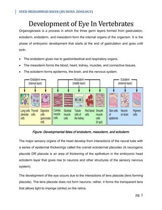
1. SYED MUHAMMAD KHAN (BS HONS. ZOOLOGY)
pg. 1
Development of Eye In Vertebrates
Organogenesis is a process in which the three germ layers formed from gastrulation,
ectoderm, endoderm, and mesoderm form the internal organs of the organism. It is the
phase of embryonic development that starts at the end of gastrulation and goes until
birth.
The endoderm gives rise to gastrointestinal and respiratory organs.
The mesoderm forms the blood, heart, kidney, muscles, and connective tissues.
The ectoderm forms epidermis, the brain, and the nervous system.
Figure: Developmental fates of endoderm, mesoderm, and ectoderm.
The major sensory organs of the head develop from interactions of the neural tube with
a series of epidermal thickenings called the cranial ectodermal placodes (A neurogenic
placode OR placode is an area of thickening of the epithelium in the embryonic head
ectoderm layer that gives rise to neurons and other structures of the sensory nervous
system).
The development of the eye occurs due to the interactions of lens placode (lens forming
placode). The lens placode does not form neurons; rather, it forms the transparent lens
that allows light to impinge (strike) on the retina.
2. SYED MUHAMMAD KHAN (BS HONS. ZOOLOGY)
pg. 2
Development of Eye
At gastrulation, the endoderm and mesoderm interact with the adjacent
prospective/future head ectoderm to give the head ectoderm a lens-forming ability. But
not all parts of the head ectoderm form lenses, the lens must have a precise spatial
(space) relationship with the retina. The optic vesicle activates the head ectoderm's
lens-forming ability and positions the lens in relation to the retina. It induces the
formation of a lens placode, which then invaginates to form the lens.
Figure: Development of eye. (a) Optic vesicle forms in head ectoderm, development of
eye starts. Optic vesicle induces the development of lens placode (which will give rise to
lens). (b) Optic vesicle becomes the optic cup (future retina), which is bi-layered (having
two layers). The outer layer will give rise to pigmented retina, whereas the inner layer
will give rise to neural retina. Lens vesicle forms, this will be converted into lens. (c)
Lens vesicle detaches from the surface epithelium and gets converted into lens. The
two layers of the optic vesicle differentiated into pigmented retina (outer) and neural
retina (inner). The eye is now fully developed.
The optic vesicle becomes the optic cup (future retina); its two layers (outer / pigmented
retina & inner / neural retina) differentiate in different ways:
3. SYED MUHAMMAD KHAN (BS HONS. ZOOLOGY)
pg. 3
Outer Layer / Pigmented Retina: The cells of the outer layer produce melanin pigment
(brown) and ultimately become the pigmented retina.
Inner Layer / Neural Retina: The cells of the inner layer proliferate rapidly (rapid cell
divisions) and generate a variety of glia (Glia/Glial cells/Neuroglia – non-neuronal cells
in the central nervous system), ganglion cells (a type of neuron located near the inner
surface of the retina), interneurons, and light-sensitive photoreceptor neurons.
Collectively, these cells constitute the neural retina.
The retinal ganglion cells are neurons whose axons send electric impulses to the brain.
Their axons meet at the base of the eye and travel down the optic stalk, which is then
called the optic nerve.
Cell Differentiation in Eye
The differentiation of cells of various parts of the eye takes place in the following ways:
(1) Neural Retina Differentiation:
The retinal precursor cells divide before they differentiate. The neural retina develops
into a layered array/arrangement of different neuronal types, these layers include:
1. Rods – Light-sensitive photoreceptor cells
2. Cones – Color-sensitive photoreceptor cells
3. Cell bodies of the ganglion cells
4. Bipolar interneurons that transmit electric stimuli from the rods and cones to the
ganglion cells
5. Numerous Muller glial cells that maintain the retina's integrity (keep it together)
6. Amacrine neurons (which lack large axons)
7. Horizontal neurons that transmit electric impulses in the plane of the retina (same
axis/direction)
4. SYED MUHAMMAD KHAN (BS HONS. ZOOLOGY)
pg. 4
The neuroblasts (neuron/nerve forming cells) of the retina are competent to generate all
seven retinal cell types (that have been mentioned above).
(2) Lens & Cornea Differentiation:
The eye can't focus on the retina unless it has a lens and a cornea.
Cornea Differentiation: Shortly after the lens vesicle has detached from the surface
ectoderm, mesenchymal cells from the neural crest (embryonic structure that gives rise
to the peripheral nervous system) migrate into the space between the lens and the
surface epithelium. These cells condense to form several flat layers of cells, which
become the corneal precursor cells. As these cells mature, they dehydrate and form
tight junctions among the cells, forming the cornea. Fluid pressure from the aqueous
humor (transparent fluid between lens and cornea) is necessary for the correct
curvature of the cornea.
Lens Differentiation: The differentiation of the lens tissue into a transparent membrane
capable of directing light onto the retina involves changes in cell structure and shape as
well as the synthesis of transparent, lens-specific proteins called crystallins. The cells at
the inner portion of the lens vesicle elongate and, under the influence of the neural
retina, become the lens fibers. As these fibers continue to grow, they synthesize
crystallins (transparent and lens-specific proteins), which eventually fill up the cell and
cause the extrusion of the nucleus (it is thrown out of the cell). The lens contains three
regions: an anterior zone of dividing epithelial cells, an equatorial zone (middle) of
cellular elongation, and a posterior and central zone of crystallin-containing fiber cells.
This arrangement persists throughout the lifetime of the animal.