
Recommended
More Related Content
What's hot
What's hot (20)
Similar to Retina embryology ppt
Similar to Retina embryology ppt (20)
Recently uploaded
Recently uploaded (20)
Retina embryology ppt
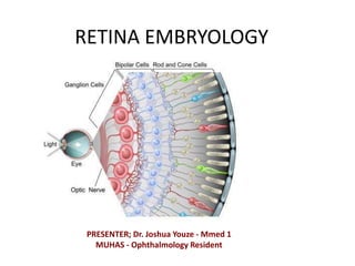
- 1. RETINA EMBRYOLOGY PRESENTER; Dr. Joshua Youze - Mmed 1 MUHAS - Ophthalmology Resident
- 2. INTRO… • The retina innermost and light sensitive of the three tunics of the eye, surrounding the vitreous body and continuous posteriorly with the optic nerve. – It consists of 10 layers.
- 4. Embryology • The embryology of the human eye is 1st seen in the 22nd day of the intra uteral life, as bilateral evagination of the neuroectoderm of the forehead. – To be precise the diencephalon.
- 6. • The bilateral envagination of the diencephalon give rise to the optic groove or optic sulci. • The groove keeps on growing to form the optic vesicles, which grows towards the ectoderm.
- 8. • At 29th day the optic vesicles, moves laterally towards the ectoderm. As they grow towards the ectoderm from the forebrain, they become attenuated to form optic stalk which will later become optic nerve. • When they reach the ectoderm the induction of the formation of the lens primodia is induced
- 10. • At 33 day the optic disc start to invaginate, forming optic cup. The ends of the optic disc remains not fused to create choroidal fissure. • The choroidal fissure transmit the Hyaloid artery and vein which later will become the central retina artery and Vein after the closure of the fissure.
- 13. • The choroidal fissure will later on fuse completely and enclose the hyaloid artery and vein in the canal within the optic stalk. • When the lens is fully formed in intrauterine the distal hyaloid artery will disintegrate and the proximal part will remain to form the central retina artery.
- 15. • The ectoderm which is anterior to the invagination enlarge and detach into the interior part of the optic cup to form the primary lens which will be floating on one end of the optic cup. • The space between the lens and the cup is filled by the mesenchyme tissue which is the primary vitreous body. LENS.
- 18. • The hollow ball become indented and the 2 layers of the cup formed(external and internal retinal layers) separated by the intraretinal space which is continuous with the optic stalk and 3rd ventricle. • The two layers are of different size as the outer being thinner than the inner.
- 20. • The outer layer become pigmented layer and the inner layer become neural layer • The two layers are separated by the intraretinal space. • The optic cup is the divided into the anterior 1/5 and 4/5 posterior.
- 22. The anterior and posterior retina
- 23. • The anterior 1/5 will later differentiate to the ciliary body and the iris. • The cells immediately to the intraretinal layer will begin differentiate to the photoreceptors. • The next cells to muller supporting cells and the bipolar neurons, and the innermost superficial to become the axons of the ganglion which will become optic nerve.
- 25. • At 8 months all layers of the retina are seen histologically, but maturation of the photoreceptors continues after birth. – It explains why visual acuity of the baby improves as the child grows.
- 26. Summary • Evagination • Formation of the optic vesicles • Vesicles moves towards the ectoderm • Formation of the lens primodia • Invagination and formation of the optic cup. • Differentiation of the optic cup layers to form the retina