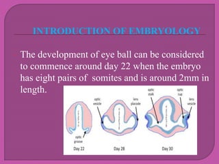
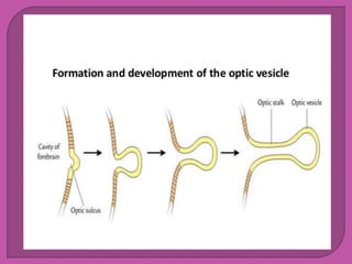
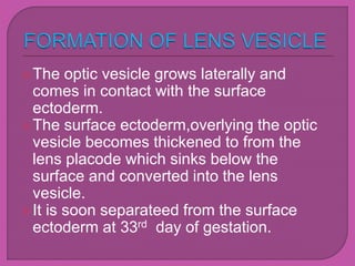
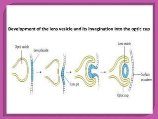
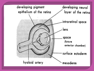
PRESENTED BY
Payal Sahu, Bikrant yadav,
Omprakash, Dhaneshwar, Nadeem
B.Optometry 2017 batch
ITM University Naya Raipur, C.G.
Eye formation in the human embryo begins
at approximiately three weeks into
embryonic development and continues
through the tenth week
cells from both the mesodermal and the
ectodermal tissues contribute to the
formation of the eye
specially,the eye is derived from the
neuroepithelium surface ectoderm and the
extracellular mesenchyme
INTRODUCTION OF EMBRYOLOGY
 The development of eye ball can be considered
to commence around day 22 when the embryo
has eight pairs of somites and is around 2mm in
length.
The eye ball and its related structure
derived following primordia.
Optic vesicle:-outer growth form
prosencephalon (a neuroectodermal
structure)
Lens placode:- a specilised area of surface
ectoderm.
Mesenchyme:-surrounding the optic
vessels
The area of neural plate which forms the
 procencephalon
develops a linear thickened area on either
side(which soon becomes depressed to
form optic sulcus)
Meanwhile neural plate gets converted into
prosencephalic vesicle
As the optic sulcus deepens,the wall of the
prosencephalon overlying the sulcus
buldge out to form the Optic vesicle.
The proximal part of the vesicle becomes
constricted and elongated to form the optic
stalk.
The optic vesicle grows laterally and
comes in contact with the surface
ectoderm.
The surface ectoderm,overlying the optic
vesicle becomes thickened to from the
lens placode which sinks below the
surface and converted into the lens
vesicle.
It is soon separateed from the surface
ectoderm at 33rd day of gestation.
The optic vesicle converted into double-
layered optic cup
This happened because the devloping lens
is invaginated it self into the optic vesicle
Infactact the conversion of the optic vesicle
to the optic cup is due to differential growth
of the walls of the vesicle
The developing neural tissue(from which
central nervous system develop) is
surrounding by mesenchyme, subsequently
condenses to form meninges
An extension of this mesenchyme also
covers the optic vesicle
DEVELOPMENT OF VARIOUS OCULAR
STRUCTURE
• Retina:-
• Retina is developed from the two walls of
the optic cup, namely:
a) nervous retina from the inner wall
b) pigment epithelium from the outer wall
It develops in the framework of optic stalk
as below:
Fibres from the nerve fibre layer of retina
grow into optic stalk by passing the
choroidal fissure and form the optic nerve
fibres
The neuroectodermal cells forming the
walls of optic stalk develop into glial
system of the nerve
The crystalline lens is developed from the
surface ectoderm as below:
Lens placode & lens vesicle formation
1.Primary lens fibres:
The cells of posterior wall of lens vesicle
elongate rapidly to form the primary lens
fibres, which obliterate the cavity of lens
vesicle
 2.Secondary lens fibres
• Are formed equatorial cells of anterior
epithelium which remain active
throughout life.
• Since the secondary lens fibres are laid
down concentrically,the lens on section
has laminated apperance
1.Epithelium is formed the surface ectoderm
2.Other layer viz,endothelium,descemets
membrane,stroma,Duas layer & bowmans
layer are derived from the fibrous layer of
mesenchyme lying anterior to the optic cup
SCLERA
Sclera is developed from the fibrous layer
of mesenchyme surrounding the optic cup
It is derived from the inner vascular layer
of mesenchyme that surround the optic
cup
CILIARY BODY
The two layers of epithelium of ciliary body
develop from the anterior part of two layers
of optic cup.
Stroma of ciliary body, ciliary muscles and
blood vessels are developed from
mesenchyme surrounding the optic cup.
Both layer of epithelium are derived from
the marginal region of optic cup
(neuroectodermal)
Sphincter & dilator pupillae muscles are
derived from the anterior epithelium
(neuroectoderm)
Stroma & blood vessels of the iris develop
from the vascular mesenchyme present
anterior to the optic cup
 Anatomy & physiology of Eye by A.K.
khurana,indu khurana(2006 edition)
 www.slideshare.net /Anatomy &
pysiology of Embryology of the Eye by
Dr.Bushra a/Rahman
 images from www.google.com/eyes
Ocular Embryology

Ocular Embryology

  • 1.
    PRESENTED BY Payal Sahu,Bikrant yadav, Omprakash, Dhaneshwar, Nadeem B.Optometry 2017 batch ITM University Naya Raipur, C.G.
  • 2.
    Eye formation inthe human embryo begins at approximiately three weeks into embryonic development and continues through the tenth week cells from both the mesodermal and the ectodermal tissues contribute to the formation of the eye specially,the eye is derived from the neuroepithelium surface ectoderm and the extracellular mesenchyme
  • 3.
    INTRODUCTION OF EMBRYOLOGY The development of eye ball can be considered to commence around day 22 when the embryo has eight pairs of somites and is around 2mm in length.
  • 4.
    The eye balland its related structure derived following primordia. Optic vesicle:-outer growth form prosencephalon (a neuroectodermal structure) Lens placode:- a specilised area of surface ectoderm. Mesenchyme:-surrounding the optic vessels
  • 6.
    The area ofneural plate which forms the  procencephalon develops a linear thickened area on either side(which soon becomes depressed to form optic sulcus) Meanwhile neural plate gets converted into prosencephalic vesicle
  • 7.
    As the opticsulcus deepens,the wall of the prosencephalon overlying the sulcus buldge out to form the Optic vesicle. The proximal part of the vesicle becomes constricted and elongated to form the optic stalk.
  • 9.
    The optic vesiclegrows laterally and comes in contact with the surface ectoderm. The surface ectoderm,overlying the optic vesicle becomes thickened to from the lens placode which sinks below the surface and converted into the lens vesicle. It is soon separateed from the surface ectoderm at 33rd day of gestation.
  • 11.
    The optic vesicleconverted into double- layered optic cup This happened because the devloping lens is invaginated it self into the optic vesicle Infactact the conversion of the optic vesicle to the optic cup is due to differential growth of the walls of the vesicle
  • 13.
    The developing neuraltissue(from which central nervous system develop) is surrounding by mesenchyme, subsequently condenses to form meninges An extension of this mesenchyme also covers the optic vesicle
  • 14.
    DEVELOPMENT OF VARIOUSOCULAR STRUCTURE • Retina:- • Retina is developed from the two walls of the optic cup, namely: a) nervous retina from the inner wall b) pigment epithelium from the outer wall
  • 15.
    It develops inthe framework of optic stalk as below: Fibres from the nerve fibre layer of retina grow into optic stalk by passing the choroidal fissure and form the optic nerve fibres The neuroectodermal cells forming the walls of optic stalk develop into glial system of the nerve
  • 16.
    The crystalline lensis developed from the surface ectoderm as below: Lens placode & lens vesicle formation 1.Primary lens fibres: The cells of posterior wall of lens vesicle elongate rapidly to form the primary lens fibres, which obliterate the cavity of lens vesicle
  • 17.
     2.Secondary lensfibres • Are formed equatorial cells of anterior epithelium which remain active throughout life. • Since the secondary lens fibres are laid down concentrically,the lens on section has laminated apperance
  • 18.
    1.Epithelium is formedthe surface ectoderm 2.Other layer viz,endothelium,descemets membrane,stroma,Duas layer & bowmans layer are derived from the fibrous layer of mesenchyme lying anterior to the optic cup SCLERA Sclera is developed from the fibrous layer of mesenchyme surrounding the optic cup
  • 20.
    It is derivedfrom the inner vascular layer of mesenchyme that surround the optic cup CILIARY BODY The two layers of epithelium of ciliary body develop from the anterior part of two layers of optic cup. Stroma of ciliary body, ciliary muscles and blood vessels are developed from mesenchyme surrounding the optic cup.
  • 21.
    Both layer ofepithelium are derived from the marginal region of optic cup (neuroectodermal) Sphincter & dilator pupillae muscles are derived from the anterior epithelium (neuroectoderm) Stroma & blood vessels of the iris develop from the vascular mesenchyme present anterior to the optic cup
  • 22.
     Anatomy &physiology of Eye by A.K. khurana,indu khurana(2006 edition)  www.slideshare.net /Anatomy & pysiology of Embryology of the Eye by Dr.Bushra a/Rahman  images from www.google.com/eyes