SlideShare a Scribd company logo
By:
                                           Sudil Paudyal
                                           B.Sc. MIT(51)
                                              IOM,MMC


11/01/12   ABDOMEN PRESENTATION BY SUDIL                   1
General Anatomy:
 The largest cavity of the body,
 Bounded
Anteriorly - by abdominal wall muscles
Posteriorly - by the vertebral column and posterior wall
 muscles
Laterally - by lower ribs and parts of muscles of abdominal
 wall
Superiorly - by the diaphragm
Inferiorly - by pelvic cavity


11/01/12         ABDOMEN PRESENTATION BY SUDIL                 2
Abdominal walls:
  Bony support of the abdomen is minimal, consisting only of lumbar
   vertebrae and portions of the pelvis (the ilium and the pubis).
  Muscles: Five pairs of muscles form anterior wall:
  Rectus abdominis
  External oblique
  Internal oblique
  Transversus abdominis
 Three pairs form the posterior wall:
  Quadratus lumborum
  Psoas major
  Iliacus
 Linea alba: A very strong midline tendinous cord, extends from xiphoid
   process to symphysis pubis. Divides the anterior abdominal wall
   longitudinally into two identical halves.
11/01/12             ABDOMEN PRESENTATION BY SUDIL                        3
Fig: Anterior abdominal wall muscles         Fig: Posterior abdominal wall muscles


11/01/12             ABDOMEN PRESENTATION BY SUDIL                              4
Planes and regions:
 Divided either into four quadrants or nine regions
 Divided into four quadrants by a transverse and a mid sagittal
  plane that intersect at the umbilicus.
 Right Upper Quadrant (RUQ),
 Right Lower Quadrant (RLQ),
 Left Upper Quadrant (LUQ), and
 Left Lower Quadrant (LLQ).


11/01/12          ABDOMEN PRESENTATION BY SUDIL                    5
Divided into nine regions by two transverse and two vertical
  planes
The upper transverse plane - The Transpyloric Plane,
 Lies midway between suprasternal notch and symphysis pubis,
  approximately midway between the upper border of
  xiphisternum and umbilicus.
 Posteriorly, passes through the body of the first lumbar
  vertebra;
 Anteriorly, passes through the tips of the right and left ninth
  costal cartilages.
The lower transverse plane - The Transtubercular Plane,
 Lies at the level of tubercles of iliac crest anteriorly, and near
  the upper border of fifth lumbar vertebra posteriorly.
11/01/12          ABDOMEN PRESENTATION BY SUDIL                        6
The two parasagittal (vertical) planes –
  Lie at right-angles to the two transverse planes.
  They run vertically, passing through a point midway between
   the anterior superior iliac spine and the symphysis pubis on
   each side.
 These planes divide the abdomen into nine regions:
  centrally from above to below epigastric, umbilical and
   hypogastric regions and
  laterally from above to below right and left hypochondriac,
   lumbar and iliac regions.

11/01/12          ABDOMEN PRESENTATION BY SUDIL                   7
11/01/12   ABDOMEN PRESENTATION BY SUDIL   8
Contents:
  contains the greater part of the alimentary tract,
  some of the accessory organs to digestion, viz. the liver,
   pancreas and spleen,
  some of the urinary organs i.e. the kidneys,upper part of the
   ureters and the suprarenal glands.
  Most of these structures, as well as the wall of the cavity are
   more or less covered by an extensive and complicated serous
   membrane, the peritoneum.


11/01/12           ABDOMEN PRESENTATION BY SUDIL                     9
Fig: organs of anterior part of abdominal cavity

11/01/12       ABDOMEN PRESENTATION BY SUDIL                  10
Fig: organs of posterior part of abdominal cavity

11/01/12       ABDOMEN PRESENTATION BY SUDIL                   11
Peritoneum:
  The serous membrane related to the viscera of the abdominal
     cavity.
  Divided into two layers:
  Parietal Layer : Lines the body wall and covers the
 retroperitoneal organs.
  Visceral Layer : Composed of two parts :
         Covering of the surface of the peritoneal organs.
         Mesentery-a double layer of peritoneum that suspends
 part of the GI tract from the body wall.

  Peritoneal cavity : The potential space located between the
 parietal and visceral layers.
11/01/12           ABDOMEN PRESENTATION BY SUDIL                 12
Abdominal Viscera

 Viscera are classified as:

 Peritoneal organs - have a mesentery and are almost
   completely
 enclosed in peritoneum. These organs are mobile.

 Retroperitoneal organs - are partially covered with peritoneum
 and are immobile or fixed organs.


11/01/12          ABDOMEN PRESENTATION BY SUDIL                    13
In a nutshell
 Major Peritoneal organs: Stomach, Liver and gallbladder,
  Spleen, Beginning of duodenum, Tail of pancreas, Jejunum,
  Ileum, Appendix , Transverse colon, Sigmoid colon
 Major Secondary Retroperitoneal organs: Most of duodenum,
  Most of pancreas, Ascending colon ,Descending colon , Upper
  rectum
 Major Primary Retroperitoneal Organs: Kidney , Adrenal
  gland, Ureter, Aorta, Inferior venacava, Lower rectum, Anal
  canal


11/01/12         ABDOMEN PRESENTATION BY SUDIL                  14
Liver:
 Lies mostly in the right hypochondrium, and protected by rib
  cage.
 Divided into two lobes of unequal size by the falciform
  ligament.
 Fissures for the ligamentum teres and the ligamentum
  venosum, the porta hepatis, and the fossa for the gallbladder
  further subdivide the right lobe into the right lobe proper, the
  quadrate lobe, and the caudate lobe.
 Has a central hilus, or porta hepatis, which receives venous
  blood from the portal vein and arterial blood from the hepatic
  artery.


11/01/12          ABDOMEN PRESENTATION BY SUDIL                      15
The central hilus also transmits the common bile duct, which
  collects bile produced by the liver.
 These structures, known collectively as the portal triad
 The hepatic veins drain the liver by collecting blood from the
  liver sinusoids and returning it to the inferior vena cava.

 Gallbladder :
  lies in a fossa on the visceral surface of the liver to the right of
  the quadrate lobe.
 It stores and concentrates bile, which enters and leaves through
  the cystic duct. The cystic duct joins the common hepatic duct
  to form the common bile duct.

11/01/12           ABDOMEN PRESENTATION BY SUDIL                          16
Fig: Liver, turned up to show posterior surface

11/01/12         ABDOMEN PRESENTATION BY SUDIL               17
Pancreas
 Most of the pancreas is secondarily retroperitoneal, but the
   distal part of the tail of the pancreas remains peritoneal . The
   tip of the tail of the pancreas reaches the hilus of the spleen.
 Both pancreatic ducts open into the second portion of the
   duodenum.
 Spleen
 a peritoneal organ in the upper left quadrant that is related to
   the left 9th, 10th, and 11th ribs. Fracture of these ribs may
   lacerate the spleen.


11/01/12          ABDOMEN PRESENTATION BY SUDIL                       18
Stomach:
 has a lesser curvature, which is connected to the porta hepatis
      of the liver by the lesser omentum, and a greater curvature
      from which the greater omentum is suspended.

 The cardiac region receives the esophagus.

 The dome-shaped upper portion of the stomach, which is
      normally filled with air, is the fundus.

 The main center portion of the stomach is the body.

 The pyloric portion of the stomach has a thick muscular wall
      and narrow lumen that leads to the duodenum.
11/01/12             ABDOMEN PRESENTATION BY SUDIL                  19
Fig: Abd. cavity showing greater and lesser        Fig:Longitudinal section of stomach
omentum
   11/01/12              ABDOMEN PRESENTATION BY SUDIL                              20
Kidneys and ureters:

 Kidney's Relation to the Posterior Abdominal Wall
 Both kidneys are in contact with the diaphragm, psoas major,
  and quadratus lumborum .
 Right kidney-contacts the above structures and the 12th rib.
 Left kidney-contacts the above structures and the 11th and
  12th ribs
 Ureter's Relation to the Posterior Abdominal Wall
 The ureter lies on the anterior surface of the psoas major.


11/01/12          ABDOMEN PRESENTATION BY SUDIL                  21
Fig: Relation of kidneys and ureters to posterior abdominal wall

11/01/12                 ABDOMEN PRESENTATION BY SUDIL                        22
Kidneys:
 A pair of bean-shaped organs approximately 12 cm long. They
 extend from vertebral level T12 to L3 when the body is in the
 erect position. The right kidney is positioned slightly lower
 than the left because of the mass of the liver.

 Internal structure
 Within the dense, connective tissue of the renal capsule, the
 kidney substance is divided into an outer cortex and an inner
 medulla


11/01/12          ABDOMEN PRESENTATION BY SUDIL                   23
Cortex-contains glomeruli, Bowman's capsules, and proximal
      and distal convoluted tubules. It forms renal columns, which
      extend between medullary pyramids.

 Medulla--consists of 10 to 18 striated pyramids and contains
      collecting ducts and loops of Henle. The apex of each pyramid
      ends as a papilla where collecting ducts open.

 Calyces-the minor calyces receive one or more papillae and
      unite to form major calyces,of which there are two to three per
      kidney.

 Renal pelvis--the dilated upper portion of the ureter that
      receives the major calyces.

11/01/12             ABDOMEN PRESENTATION BY SUDIL                      24
Fig: cross section of a kidney

11/01/12   ABDOMEN PRESENTATION BY SUDIL          25
Ureters : are fibro-muscular tubes that connect the kidneys to
  the urinary bladder in the pelvis.

 Urinary Bladder:
 The urinary bladder is covered superiorly by peritoneum.
 The body is a hollow muscular cavity.
 The neck is continuous with the urethra.
 The trigone is a smooth triangular area of mucosa located
 internally at the base of the bladder.
 The base of the triangle is superior and bounded by the two
   openings of the ureters.
  The apex of the trigone points inferiorly and is the opening
   for the urethra.
11/01/12          ABDOMEN PRESENTATION BY SUDIL                   26
Fig: Kidneys, Ureters and
           Bladder
11/01/12        ABDOMEN PRESENTATION BY SUDIL   27
For any body habitus whether hypersthenic or asthenic,
   abdominal viscera occupy a lower position:
  in inspiration compared with expiration;
  in the erect position compared with the recumbent position;
  with age and the associated loss of muscle tone.


11/01/12          ABDOMEN PRESENTATION BY SUDIL                  28
11/01/12   ABDOMEN PRESENTATION BY SUDIL   29
Radiography:
Preparation:
  Careful preliminary patient preparation of the intestinal and
   gastric contents is important for a clear view of all the
   abdominal structures.
  For non-acute conditions, patient preparation is as follows:
   (1) Patient placed on a low-residue diet for (2 days) prior to x-
   ray examination to prevent formation of gas due to excessive
   fermentation of the intestinal contents
   (2) Patient should be instructed to take some laxative the night
   before the examination.


11/01/12          ABDOMEN PRESENTATION BY SUDIL                        30
Exposure technique:
  In examinations of the abdomen without a contrast medium, it
   is necessary to obtain maximum soft tissue differentiation
   throughout its different regions.
  Because of the wide range in thickness of the abdomen and the
   delicate differences in physical density between the contained
   viscera, it is necessary to use a more critical exposure
   technique than is required to demonstrate the difference in
   density between an opacified organ and the structures adjacent
   to it.
  The exposure factors should thus be adjusted to produce a
   radiograph with moderate gray tones and less black and white
   contrast.

11/01/12         ABDOMEN PRESENTATION BY SUDIL                    31
 A sharply demonstrated outline of the psoas muscles, lower
      border of liver, kidneys ribs and spinous processes of the
      lumbar vertebra are the best criteria for judging the quality of
      an abdominal radiograph.

 High mA and shorter exposure times must be used to freeze
  voluntary and involuntary organ movements (breathing and
  bowel peristalsis).
 Exposure is taken on second full arrested expiration (to
  displace diaphragm upward ) to give a better view of the
  abdominal structures.
11/01/12             ABDOMEN PRESENTATION BY SUDIL                       32
Immobilization:
      One of the prime requisite in abdominal examinations is the
        prevention of movement, both voluntary and involuntary.
       To prevent muscle contraction, the patient must be adjusted
        in a comfortable position so that he can relax.
       A compression band may be applied across the abdomen for
        immobilization but not compression.
       The exposure should be made 1-2 sec after suspension of
        respiration to allow involuntary movement of viscera to
        subside.


11/01/12            ABDOMEN PRESENTATION BY SUDIL                 33
Radiation protection:
  Gonadal shields should often be used on males (upper edge of
   the shield at the symphysis pubis). For females, shields are
   used only where they could not obscure essential anatomical
   structures (the lower border of the shield should be at the
   symphysis pubis).
  For potential early pregnancy, the ‘10-day Rule’ (the LMP)
   must always be observed, unless permission has been given by
   the medical specialist as to ‘ignore’ it, e.g., in the case of an
   emergency (e.g., trauma), or in case of a female with a
   removed uterus.


11/01/12          ABDOMEN PRESENTATION BY SUDIL                        34
Radiographic projections:
 Basic : Antero-posterior – supine (KUB) (so named because it
  includes the kidneys, ureters and bladder).
 Alternative: Postero-anterior – prone
 Supplementary: Antero-posterior –erect
                   Anteroposterior – left lateral decubitus
                    Lateral
                    Lateral- dorsal decubitus
                    Anterior and posterior obliques ( for
                                              contrast studies)


11/01/12          ABDOMEN PRESENTATION BY SUDIL                   35
Indications:
  Bowel obstruction
  Perforation
  Renal pathology
  Acute abdomen
  Foreign body localization
  Toxic megacolon
  Aortic aneurysm
  Control or preliminary films for contrast studies
  Detection of calcification or abnormal gas collection


11/01/12          ABDOMEN PRESENTATION BY SUDIL            36
AP-supine (KUB)
Patient position:
 Patient supine, with the median sagittal plane at right angles
 Pelvis adjusted so that the ASIS are equidistant from the table
 Cassette placed longitudinally and positioned so that the symphysis pubis is
  included
 Arms placed alongside the trunk or above the head.


11/01/12             ABDOMEN PRESENTATION BY SUDIL                               37
Centering of beam:
  Vertical central ray directed approx. at the level of a point 1 cm below the
      line joining the iliac crests.
 Equipment setting: ( for screen film combination)

     Kv         mA         S           mAs       FFD      Film      Grid   focus
                                                          size
     65         300        0.12        36        100 cm   35 X 43   Yes    large
                                                          cm


11/01/12                  ABDOMEN PRESENTATION BY SUDIL                            38
Picture criteria:
  Whole of abdomen from upper abdomen to symphysis pubis.
  Lateral abdominal wall and the properitoneal fat layer.
  Psoas muscle, lower border of liver and the kidneys.
  Ribs and spinous processes of the lumbar vertebra.
  Whole of the urinary tract should be visualized.
  Bowel gas pattern with minimal unsharpness.


11/01/12           ABDOMEN PRESENTATION BY SUDIL             39
11/01/12   ABDOMEN PRESENTATION BY SUDIL   40
PA- prone
  When kidneys are not of primary interest, PA projection should be used.
  It reduces patient gonad dose compared to the AP projection
 Patient position:
  Patient prone, with median sagittal plane at right angles to the table
  Arms up beside the head and both legs extended


11/01/12              ABDOMEN PRESENTATION BY SUDIL                          41
 CR, equipment setting, picture criteria same as supine projection.


11/01/12            ABDOMEN PRESENTATION BY SUDIL                       42
Lateral:
Position of patient:
 Patient turned onto the side of examination, with hands resting near the
  head. The hips and knees are flexed for stability.
 With the MSP parallel to the table, the vertebral column( abt 8 cm anterior
  to the posterior skin surface) positioned over the midline of the table
 Immobilization band applied across the pelvis.
 Cassette centered at the level of iliac crests.


11/01/12             ABDOMEN PRESENTATION BY SUDIL                              43
Centring of the beam:
  Vertical central ray directed to the centre of the cassette


 Equipment setting:
  Kv        mA        S         mAs        FFD        Film       Grid   focus
                                                      size
  75        300       0.12      64         100 cm     35 X 43    Yes    large
                                                      cm


11/01/12              ABDOMEN PRESENTATION BY SUDIL                             44
Picture criteria:
  The prevertebral space along with abdominal aorta
  Any other intra abdominal calcifications or tumour masses should be
      clearly visible.


11/01/12                 ABDOMEN PRESENTATION BY SUDIL                   45
AP -erect
 Patient position:
  Patient stands with the back against the
   vertical bucky.
  Patient’s legs separated well apart to
   maintain a comfortable position.
  The median sagittal plane is adjusted at
   right angles and coincident with the midline
   of the table.
  The pelvis is adjusted so that the anterior
   superior iliac spines are equidistant.


11/01/12             ABDOMEN PRESENTATION BY SUDIL   46
Centring of beam:
  The horizontal central ray is directed perpendicular to midpoint at the level
      of iliac crests.
 Equipment setting:
    Kv         mA        S         mAs        FFD        Film      Grid   focus
                                                         size
    65         300       0.12      36         100 cm     35 X 43   Yes    large
                                                         cm


11/01/12                 ABDOMEN PRESENTATION BY SUDIL                             47
Picture criteria:
  Both domes of diaphragm to ensure that any free air in the peritoneal cavity
      is demonstrated.
     Lateral abdominal wall and properitoneal fat
      Psoas muscle, lower border of liver and kidney shadows
     Vertebra in center of film.
     Side identification marker placed properly.


11/01/12               ABDOMEN PRESENTATION BY SUDIL                          48
11/01/12   ABDOMEN PRESENTATION BY SUDIL   49
Lateral Decubitus -AP
Lateral decubitus is done instead of abdomen erect if
 patient is unable to stand or sit.
Patient position:
 Patient in lateral recumbent position
 Elbows and arms flexed and hand resting near head
 Cassette positioned in vertical bucky against the posterior aspect of the
    trunk


11/01/12             ABDOMEN PRESENTATION BY SUDIL                            50
Centring of beam:
  The central ray is directed perpendicular to midpoint at the level of iliac
      crest with x-ray tube horizontally.
 Equipment setting:
     Kv         mA         S         mAs        FFD      Film      Grid    focus
                                                         size
     65         300        0.12      36         100 cm   35 X 43   Yes     large
                                                         cm


  Note: Patient should be placed in lateral decubitus position for 5-10 mins to
      allow the free air to rise

11/01/12                 ABDOMEN PRESENTATION BY SUDIL                             51
Picture criteria:
  Air fluid levels when an erect abdomen cannot be obtained.
  Lung area above dome of diaphragm
  Lateral abdominal wall and properitoneal fat
  Psoas muscle, lower border of liver and kidney shadows
  No rotation


11/01/12             ABDOMEN PRESENTATION BY SUDIL              52
11/01/12   ABDOMEN PRESENTATION BY SUDIL   53
Lateral dorsal decubitus (supine):
Occasionally, the patient cannot sit or even be rolled on to the
 side, in which case the patient remains supine and a lateral
 projection is taken using a horizontal central ray.
Patient position:
 Patient supine
 Arms raised away from the abdomen and thorax.
 Cassette positioned vertically against patient’s side


11/01/12            ABDOMEN PRESENTATION BY SUDIL                  54
Centring of the beam:
  The horizontal central ray is directed to the lateral aspect of the trunk so
      that it is at right-angles to the cassette and centred to it.
 Equipment setting:
    Kv         mA         S          mAs        FFD        Film       Grid   focus
                                                           size
    75         300        0.12       36         100 cm     35 X 43    Yes    large
                                                           cm


11/01/12                  ABDOMEN PRESENTATION BY SUDIL                              55
Picture criteria:
  Thorax to the level of mid-sternum and as much of the abdomen as
   possible.
  Pre-vertebral space for determining the air fluid levels in abdomen.
  Lung area above dome of diaphragm, without motion.
  Patient elevated to demonstrate entire abdomen


11/01/12             ABDOMEN PRESENTATION BY SUDIL                        56
References:
 Clark’s positioning in radiography, 12th edition
 Merrill’s atlas of radiographic positions and radiologic
  procedures, 12th edition
 Different other books and websites


11/01/12        ABDOMEN PRESENTATION BY SUDIL            57
THANK YOU
11/01/12       ABDOMEN PRESENTATION BY SUDIL   58

More Related Content

What's hot

Learn Barium Meal & Follow Through
Learn Barium Meal & Follow ThroughLearn Barium Meal & Follow Through
Learn Barium Meal & Follow Through
Dr.Santosh Atreya
 
Pediatric radiography
Pediatric radiographyPediatric radiography
Pediatric radiography
Julie Parsons
 
Presentation1.pptx, radiological anatomy of the abdomen and pelvis.
Presentation1.pptx, radiological anatomy of the abdomen and pelvis.Presentation1.pptx, radiological anatomy of the abdomen and pelvis.
Presentation1.pptx, radiological anatomy of the abdomen and pelvis.
Abdellah Nazeer
 
Barium meal PPT Slide PK
Barium meal PPT Slide  PKBarium meal PPT Slide  PK
Barium meal PPT Slide PK
Dr pradeep Kumar
 
Upper limb
Upper limbUpper limb
Upper limb
Rituraj Mishra
 
Basic anatomy Views -importance and positioning Interpretation Skull radiography
Basic anatomy Views -importance and positioning Interpretation Skull radiographyBasic anatomy Views -importance and positioning Interpretation Skull radiography
Basic anatomy Views -importance and positioning Interpretation Skull radiography
airwave12
 
Loopogram
LoopogramLoopogram
Loopogram
Sam Shaikh
 
Radiographic views of lumbar spine
Radiographic views of lumbar spineRadiographic views of lumbar spine
Radiographic views of lumbar spine
Chandan Prasad
 
Contrast media
Contrast mediaContrast media
Contrast media
mr_koky
 
Positioning and radiographic anatomy of the skull
Positioning and radiographic anatomy of the skullPositioning and radiographic anatomy of the skull
Positioning and radiographic anatomy of the skull
mr_koky
 
Radiography Positioning Spine
Radiography Positioning SpineRadiography Positioning Spine
Radiography Positioning Spine
Deepak Prasath
 
Learn Chest X-Ray With Its Normal Positioning & Radio-Anatomy
Learn Chest X-Ray With Its Normal Positioning & Radio-AnatomyLearn Chest X-Ray With Its Normal Positioning & Radio-Anatomy
Learn Chest X-Ray With Its Normal Positioning & Radio-Anatomy
Dr.Santosh Atreya
 
RADIOGRAPHIC VIEWS FOR HIP JOINT
RADIOGRAPHIC VIEWS FOR HIP JOINTRADIOGRAPHIC VIEWS FOR HIP JOINT
RADIOGRAPHIC VIEWS FOR HIP JOINT
Ganesan Yogananthem
 
Barium follow through & small bowel enema ranju
Barium follow through & small bowel enema   ranjuBarium follow through & small bowel enema   ranju
Barium follow through & small bowel enema ranju
RABIN PAUDEL
 
Enteroclysis
EnteroclysisEnteroclysis
Radiographic views of thoracic spine
Radiographic views of thoracic spineRadiographic views of thoracic spine
Radiographic views of thoracic spine
Chandan Prasad
 
Emegency drugs in radiology department
Emegency drugs in radiology department Emegency drugs in radiology department
Emegency drugs in radiology department
AbubakarMustaphaAman
 
Magnification(macro and micro radiography), distortion
Magnification(macro and micro radiography), distortionMagnification(macro and micro radiography), distortion
Magnification(macro and micro radiography), distortion
parthajyotidas11
 
Basics of contrast media
Basics of contrast mediaBasics of contrast media
Basics of contrast media
Mohammad Naufal
 
Barium swallow,,
Barium swallow,,Barium swallow,,
Barium swallow,,
Varsha Pathkala
 

What's hot (20)

Learn Barium Meal & Follow Through
Learn Barium Meal & Follow ThroughLearn Barium Meal & Follow Through
Learn Barium Meal & Follow Through
 
Pediatric radiography
Pediatric radiographyPediatric radiography
Pediatric radiography
 
Presentation1.pptx, radiological anatomy of the abdomen and pelvis.
Presentation1.pptx, radiological anatomy of the abdomen and pelvis.Presentation1.pptx, radiological anatomy of the abdomen and pelvis.
Presentation1.pptx, radiological anatomy of the abdomen and pelvis.
 
Barium meal PPT Slide PK
Barium meal PPT Slide  PKBarium meal PPT Slide  PK
Barium meal PPT Slide PK
 
Upper limb
Upper limbUpper limb
Upper limb
 
Basic anatomy Views -importance and positioning Interpretation Skull radiography
Basic anatomy Views -importance and positioning Interpretation Skull radiographyBasic anatomy Views -importance and positioning Interpretation Skull radiography
Basic anatomy Views -importance and positioning Interpretation Skull radiography
 
Loopogram
LoopogramLoopogram
Loopogram
 
Radiographic views of lumbar spine
Radiographic views of lumbar spineRadiographic views of lumbar spine
Radiographic views of lumbar spine
 
Contrast media
Contrast mediaContrast media
Contrast media
 
Positioning and radiographic anatomy of the skull
Positioning and radiographic anatomy of the skullPositioning and radiographic anatomy of the skull
Positioning and radiographic anatomy of the skull
 
Radiography Positioning Spine
Radiography Positioning SpineRadiography Positioning Spine
Radiography Positioning Spine
 
Learn Chest X-Ray With Its Normal Positioning & Radio-Anatomy
Learn Chest X-Ray With Its Normal Positioning & Radio-AnatomyLearn Chest X-Ray With Its Normal Positioning & Radio-Anatomy
Learn Chest X-Ray With Its Normal Positioning & Radio-Anatomy
 
RADIOGRAPHIC VIEWS FOR HIP JOINT
RADIOGRAPHIC VIEWS FOR HIP JOINTRADIOGRAPHIC VIEWS FOR HIP JOINT
RADIOGRAPHIC VIEWS FOR HIP JOINT
 
Barium follow through & small bowel enema ranju
Barium follow through & small bowel enema   ranjuBarium follow through & small bowel enema   ranju
Barium follow through & small bowel enema ranju
 
Enteroclysis
EnteroclysisEnteroclysis
Enteroclysis
 
Radiographic views of thoracic spine
Radiographic views of thoracic spineRadiographic views of thoracic spine
Radiographic views of thoracic spine
 
Emegency drugs in radiology department
Emegency drugs in radiology department Emegency drugs in radiology department
Emegency drugs in radiology department
 
Magnification(macro and micro radiography), distortion
Magnification(macro and micro radiography), distortionMagnification(macro and micro radiography), distortion
Magnification(macro and micro radiography), distortion
 
Basics of contrast media
Basics of contrast mediaBasics of contrast media
Basics of contrast media
 
Barium swallow,,
Barium swallow,,Barium swallow,,
Barium swallow,,
 

Similar to Abdomen radiography

abdomenradiography-121101102111-phpapp02 (1).pdf
abdomenradiography-121101102111-phpapp02 (1).pdfabdomenradiography-121101102111-phpapp02 (1).pdf
abdomenradiography-121101102111-phpapp02 (1).pdf
VanshikaGarg76
 
Ct abdominal ………………………………………………………...pdf
Ct abdominal ………………………………………………………...pdfCt abdominal ………………………………………………………...pdf
Ct abdominal ………………………………………………………...pdf
hdhdufyfuei78
 
Mj final seminar 20 01-17
Mj final seminar 20 01-17Mj final seminar 20 01-17
Mj final seminar 20 01-17
Dr. madan jakhar
 
Anatomy of abdomen for HO.pdf
Anatomy of abdomen for HO.pdfAnatomy of abdomen for HO.pdf
Anatomy of abdomen for HO.pdf
AxmedAbdiHasen
 
RENAL ANATOMY.pptx
RENAL ANATOMY.pptxRENAL ANATOMY.pptx
RENAL ANATOMY.pptx
Anees Puthawala
 
small &large intestines .ppsx
small &large intestines  .ppsxsmall &large intestines  .ppsx
small &large intestines .ppsx
ssuser3cccba
 
Anatomy of abdomen and regions of trunk
Anatomy of abdomen and regions of trunkAnatomy of abdomen and regions of trunk
Anatomy of abdomen and regions of trunk
Faarah Yusuf
 
anatomyofabdominalorgans-140520021826-phpapp01.pdf
anatomyofabdominalorgans-140520021826-phpapp01.pdfanatomyofabdominalorgans-140520021826-phpapp01.pdf
anatomyofabdominalorgans-140520021826-phpapp01.pdf
AxmedAbdiHasen
 
anatomyofabdominalorgans-140520021826-phpapp01.pdf
anatomyofabdominalorgans-140520021826-phpapp01.pdfanatomyofabdominalorgans-140520021826-phpapp01.pdf
anatomyofabdominalorgans-140520021826-phpapp01.pdf
AxmedAbdiHasen
 
Anatomy of the stomach
Anatomy of the stomachAnatomy of the stomach
Anatomy of the stomach
drsukriti1
 
Kidney
KidneyKidney
THE UPPER FLOOR OF ABDOMINAL CAVITY.pptx
THE UPPER FLOOR OF ABDOMINAL CAVITY.pptxTHE UPPER FLOOR OF ABDOMINAL CAVITY.pptx
THE UPPER FLOOR OF ABDOMINAL CAVITY.pptx
shahajipawale0
 
peritoneum
 peritoneum peritoneum
peritoneum
husen123h
 
The Small & Large Intestine.pptx
The Small & Large Intestine.pptxThe Small & Large Intestine.pptx
The Small & Large Intestine.pptx
Dr Ndayisaba Corneille
 
Kidney
KidneyKidney
Spleen
SpleenSpleen
Spleen
DibuDivesh1
 
RENAL ANATOMY2.pptx
RENAL ANATOMY2.pptxRENAL ANATOMY2.pptx
RENAL ANATOMY2.pptx
Anees Puthawala
 
kidney.pptx
kidney.pptxkidney.pptx
kidney.pptx
DrMohammed43
 
LIVER.pptx
LIVER.pptxLIVER.pptx
LIVER.pptx
JovialLife
 
Abdomen & pelvis part I
Abdomen & pelvis part IAbdomen & pelvis part I
Abdomen & pelvis part I
SaruGosain
 

Similar to Abdomen radiography (20)

abdomenradiography-121101102111-phpapp02 (1).pdf
abdomenradiography-121101102111-phpapp02 (1).pdfabdomenradiography-121101102111-phpapp02 (1).pdf
abdomenradiography-121101102111-phpapp02 (1).pdf
 
Ct abdominal ………………………………………………………...pdf
Ct abdominal ………………………………………………………...pdfCt abdominal ………………………………………………………...pdf
Ct abdominal ………………………………………………………...pdf
 
Mj final seminar 20 01-17
Mj final seminar 20 01-17Mj final seminar 20 01-17
Mj final seminar 20 01-17
 
Anatomy of abdomen for HO.pdf
Anatomy of abdomen for HO.pdfAnatomy of abdomen for HO.pdf
Anatomy of abdomen for HO.pdf
 
RENAL ANATOMY.pptx
RENAL ANATOMY.pptxRENAL ANATOMY.pptx
RENAL ANATOMY.pptx
 
small &large intestines .ppsx
small &large intestines  .ppsxsmall &large intestines  .ppsx
small &large intestines .ppsx
 
Anatomy of abdomen and regions of trunk
Anatomy of abdomen and regions of trunkAnatomy of abdomen and regions of trunk
Anatomy of abdomen and regions of trunk
 
anatomyofabdominalorgans-140520021826-phpapp01.pdf
anatomyofabdominalorgans-140520021826-phpapp01.pdfanatomyofabdominalorgans-140520021826-phpapp01.pdf
anatomyofabdominalorgans-140520021826-phpapp01.pdf
 
anatomyofabdominalorgans-140520021826-phpapp01.pdf
anatomyofabdominalorgans-140520021826-phpapp01.pdfanatomyofabdominalorgans-140520021826-phpapp01.pdf
anatomyofabdominalorgans-140520021826-phpapp01.pdf
 
Anatomy of the stomach
Anatomy of the stomachAnatomy of the stomach
Anatomy of the stomach
 
Kidney
KidneyKidney
Kidney
 
THE UPPER FLOOR OF ABDOMINAL CAVITY.pptx
THE UPPER FLOOR OF ABDOMINAL CAVITY.pptxTHE UPPER FLOOR OF ABDOMINAL CAVITY.pptx
THE UPPER FLOOR OF ABDOMINAL CAVITY.pptx
 
peritoneum
 peritoneum peritoneum
peritoneum
 
The Small & Large Intestine.pptx
The Small & Large Intestine.pptxThe Small & Large Intestine.pptx
The Small & Large Intestine.pptx
 
Kidney
KidneyKidney
Kidney
 
Spleen
SpleenSpleen
Spleen
 
RENAL ANATOMY2.pptx
RENAL ANATOMY2.pptxRENAL ANATOMY2.pptx
RENAL ANATOMY2.pptx
 
kidney.pptx
kidney.pptxkidney.pptx
kidney.pptx
 
LIVER.pptx
LIVER.pptxLIVER.pptx
LIVER.pptx
 
Abdomen & pelvis part I
Abdomen & pelvis part IAbdomen & pelvis part I
Abdomen & pelvis part I
 

More from Sudil Paudyal

Doppler Ultrasonography And Advancements in USG
Doppler Ultrasonography And Advancements in USGDoppler Ultrasonography And Advancements in USG
Doppler Ultrasonography And Advancements in USG
Sudil Paudyal
 
Dual Energy CT
Dual Energy CTDual Energy CT
Dual Energy CT
Sudil Paudyal
 
Cardiac CT
Cardiac CT Cardiac CT
Cardiac CT
Sudil Paudyal
 
Darkroom procedures during Radiography
Darkroom procedures during RadiographyDarkroom procedures during Radiography
Darkroom procedures during Radiography
Sudil Paudyal
 
MRI Procedure of Brain
MRI Procedure of BrainMRI Procedure of Brain
MRI Procedure of Brain
Sudil Paudyal
 
Excretion Urography / Intravenous Urography (IVU)
Excretion Urography / Intravenous Urography (IVU)Excretion Urography / Intravenous Urography (IVU)
Excretion Urography / Intravenous Urography (IVU)
Sudil Paudyal
 
MRI artifacts
MRI artifactsMRI artifacts
MRI artifacts
Sudil Paudyal
 
Radiographic cassettes
Radiographic cassettesRadiographic cassettes
Radiographic cassettes
Sudil Paudyal
 
Portable n mobile unit
Portable n mobile unitPortable n mobile unit
Portable n mobile unit
Sudil Paudyal
 

More from Sudil Paudyal (9)

Doppler Ultrasonography And Advancements in USG
Doppler Ultrasonography And Advancements in USGDoppler Ultrasonography And Advancements in USG
Doppler Ultrasonography And Advancements in USG
 
Dual Energy CT
Dual Energy CTDual Energy CT
Dual Energy CT
 
Cardiac CT
Cardiac CT Cardiac CT
Cardiac CT
 
Darkroom procedures during Radiography
Darkroom procedures during RadiographyDarkroom procedures during Radiography
Darkroom procedures during Radiography
 
MRI Procedure of Brain
MRI Procedure of BrainMRI Procedure of Brain
MRI Procedure of Brain
 
Excretion Urography / Intravenous Urography (IVU)
Excretion Urography / Intravenous Urography (IVU)Excretion Urography / Intravenous Urography (IVU)
Excretion Urography / Intravenous Urography (IVU)
 
MRI artifacts
MRI artifactsMRI artifacts
MRI artifacts
 
Radiographic cassettes
Radiographic cassettesRadiographic cassettes
Radiographic cassettes
 
Portable n mobile unit
Portable n mobile unitPortable n mobile unit
Portable n mobile unit
 

Recently uploaded

CBL Seminar 2024_Preliminary Program.pdf
CBL Seminar 2024_Preliminary Program.pdfCBL Seminar 2024_Preliminary Program.pdf
CBL Seminar 2024_Preliminary Program.pdf
suvadeepdas911
 
Hemodialysis: Chapter 4, Dialysate Circuit - Dr.Gawad
Hemodialysis: Chapter 4, Dialysate Circuit - Dr.GawadHemodialysis: Chapter 4, Dialysate Circuit - Dr.Gawad
Hemodialysis: Chapter 4, Dialysate Circuit - Dr.Gawad
NephroTube - Dr.Gawad
 
Journal Article Review on Rasamanikya
Journal Article Review on RasamanikyaJournal Article Review on Rasamanikya
Journal Article Review on Rasamanikya
Dr. Jyothirmai Paindla
 
pathology MCQS introduction to pathology general pathology
pathology MCQS introduction to pathology general pathologypathology MCQS introduction to pathology general pathology
pathology MCQS introduction to pathology general pathology
ZayedKhan38
 
Top Travel Vaccinations in Manchester
Top Travel Vaccinations in ManchesterTop Travel Vaccinations in Manchester
Top Travel Vaccinations in Manchester
NX Healthcare
 
The Electrocardiogram - Physiologic Principles
The Electrocardiogram - Physiologic PrinciplesThe Electrocardiogram - Physiologic Principles
The Electrocardiogram - Physiologic Principles
MedicoseAcademics
 
Cell Therapy Expansion and Challenges in Autoimmune Disease
Cell Therapy Expansion and Challenges in Autoimmune DiseaseCell Therapy Expansion and Challenges in Autoimmune Disease
Cell Therapy Expansion and Challenges in Autoimmune Disease
Health Advances
 
Outbreak management including quarantine, isolation, contact.pptx
Outbreak management including quarantine, isolation, contact.pptxOutbreak management including quarantine, isolation, contact.pptx
Outbreak management including quarantine, isolation, contact.pptx
Pratik328635
 
Histopathology of Rheumatoid Arthritis: Visual treat
Histopathology of Rheumatoid Arthritis: Visual treatHistopathology of Rheumatoid Arthritis: Visual treat
Histopathology of Rheumatoid Arthritis: Visual treat
DIVYANSHU740006
 
CHEMOTHERAPY_RDP_CHAPTER 1_ANTI TB DRUGS.pdf
CHEMOTHERAPY_RDP_CHAPTER 1_ANTI TB DRUGS.pdfCHEMOTHERAPY_RDP_CHAPTER 1_ANTI TB DRUGS.pdf
CHEMOTHERAPY_RDP_CHAPTER 1_ANTI TB DRUGS.pdf
rishi2789
 
NARCOTICS- POLICY AND PROCEDURES FOR ITS USE
NARCOTICS- POLICY AND PROCEDURES FOR ITS USENARCOTICS- POLICY AND PROCEDURES FOR ITS USE
NARCOTICS- POLICY AND PROCEDURES FOR ITS USE
Dr. Ahana Haroon
 
CHEMOTHERAPY_RDP_CHAPTER 2 _LEPROSY.pdf1
CHEMOTHERAPY_RDP_CHAPTER 2 _LEPROSY.pdf1CHEMOTHERAPY_RDP_CHAPTER 2 _LEPROSY.pdf1
CHEMOTHERAPY_RDP_CHAPTER 2 _LEPROSY.pdf1
rishi2789
 
Does Over-Masturbation Contribute to Chronic Prostatitis.pptx
Does Over-Masturbation Contribute to Chronic Prostatitis.pptxDoes Over-Masturbation Contribute to Chronic Prostatitis.pptx
Does Over-Masturbation Contribute to Chronic Prostatitis.pptx
walterHu5
 
Local Advanced Lung Cancer: Artificial Intelligence, Synergetics, Complex Sys...
Local Advanced Lung Cancer: Artificial Intelligence, Synergetics, Complex Sys...Local Advanced Lung Cancer: Artificial Intelligence, Synergetics, Complex Sys...
Local Advanced Lung Cancer: Artificial Intelligence, Synergetics, Complex Sys...
Oleg Kshivets
 
Muscles of Mastication by Dr. Rabia Inam Gandapore.pptx
Muscles of Mastication by Dr. Rabia Inam Gandapore.pptxMuscles of Mastication by Dr. Rabia Inam Gandapore.pptx
Muscles of Mastication by Dr. Rabia Inam Gandapore.pptx
Dr. Rabia Inam Gandapore
 
Chapter 11 Nutrition and Chronic Diseases.pptx
Chapter 11 Nutrition and Chronic Diseases.pptxChapter 11 Nutrition and Chronic Diseases.pptx
Chapter 11 Nutrition and Chronic Diseases.pptx
Earlene McNair
 
Netter's Atlas of Human Anatomy 7.ed.pdf
Netter's Atlas of Human Anatomy 7.ed.pdfNetter's Atlas of Human Anatomy 7.ed.pdf
Netter's Atlas of Human Anatomy 7.ed.pdf
BrissaOrtiz3
 
Identifying Major Symptoms of Slip Disc.
 Identifying Major Symptoms of Slip Disc. Identifying Major Symptoms of Slip Disc.
Identifying Major Symptoms of Slip Disc.
Gokuldas Hospital
 
Ear and its clinical correlations By Dr. Rabia Inam Gandapore.pptx
Ear and its clinical correlations By Dr. Rabia Inam Gandapore.pptxEar and its clinical correlations By Dr. Rabia Inam Gandapore.pptx
Ear and its clinical correlations By Dr. Rabia Inam Gandapore.pptx
Dr. Rabia Inam Gandapore
 
Adhd Medication Shortage Uk - trinexpharmacy.com
Adhd Medication Shortage Uk - trinexpharmacy.comAdhd Medication Shortage Uk - trinexpharmacy.com
Adhd Medication Shortage Uk - trinexpharmacy.com
reignlana06
 

Recently uploaded (20)

CBL Seminar 2024_Preliminary Program.pdf
CBL Seminar 2024_Preliminary Program.pdfCBL Seminar 2024_Preliminary Program.pdf
CBL Seminar 2024_Preliminary Program.pdf
 
Hemodialysis: Chapter 4, Dialysate Circuit - Dr.Gawad
Hemodialysis: Chapter 4, Dialysate Circuit - Dr.GawadHemodialysis: Chapter 4, Dialysate Circuit - Dr.Gawad
Hemodialysis: Chapter 4, Dialysate Circuit - Dr.Gawad
 
Journal Article Review on Rasamanikya
Journal Article Review on RasamanikyaJournal Article Review on Rasamanikya
Journal Article Review on Rasamanikya
 
pathology MCQS introduction to pathology general pathology
pathology MCQS introduction to pathology general pathologypathology MCQS introduction to pathology general pathology
pathology MCQS introduction to pathology general pathology
 
Top Travel Vaccinations in Manchester
Top Travel Vaccinations in ManchesterTop Travel Vaccinations in Manchester
Top Travel Vaccinations in Manchester
 
The Electrocardiogram - Physiologic Principles
The Electrocardiogram - Physiologic PrinciplesThe Electrocardiogram - Physiologic Principles
The Electrocardiogram - Physiologic Principles
 
Cell Therapy Expansion and Challenges in Autoimmune Disease
Cell Therapy Expansion and Challenges in Autoimmune DiseaseCell Therapy Expansion and Challenges in Autoimmune Disease
Cell Therapy Expansion and Challenges in Autoimmune Disease
 
Outbreak management including quarantine, isolation, contact.pptx
Outbreak management including quarantine, isolation, contact.pptxOutbreak management including quarantine, isolation, contact.pptx
Outbreak management including quarantine, isolation, contact.pptx
 
Histopathology of Rheumatoid Arthritis: Visual treat
Histopathology of Rheumatoid Arthritis: Visual treatHistopathology of Rheumatoid Arthritis: Visual treat
Histopathology of Rheumatoid Arthritis: Visual treat
 
CHEMOTHERAPY_RDP_CHAPTER 1_ANTI TB DRUGS.pdf
CHEMOTHERAPY_RDP_CHAPTER 1_ANTI TB DRUGS.pdfCHEMOTHERAPY_RDP_CHAPTER 1_ANTI TB DRUGS.pdf
CHEMOTHERAPY_RDP_CHAPTER 1_ANTI TB DRUGS.pdf
 
NARCOTICS- POLICY AND PROCEDURES FOR ITS USE
NARCOTICS- POLICY AND PROCEDURES FOR ITS USENARCOTICS- POLICY AND PROCEDURES FOR ITS USE
NARCOTICS- POLICY AND PROCEDURES FOR ITS USE
 
CHEMOTHERAPY_RDP_CHAPTER 2 _LEPROSY.pdf1
CHEMOTHERAPY_RDP_CHAPTER 2 _LEPROSY.pdf1CHEMOTHERAPY_RDP_CHAPTER 2 _LEPROSY.pdf1
CHEMOTHERAPY_RDP_CHAPTER 2 _LEPROSY.pdf1
 
Does Over-Masturbation Contribute to Chronic Prostatitis.pptx
Does Over-Masturbation Contribute to Chronic Prostatitis.pptxDoes Over-Masturbation Contribute to Chronic Prostatitis.pptx
Does Over-Masturbation Contribute to Chronic Prostatitis.pptx
 
Local Advanced Lung Cancer: Artificial Intelligence, Synergetics, Complex Sys...
Local Advanced Lung Cancer: Artificial Intelligence, Synergetics, Complex Sys...Local Advanced Lung Cancer: Artificial Intelligence, Synergetics, Complex Sys...
Local Advanced Lung Cancer: Artificial Intelligence, Synergetics, Complex Sys...
 
Muscles of Mastication by Dr. Rabia Inam Gandapore.pptx
Muscles of Mastication by Dr. Rabia Inam Gandapore.pptxMuscles of Mastication by Dr. Rabia Inam Gandapore.pptx
Muscles of Mastication by Dr. Rabia Inam Gandapore.pptx
 
Chapter 11 Nutrition and Chronic Diseases.pptx
Chapter 11 Nutrition and Chronic Diseases.pptxChapter 11 Nutrition and Chronic Diseases.pptx
Chapter 11 Nutrition and Chronic Diseases.pptx
 
Netter's Atlas of Human Anatomy 7.ed.pdf
Netter's Atlas of Human Anatomy 7.ed.pdfNetter's Atlas of Human Anatomy 7.ed.pdf
Netter's Atlas of Human Anatomy 7.ed.pdf
 
Identifying Major Symptoms of Slip Disc.
 Identifying Major Symptoms of Slip Disc. Identifying Major Symptoms of Slip Disc.
Identifying Major Symptoms of Slip Disc.
 
Ear and its clinical correlations By Dr. Rabia Inam Gandapore.pptx
Ear and its clinical correlations By Dr. Rabia Inam Gandapore.pptxEar and its clinical correlations By Dr. Rabia Inam Gandapore.pptx
Ear and its clinical correlations By Dr. Rabia Inam Gandapore.pptx
 
Adhd Medication Shortage Uk - trinexpharmacy.com
Adhd Medication Shortage Uk - trinexpharmacy.comAdhd Medication Shortage Uk - trinexpharmacy.com
Adhd Medication Shortage Uk - trinexpharmacy.com
 

Abdomen radiography

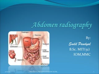
  • 1. By: Sudil Paudyal B.Sc. MIT(51) IOM,MMC 11/01/12 ABDOMEN PRESENTATION BY SUDIL 1
  • 2. General Anatomy:  The largest cavity of the body,  Bounded Anteriorly - by abdominal wall muscles Posteriorly - by the vertebral column and posterior wall muscles Laterally - by lower ribs and parts of muscles of abdominal wall Superiorly - by the diaphragm Inferiorly - by pelvic cavity 11/01/12 ABDOMEN PRESENTATION BY SUDIL 2
  • 3. Abdominal walls:  Bony support of the abdomen is minimal, consisting only of lumbar vertebrae and portions of the pelvis (the ilium and the pubis).  Muscles: Five pairs of muscles form anterior wall:  Rectus abdominis  External oblique  Internal oblique  Transversus abdominis Three pairs form the posterior wall:  Quadratus lumborum  Psoas major  Iliacus Linea alba: A very strong midline tendinous cord, extends from xiphoid process to symphysis pubis. Divides the anterior abdominal wall longitudinally into two identical halves. 11/01/12 ABDOMEN PRESENTATION BY SUDIL 3
  • 4. Fig: Anterior abdominal wall muscles Fig: Posterior abdominal wall muscles 11/01/12 ABDOMEN PRESENTATION BY SUDIL 4
  • 5. Planes and regions: Divided either into four quadrants or nine regions Divided into four quadrants by a transverse and a mid sagittal plane that intersect at the umbilicus. Right Upper Quadrant (RUQ), Right Lower Quadrant (RLQ), Left Upper Quadrant (LUQ), and Left Lower Quadrant (LLQ). 11/01/12 ABDOMEN PRESENTATION BY SUDIL 5
  • 6. Divided into nine regions by two transverse and two vertical planes The upper transverse plane - The Transpyloric Plane,  Lies midway between suprasternal notch and symphysis pubis, approximately midway between the upper border of xiphisternum and umbilicus.  Posteriorly, passes through the body of the first lumbar vertebra;  Anteriorly, passes through the tips of the right and left ninth costal cartilages. The lower transverse plane - The Transtubercular Plane,  Lies at the level of tubercles of iliac crest anteriorly, and near the upper border of fifth lumbar vertebra posteriorly. 11/01/12 ABDOMEN PRESENTATION BY SUDIL 6
  • 7. The two parasagittal (vertical) planes –  Lie at right-angles to the two transverse planes.  They run vertically, passing through a point midway between the anterior superior iliac spine and the symphysis pubis on each side. These planes divide the abdomen into nine regions:  centrally from above to below epigastric, umbilical and hypogastric regions and  laterally from above to below right and left hypochondriac, lumbar and iliac regions. 11/01/12 ABDOMEN PRESENTATION BY SUDIL 7
  • 8. 11/01/12 ABDOMEN PRESENTATION BY SUDIL 8
  • 9. Contents:  contains the greater part of the alimentary tract,  some of the accessory organs to digestion, viz. the liver, pancreas and spleen,  some of the urinary organs i.e. the kidneys,upper part of the ureters and the suprarenal glands.  Most of these structures, as well as the wall of the cavity are more or less covered by an extensive and complicated serous membrane, the peritoneum. 11/01/12 ABDOMEN PRESENTATION BY SUDIL 9
  • 10. Fig: organs of anterior part of abdominal cavity 11/01/12 ABDOMEN PRESENTATION BY SUDIL 10
  • 11. Fig: organs of posterior part of abdominal cavity 11/01/12 ABDOMEN PRESENTATION BY SUDIL 11
  • 12. Peritoneum: The serous membrane related to the viscera of the abdominal cavity.  Divided into two layers:  Parietal Layer : Lines the body wall and covers the retroperitoneal organs.  Visceral Layer : Composed of two parts : Covering of the surface of the peritoneal organs. Mesentery-a double layer of peritoneum that suspends part of the GI tract from the body wall.  Peritoneal cavity : The potential space located between the parietal and visceral layers. 11/01/12 ABDOMEN PRESENTATION BY SUDIL 12
  • 13. Abdominal Viscera Viscera are classified as: Peritoneal organs - have a mesentery and are almost completely enclosed in peritoneum. These organs are mobile. Retroperitoneal organs - are partially covered with peritoneum and are immobile or fixed organs. 11/01/12 ABDOMEN PRESENTATION BY SUDIL 13
  • 14. In a nutshell Major Peritoneal organs: Stomach, Liver and gallbladder, Spleen, Beginning of duodenum, Tail of pancreas, Jejunum, Ileum, Appendix , Transverse colon, Sigmoid colon Major Secondary Retroperitoneal organs: Most of duodenum, Most of pancreas, Ascending colon ,Descending colon , Upper rectum Major Primary Retroperitoneal Organs: Kidney , Adrenal gland, Ureter, Aorta, Inferior venacava, Lower rectum, Anal canal 11/01/12 ABDOMEN PRESENTATION BY SUDIL 14
  • 15. Liver: Lies mostly in the right hypochondrium, and protected by rib cage. Divided into two lobes of unequal size by the falciform ligament. Fissures for the ligamentum teres and the ligamentum venosum, the porta hepatis, and the fossa for the gallbladder further subdivide the right lobe into the right lobe proper, the quadrate lobe, and the caudate lobe. Has a central hilus, or porta hepatis, which receives venous blood from the portal vein and arterial blood from the hepatic artery. 11/01/12 ABDOMEN PRESENTATION BY SUDIL 15
  • 16. The central hilus also transmits the common bile duct, which collects bile produced by the liver. These structures, known collectively as the portal triad The hepatic veins drain the liver by collecting blood from the liver sinusoids and returning it to the inferior vena cava. Gallbladder :  lies in a fossa on the visceral surface of the liver to the right of the quadrate lobe. It stores and concentrates bile, which enters and leaves through the cystic duct. The cystic duct joins the common hepatic duct to form the common bile duct. 11/01/12 ABDOMEN PRESENTATION BY SUDIL 16
  • 17. Fig: Liver, turned up to show posterior surface 11/01/12 ABDOMEN PRESENTATION BY SUDIL 17
  • 18. Pancreas Most of the pancreas is secondarily retroperitoneal, but the distal part of the tail of the pancreas remains peritoneal . The tip of the tail of the pancreas reaches the hilus of the spleen. Both pancreatic ducts open into the second portion of the duodenum. Spleen a peritoneal organ in the upper left quadrant that is related to the left 9th, 10th, and 11th ribs. Fracture of these ribs may lacerate the spleen. 11/01/12 ABDOMEN PRESENTATION BY SUDIL 18
  • 19. Stomach: has a lesser curvature, which is connected to the porta hepatis of the liver by the lesser omentum, and a greater curvature from which the greater omentum is suspended. The cardiac region receives the esophagus. The dome-shaped upper portion of the stomach, which is normally filled with air, is the fundus. The main center portion of the stomach is the body. The pyloric portion of the stomach has a thick muscular wall and narrow lumen that leads to the duodenum. 11/01/12 ABDOMEN PRESENTATION BY SUDIL 19
  • 20. Fig: Abd. cavity showing greater and lesser Fig:Longitudinal section of stomach omentum 11/01/12 ABDOMEN PRESENTATION BY SUDIL 20
  • 21. Kidneys and ureters: Kidney's Relation to the Posterior Abdominal Wall Both kidneys are in contact with the diaphragm, psoas major, and quadratus lumborum . Right kidney-contacts the above structures and the 12th rib. Left kidney-contacts the above structures and the 11th and 12th ribs Ureter's Relation to the Posterior Abdominal Wall The ureter lies on the anterior surface of the psoas major. 11/01/12 ABDOMEN PRESENTATION BY SUDIL 21
  • 22. Fig: Relation of kidneys and ureters to posterior abdominal wall 11/01/12 ABDOMEN PRESENTATION BY SUDIL 22
  • 23. Kidneys: A pair of bean-shaped organs approximately 12 cm long. They extend from vertebral level T12 to L3 when the body is in the erect position. The right kidney is positioned slightly lower than the left because of the mass of the liver. Internal structure Within the dense, connective tissue of the renal capsule, the kidney substance is divided into an outer cortex and an inner medulla 11/01/12 ABDOMEN PRESENTATION BY SUDIL 23
  • 24. Cortex-contains glomeruli, Bowman's capsules, and proximal and distal convoluted tubules. It forms renal columns, which extend between medullary pyramids. Medulla--consists of 10 to 18 striated pyramids and contains collecting ducts and loops of Henle. The apex of each pyramid ends as a papilla where collecting ducts open. Calyces-the minor calyces receive one or more papillae and unite to form major calyces,of which there are two to three per kidney. Renal pelvis--the dilated upper portion of the ureter that receives the major calyces. 11/01/12 ABDOMEN PRESENTATION BY SUDIL 24
  • 25. Fig: cross section of a kidney 11/01/12 ABDOMEN PRESENTATION BY SUDIL 25
  • 26. Ureters : are fibro-muscular tubes that connect the kidneys to the urinary bladder in the pelvis. Urinary Bladder: The urinary bladder is covered superiorly by peritoneum. The body is a hollow muscular cavity. The neck is continuous with the urethra. The trigone is a smooth triangular area of mucosa located internally at the base of the bladder. The base of the triangle is superior and bounded by the two openings of the ureters.  The apex of the trigone points inferiorly and is the opening for the urethra. 11/01/12 ABDOMEN PRESENTATION BY SUDIL 26
  • 27. Fig: Kidneys, Ureters and Bladder 11/01/12 ABDOMEN PRESENTATION BY SUDIL 27
  • 28. For any body habitus whether hypersthenic or asthenic, abdominal viscera occupy a lower position:  in inspiration compared with expiration;  in the erect position compared with the recumbent position;  with age and the associated loss of muscle tone. 11/01/12 ABDOMEN PRESENTATION BY SUDIL 28
  • 29. 11/01/12 ABDOMEN PRESENTATION BY SUDIL 29
  • 30. Radiography: Preparation:  Careful preliminary patient preparation of the intestinal and gastric contents is important for a clear view of all the abdominal structures.  For non-acute conditions, patient preparation is as follows: (1) Patient placed on a low-residue diet for (2 days) prior to x- ray examination to prevent formation of gas due to excessive fermentation of the intestinal contents (2) Patient should be instructed to take some laxative the night before the examination. 11/01/12 ABDOMEN PRESENTATION BY SUDIL 30
  • 31. Exposure technique:  In examinations of the abdomen without a contrast medium, it is necessary to obtain maximum soft tissue differentiation throughout its different regions.  Because of the wide range in thickness of the abdomen and the delicate differences in physical density between the contained viscera, it is necessary to use a more critical exposure technique than is required to demonstrate the difference in density between an opacified organ and the structures adjacent to it.  The exposure factors should thus be adjusted to produce a radiograph with moderate gray tones and less black and white contrast. 11/01/12 ABDOMEN PRESENTATION BY SUDIL 31
  • 32.  A sharply demonstrated outline of the psoas muscles, lower border of liver, kidneys ribs and spinous processes of the lumbar vertebra are the best criteria for judging the quality of an abdominal radiograph. High mA and shorter exposure times must be used to freeze voluntary and involuntary organ movements (breathing and bowel peristalsis). Exposure is taken on second full arrested expiration (to displace diaphragm upward ) to give a better view of the abdominal structures. 11/01/12 ABDOMEN PRESENTATION BY SUDIL 32
  • 33. Immobilization: One of the prime requisite in abdominal examinations is the prevention of movement, both voluntary and involuntary.  To prevent muscle contraction, the patient must be adjusted in a comfortable position so that he can relax.  A compression band may be applied across the abdomen for immobilization but not compression.  The exposure should be made 1-2 sec after suspension of respiration to allow involuntary movement of viscera to subside. 11/01/12 ABDOMEN PRESENTATION BY SUDIL 33
  • 34. Radiation protection:  Gonadal shields should often be used on males (upper edge of the shield at the symphysis pubis). For females, shields are used only where they could not obscure essential anatomical structures (the lower border of the shield should be at the symphysis pubis).  For potential early pregnancy, the ‘10-day Rule’ (the LMP) must always be observed, unless permission has been given by the medical specialist as to ‘ignore’ it, e.g., in the case of an emergency (e.g., trauma), or in case of a female with a removed uterus. 11/01/12 ABDOMEN PRESENTATION BY SUDIL 34
  • 35. Radiographic projections: Basic : Antero-posterior – supine (KUB) (so named because it includes the kidneys, ureters and bladder). Alternative: Postero-anterior – prone Supplementary: Antero-posterior –erect Anteroposterior – left lateral decubitus Lateral Lateral- dorsal decubitus Anterior and posterior obliques ( for contrast studies) 11/01/12 ABDOMEN PRESENTATION BY SUDIL 35
  • 36. Indications:  Bowel obstruction  Perforation  Renal pathology  Acute abdomen  Foreign body localization  Toxic megacolon  Aortic aneurysm  Control or preliminary films for contrast studies  Detection of calcification or abnormal gas collection 11/01/12 ABDOMEN PRESENTATION BY SUDIL 36
  • 37. AP-supine (KUB) Patient position:  Patient supine, with the median sagittal plane at right angles  Pelvis adjusted so that the ASIS are equidistant from the table  Cassette placed longitudinally and positioned so that the symphysis pubis is included  Arms placed alongside the trunk or above the head. 11/01/12 ABDOMEN PRESENTATION BY SUDIL 37
  • 38. Centering of beam:  Vertical central ray directed approx. at the level of a point 1 cm below the line joining the iliac crests. Equipment setting: ( for screen film combination) Kv mA S mAs FFD Film Grid focus size 65 300 0.12 36 100 cm 35 X 43 Yes large cm 11/01/12 ABDOMEN PRESENTATION BY SUDIL 38
  • 39. Picture criteria:  Whole of abdomen from upper abdomen to symphysis pubis.  Lateral abdominal wall and the properitoneal fat layer.  Psoas muscle, lower border of liver and the kidneys.  Ribs and spinous processes of the lumbar vertebra.  Whole of the urinary tract should be visualized.  Bowel gas pattern with minimal unsharpness. 11/01/12 ABDOMEN PRESENTATION BY SUDIL 39
  • 40. 11/01/12 ABDOMEN PRESENTATION BY SUDIL 40
  • 41. PA- prone  When kidneys are not of primary interest, PA projection should be used.  It reduces patient gonad dose compared to the AP projection Patient position:  Patient prone, with median sagittal plane at right angles to the table  Arms up beside the head and both legs extended 11/01/12 ABDOMEN PRESENTATION BY SUDIL 41
  • 42.  CR, equipment setting, picture criteria same as supine projection. 11/01/12 ABDOMEN PRESENTATION BY SUDIL 42
  • 43. Lateral: Position of patient:  Patient turned onto the side of examination, with hands resting near the head. The hips and knees are flexed for stability.  With the MSP parallel to the table, the vertebral column( abt 8 cm anterior to the posterior skin surface) positioned over the midline of the table  Immobilization band applied across the pelvis.  Cassette centered at the level of iliac crests. 11/01/12 ABDOMEN PRESENTATION BY SUDIL 43
  • 44. Centring of the beam:  Vertical central ray directed to the centre of the cassette Equipment setting: Kv mA S mAs FFD Film Grid focus size 75 300 0.12 64 100 cm 35 X 43 Yes large cm 11/01/12 ABDOMEN PRESENTATION BY SUDIL 44
  • 45. Picture criteria:  The prevertebral space along with abdominal aorta  Any other intra abdominal calcifications or tumour masses should be clearly visible. 11/01/12 ABDOMEN PRESENTATION BY SUDIL 45
  • 46. AP -erect Patient position:  Patient stands with the back against the vertical bucky.  Patient’s legs separated well apart to maintain a comfortable position.  The median sagittal plane is adjusted at right angles and coincident with the midline of the table.  The pelvis is adjusted so that the anterior superior iliac spines are equidistant. 11/01/12 ABDOMEN PRESENTATION BY SUDIL 46
  • 47. Centring of beam:  The horizontal central ray is directed perpendicular to midpoint at the level of iliac crests. Equipment setting: Kv mA S mAs FFD Film Grid focus size 65 300 0.12 36 100 cm 35 X 43 Yes large cm 11/01/12 ABDOMEN PRESENTATION BY SUDIL 47
  • 48. Picture criteria:  Both domes of diaphragm to ensure that any free air in the peritoneal cavity is demonstrated.  Lateral abdominal wall and properitoneal fat  Psoas muscle, lower border of liver and kidney shadows  Vertebra in center of film.  Side identification marker placed properly. 11/01/12 ABDOMEN PRESENTATION BY SUDIL 48
  • 49. 11/01/12 ABDOMEN PRESENTATION BY SUDIL 49
  • 50. Lateral Decubitus -AP Lateral decubitus is done instead of abdomen erect if patient is unable to stand or sit. Patient position:  Patient in lateral recumbent position  Elbows and arms flexed and hand resting near head  Cassette positioned in vertical bucky against the posterior aspect of the trunk 11/01/12 ABDOMEN PRESENTATION BY SUDIL 50
  • 51. Centring of beam:  The central ray is directed perpendicular to midpoint at the level of iliac crest with x-ray tube horizontally. Equipment setting: Kv mA S mAs FFD Film Grid focus size 65 300 0.12 36 100 cm 35 X 43 Yes large cm  Note: Patient should be placed in lateral decubitus position for 5-10 mins to allow the free air to rise 11/01/12 ABDOMEN PRESENTATION BY SUDIL 51
  • 52. Picture criteria:  Air fluid levels when an erect abdomen cannot be obtained.  Lung area above dome of diaphragm  Lateral abdominal wall and properitoneal fat  Psoas muscle, lower border of liver and kidney shadows  No rotation 11/01/12 ABDOMEN PRESENTATION BY SUDIL 52
  • 53. 11/01/12 ABDOMEN PRESENTATION BY SUDIL 53
  • 54. Lateral dorsal decubitus (supine): Occasionally, the patient cannot sit or even be rolled on to the side, in which case the patient remains supine and a lateral projection is taken using a horizontal central ray. Patient position:  Patient supine  Arms raised away from the abdomen and thorax.  Cassette positioned vertically against patient’s side 11/01/12 ABDOMEN PRESENTATION BY SUDIL 54
  • 55. Centring of the beam:  The horizontal central ray is directed to the lateral aspect of the trunk so that it is at right-angles to the cassette and centred to it. Equipment setting: Kv mA S mAs FFD Film Grid focus size 75 300 0.12 36 100 cm 35 X 43 Yes large cm 11/01/12 ABDOMEN PRESENTATION BY SUDIL 55
  • 56. Picture criteria:  Thorax to the level of mid-sternum and as much of the abdomen as possible.  Pre-vertebral space for determining the air fluid levels in abdomen.  Lung area above dome of diaphragm, without motion.  Patient elevated to demonstrate entire abdomen 11/01/12 ABDOMEN PRESENTATION BY SUDIL 56
  • 57. References: Clark’s positioning in radiography, 12th edition Merrill’s atlas of radiographic positions and radiologic procedures, 12th edition Different other books and websites 11/01/12 ABDOMEN PRESENTATION BY SUDIL 57
  • 58. THANK YOU 11/01/12 ABDOMEN PRESENTATION BY SUDIL 58