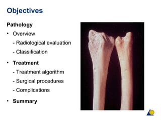
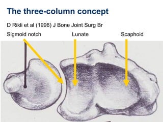
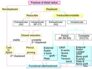
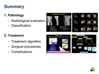
1) The document provides an overview of distal radius fractures, including classification systems, treatment algorithms, and surgical procedures.
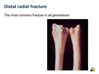
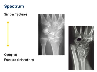
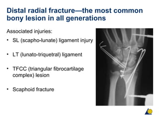
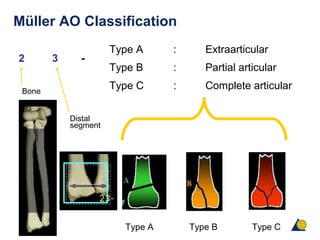
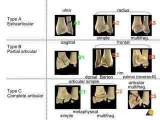
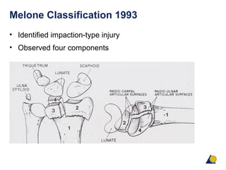
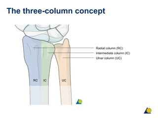
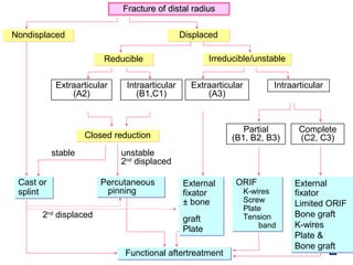
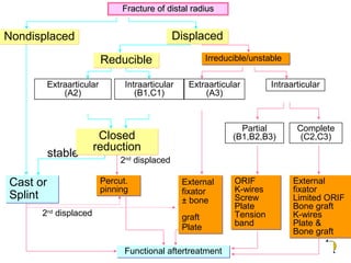
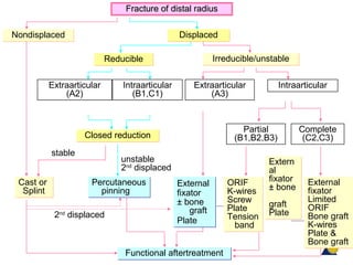
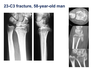
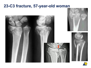
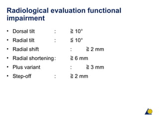
2) Key points include that distal radius fractures are the most common fractures, with a spectrum ranging from simple to complex fractures. Treatment depends on factors like displacement and stability.
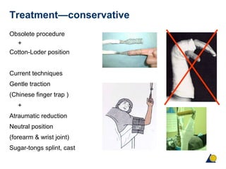
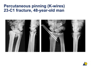
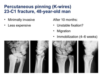
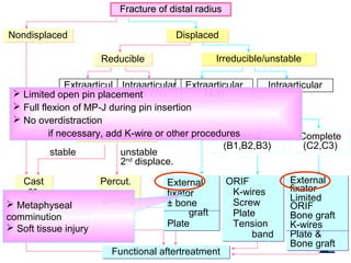
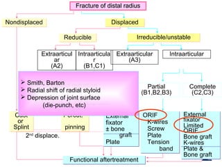
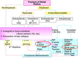
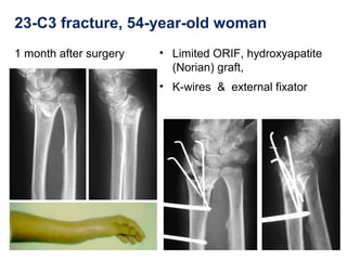
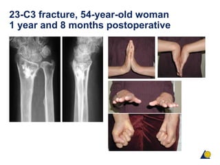
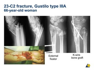
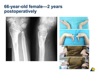
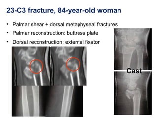
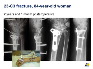
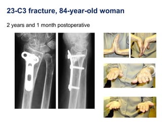
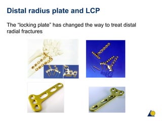
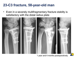
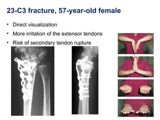
3) Surgical techniques discussed include percutaneous pinning, external fixation, limited open reduction with internal fixation using plates, screws, or bone grafts. The choice of technique depends on the fracture pattern and stability.