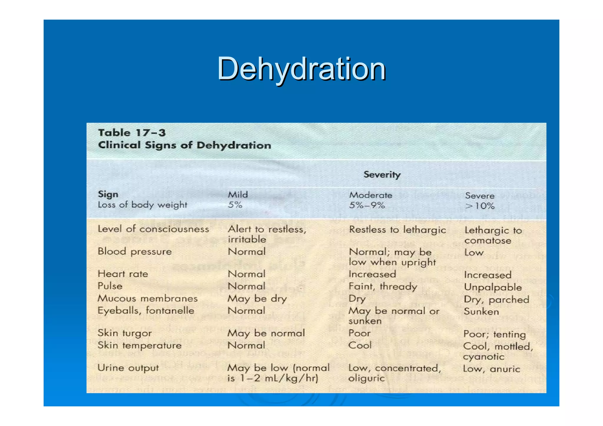
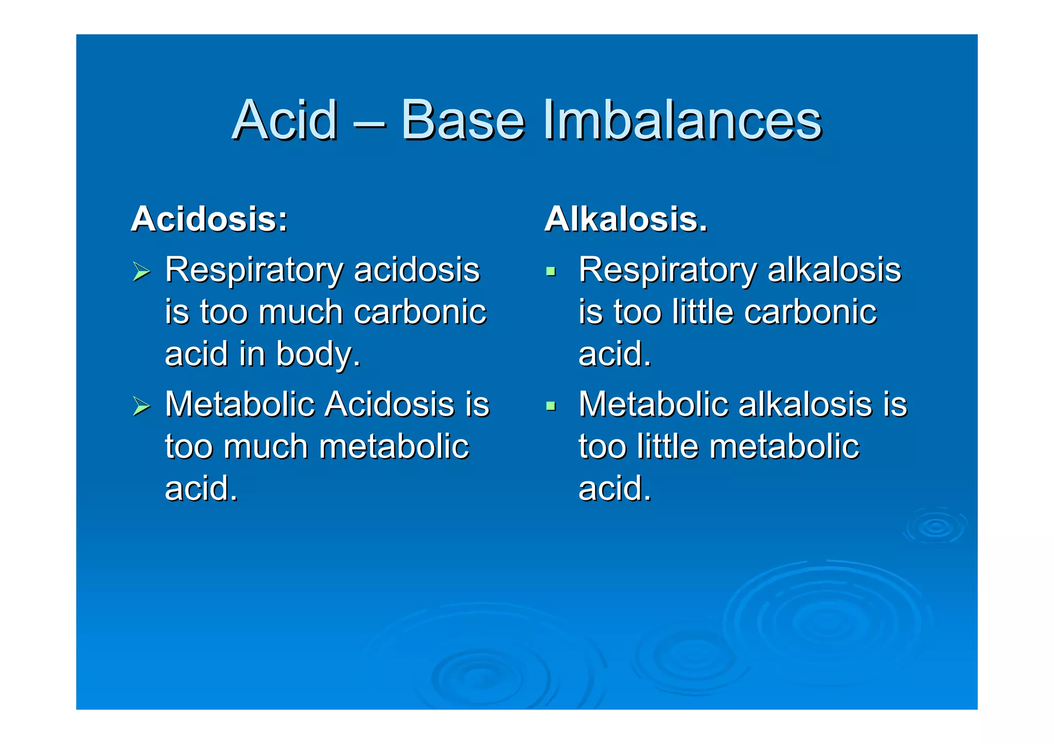
Fluid and Electrolytes
     Infants and children
Alteration in Fluid and
           Electrolyte Status
                               Lungs


                                                    Ball &
                                                    Bender


              Urine & faeces     Skin

Normal routes of fluid excretion in infants and children.
Developmental and Biological
        Variances
Infants younger than 6 weeks do not
produce tears.
In an infant a sunken fontanel may
indicate dehydration.
Infants are dependant on others to meet
their fluid needs.
Infants have limited ability to dilute and
concentrate urine.
Developmental and Biological
Smaller the child, greater proportion of
body water to weight and proportion of
extracellular fluid to intracellular fluid.
Infants larger proportional surface area of
GI tract than adults.
Infants greater body surface area and
higher metabolic rate than adults.
Water Balance
Regulated by Anti-diuretic Hormone ADH.
Acts on kidney tubules to reabsorb water.
The young infant is highly susceptible to
dehydration.
Increased Water Needs
Fever / sepsis
Vomiting and Diarrhoea
High-output in renal failure
Diabetes insipidus
Burns
Shock
Tachypnea
Decreased Water Needs
Congestive Heart Failure
Mechanical Ventilation
Renal failure
Head trauma / meningitis
General Appearance
How does the child look?
  Skin:
   • Temperature
   • Dry skin and mucous membranes
   • Poor turgor, tenting, dough-like feel
   • Sunken eyeballs; no tears
   • Pale, ashen, cyanotic nail beds or mucous
     membranes.
   • Delayed capillary refill > 3 seconds
Loss of Skin Elasticity

            Loss of skin elasticity
            Due to dehydration.

            Whaley & Wong Text
Cardiovascular
Pulse rate change:
  Note rate and quality
  Rapid, weak, or thready - inappropriate
  Bounding or arrhythmias


Blood Pressure (poor indicator)
  Note increase or decrease
Respiratory
Change in rate or quality
Dehydration of hypovolemia
  Tachypnea
  Apnea
  Deep shallow respirations
Fluid overload
  Moist breath sounds
  Cough
Diagnostic Tests
Make sure free flowing specimen is
obtained, a hemolysed or clotted blood
specimen may give false values.
Hemoglobin and Hematocrit
Measures hemoglobin, main component of
erythrocytes, vehicle for transporting
oxygen.
 Hb and hct will be increased in extracellular
 fluid volume loss.

 Hb and hct will be decreased in extracellular
 fluid volume excess.
Electrolytes
Electrolytes account for approximately
95% solute molecules in body water.
Sodium Na+ predominant extracellular
cation.
Potassium K+ is the predominant
intracellular cation.
Potassium
High or low values can lead to cardiac
arrest.
With adequate kidney function excess
potassium is excreted in the kidneys.
If kidneys are not functioning, the
potassium will accumulate in the
intravascular fluid
Potassium
 Adults: 3.5 to 5.3 mEq /L
 Child: 3.5 to 5.5 mEq / L
 Infant: 3.6 to 5.8 mEq / L

 Panic Values:
< 2.5 mEq /L or > 7.0 mEq / L
Hyperkalemia
Potassium level above 5.0 mEq / L
Significant dysrhythmias and cardiac
arrest may result when potassium levels
arise above 6.0 mEq/L
Adequate intake of fluids to insure
excretion of potassium through the
kidneys.
CM: Hyperkalemia
Nausea
Irregular heart rate
Pulse slow / irregular
Causes of Hyperkalemia
Acute renal failure
Chronic renal failure
Glomerulonephritis
Diagnostic tests:
Serum potassium
ECG
 Bradycardia
 Heart block
 Ventricular fibrillation
Hypokalemia
Potassium level below 3.5 mEq / L
Before administering make sure child is
producing urine.
A child on potassium wasting diuretics is
at risk – Lasix
CM: Hypokalemia
Neuromuscular manifestations are: neck
 flop, diminished bowel sounds, truncal
 weakness, limb weakness, lethargy, and
 abdominal distention.
Causes of Hypokalemia
Vomiting / diarrhea
Malnutrition / starvation
Stress due to trauma from injury or
surgery.
Gastric suction / intestinal fistula
Potassium wasting diuretics
Ingestion of large amounts of ASA
Foods high in potassium
Apricots, bananas, oranges,
pomegranates, prunes
Baked potato with skin, spinach, tomato,
lima beans, squash
Milk and yogurt
Pork, veal and fish
Monitor Potassium Levels


A child with a nasogastric tube in place that is set to suction,
needs to have potassium levels monitored.
Sodium
Sodium is the most abundant cation and
chief base of the blood.
The primary function is to chemically
maintain osmotic pressure and acid-base
balance and to transmit nerve impulses.
Normal values: 135 to 148 mEq / L
Treatment Modalities


Peripheral IV
IV Therapy


         Ball & Bender
Intraosseous Therapy


Intraosseous needle in place for emergency vascular access.
Central Venous Catheter
Total Parental Nutrition


                A tunneled catheter should have
Whaley & Wong   An occlusive dressing in place.
TPN Therapy
TPN provides complete nutrition for
children who cannot consume sufficient
nutrients through gastrointestinal tract to
meet and sustain metabolic requirements.
TPN solutions provide protein,
carbohydrates, electrolytes, vitamins,
minerals, trace elements and fats.
Complications of TPN
Sepsis: infection
Liver dysfunction
Respiratory distress from too –rapid
infusion of fluids
TPN: care reminder

The TPN infusion rate should remain fairly
constant to avoid glucose overload. The
infusion rate should never be abruptly
increased or decreased.
Dehydration
Skin Turgor
In moderate dehydration the skin may
have a doughy texture and appearance.

In severe dehydration the more typical
“tenting” of skin is observed.
Skin Turgor
Treatment of Mild to Moderate
ORT – oral re-hydration therapy
  50 ml / kg every 4 hours
  Increase to 100 ml / kg every 4 hours
  Non carbonated soda, jelly, fruit juices
  Commercially prepared solutions are the
  best.
Re-hydration Therapy
Increase po fluids if diarrhea increases.
Give po fluids slowly if vomiting.
Stop ORT when hydration status is normal
Start on BRAT diet
  Bananas
  Rice
  Applesauce
  Toast
Teaching / Parent Instruction
Call H/S
If diarrhea or vomiting increases
No improvement seen in child’s hydration
status.
Child appears worse.
Child will not take fluids.
NO URINE OUTPUT
Moderate to Severe Dehydration


                       IV Therapy
                       needed
Fluid replacement

Isotonic fluids initially:

  Normal Saline 0.9%

Potassium is added only after child has voided.
Nursing Interventions
Assess child’s hydration status
Accurate intake and output
Daily weights
  most accurate way to monitor fluid levels
Hourly monitoring of IV rate and site of infusion.
  Increase fluids if increase in vomiting or diarrhea.
  Decrease fluids when taking po fluids or signs of
  odema.
Care Reminder
A child with severe dehydration will need
more than maintenance to replace lost
fluids. 1 ½ to 2 times maintenance.
Adding potassium to IV solution.
  Never add in cases of oliguria / anuria
   • Urine output less than 0.5 mg/kg/hour
  Never give IV push
  Double check dosage
Over hydration
Occurs when child receives more IV fluids
that needed for maintenance.
In pre-existing conditions such as
meningitis, head trauma, kidney shutdown,
nephrotic syndrome, congestive heart
failure, or pulmonary congestion.
Signs and Symptoms
Tachypnea
Dyspnea
Cough
Moist breath sounds
Weight gain from edema
Jugular vein distention
Congestive Heart Failure


             Ball & Bender
Safety Precautions
Use buretrol to control fluid volume.
Check IV solution infusion against physician
orders.
Always use infusion pump so that the rate can
be programmed and monitored.
Even mechanical pumps can fail, so check the
intravenous bag and rate frequently.
Record IV rate hourly
Acid – Base Imbalances
Acidosis:                Alkalosis.
 Respiratory acidosis      Respiratory alkalosis
 is too much carbonic      is too little carbonic
 acid in body.             acid.
 Metabolic Acidosis is     Metabolic alkalosis is
 too much metabolic        too little metabolic
 acid.                     acid.
Respiratory Acidosis
Caused by the accumulation of carbon
dioxide in the blood.
Acute respiratory acidosis can lead to
tachycardia and cardiac arrhythmias.
Causes of Respiratory Acidosis
Any factor that interferes with the ability of
the lungs to excrete carbon dioxide can
cause respiratory acidosis.
Aspiration, spasm of airway, laryngeal
odema, epiglottitis, croup, pulmonary
odema, cystic fibrosis, and
Bronchopulmonary dysplasia.
Sedation overdose, head injury, or sleep
apnoea.
Medical Management
Correction of underlying cause.
Bronchodilators: asthma
Antibiotics: infection
Mechanical ventilation
Decreasing sedative use.
Ventilation Assist


          Ball & Bender
Respiratory Alkalosis
Occurs when the blood contains too little
carbon dioxide.
Excess carbon dioxide loss is caused by
hyperventilation.
Causes of hyperventilation

Hypoxemia
Anxiety
Pain
Fever
Salicylate poisoning: ASA
Meningitis
Over-ventilation
Management
Stress management if caused by
hyperventilation.
Pain control.
Adjust ventilation rate.
Treat underlying disease process.
Metabolic Acidosis
Caused by an imbalance in production and
excretion of acid or by excess loss of
bicarbonate.
Causes:
Gain in acid: ingestion of acids, oliguria,
starvation (anorexia), DKA or diabetic
ketoacidosis, tissue hypoxia.
Loss of bicarbonate:
diarrhea, intestinal or pancreatic fistula, or
renal anomaly.
Ingestion of large doses of Aspirin
Management
Treat and identify underlying cause.
IV sodium bicarbonate in severe cases.
Assess rate and depth of respirations and
level of consciousness.
Metabolic Alkalosis
A gain in bicarbonate or a loss of
metabolic acid can cause metabolic
alkalosis.
Causes:
  Gain in bicarbonate:
Ingestion of baking soda or antacids.
Loss of acid:
Vomiting, nasogastric suctioning, diuretics
  massive blood transfusion
Clinical Manifestations
Hypertonicity or tetany
Management: Correct the underlying
condition

Fluids and Electrolytes in Infants and Children

  • 1.
    Fluid and Electrolytes Infants and children
  • 2.
    Alteration in Fluidand Electrolyte Status Lungs Ball & Bender Urine & faeces Skin Normal routes of fluid excretion in infants and children.
  • 3.
    Developmental and Biological Variances Infants younger than 6 weeks do not produce tears. In an infant a sunken fontanel may indicate dehydration. Infants are dependant on others to meet their fluid needs. Infants have limited ability to dilute and concentrate urine.
  • 4.
    Developmental and Biological Smallerthe child, greater proportion of body water to weight and proportion of extracellular fluid to intracellular fluid. Infants larger proportional surface area of GI tract than adults. Infants greater body surface area and higher metabolic rate than adults.
  • 5.
    Water Balance Regulated byAnti-diuretic Hormone ADH. Acts on kidney tubules to reabsorb water. The young infant is highly susceptible to dehydration.
  • 6.
    Increased Water Needs Fever/ sepsis Vomiting and Diarrhoea High-output in renal failure Diabetes insipidus Burns Shock Tachypnea
  • 7.
    Decreased Water Needs CongestiveHeart Failure Mechanical Ventilation Renal failure Head trauma / meningitis
  • 8.
    General Appearance How doesthe child look? Skin: • Temperature • Dry skin and mucous membranes • Poor turgor, tenting, dough-like feel • Sunken eyeballs; no tears • Pale, ashen, cyanotic nail beds or mucous membranes. • Delayed capillary refill > 3 seconds
  • 9.
    Loss of SkinElasticity Loss of skin elasticity Due to dehydration. Whaley & Wong Text
  • 10.
    Cardiovascular Pulse rate change: Note rate and quality Rapid, weak, or thready - inappropriate Bounding or arrhythmias Blood Pressure (poor indicator) Note increase or decrease
  • 11.
    Respiratory Change in rateor quality Dehydration of hypovolemia Tachypnea Apnea Deep shallow respirations Fluid overload Moist breath sounds Cough
  • 12.
    Diagnostic Tests Make surefree flowing specimen is obtained, a hemolysed or clotted blood specimen may give false values.
  • 13.
    Hemoglobin and Hematocrit Measureshemoglobin, main component of erythrocytes, vehicle for transporting oxygen. Hb and hct will be increased in extracellular fluid volume loss. Hb and hct will be decreased in extracellular fluid volume excess.
  • 14.
    Electrolytes Electrolytes account forapproximately 95% solute molecules in body water. Sodium Na+ predominant extracellular cation. Potassium K+ is the predominant intracellular cation.
  • 15.
    Potassium High or lowvalues can lead to cardiac arrest. With adequate kidney function excess potassium is excreted in the kidneys. If kidneys are not functioning, the potassium will accumulate in the intravascular fluid
  • 16.
    Potassium Adults: 3.5to 5.3 mEq /L Child: 3.5 to 5.5 mEq / L Infant: 3.6 to 5.8 mEq / L Panic Values: < 2.5 mEq /L or > 7.0 mEq / L
  • 17.
    Hyperkalemia Potassium level above5.0 mEq / L Significant dysrhythmias and cardiac arrest may result when potassium levels arise above 6.0 mEq/L Adequate intake of fluids to insure excretion of potassium through the kidneys.
  • 18.
    CM: Hyperkalemia Nausea Irregular heartrate Pulse slow / irregular
  • 19.
    Causes of Hyperkalemia Acuterenal failure Chronic renal failure Glomerulonephritis
  • 20.
    Diagnostic tests: Serum potassium ECG Bradycardia Heart block Ventricular fibrillation
  • 21.
    Hypokalemia Potassium level below3.5 mEq / L Before administering make sure child is producing urine. A child on potassium wasting diuretics is at risk – Lasix
  • 22.
    CM: Hypokalemia Neuromuscular manifestationsare: neck flop, diminished bowel sounds, truncal weakness, limb weakness, lethargy, and abdominal distention.
  • 23.
    Causes of Hypokalemia Vomiting/ diarrhea Malnutrition / starvation Stress due to trauma from injury or surgery. Gastric suction / intestinal fistula Potassium wasting diuretics Ingestion of large amounts of ASA
  • 24.
    Foods high inpotassium Apricots, bananas, oranges, pomegranates, prunes Baked potato with skin, spinach, tomato, lima beans, squash Milk and yogurt Pork, veal and fish
  • 25.
    Monitor Potassium Levels Achild with a nasogastric tube in place that is set to suction, needs to have potassium levels monitored.
  • 26.
    Sodium Sodium is themost abundant cation and chief base of the blood. The primary function is to chemically maintain osmotic pressure and acid-base balance and to transmit nerve impulses. Normal values: 135 to 148 mEq / L
  • 27.
  • 28.
    IV Therapy Ball & Bender
  • 29.
    Intraosseous Therapy Intraosseous needlein place for emergency vascular access.
  • 30.
  • 31.
    Total Parental Nutrition A tunneled catheter should have Whaley & Wong An occlusive dressing in place.
  • 32.
    TPN Therapy TPN providescomplete nutrition for children who cannot consume sufficient nutrients through gastrointestinal tract to meet and sustain metabolic requirements. TPN solutions provide protein, carbohydrates, electrolytes, vitamins, minerals, trace elements and fats.
  • 33.
    Complications of TPN Sepsis:infection Liver dysfunction Respiratory distress from too –rapid infusion of fluids
  • 34.
    TPN: care reminder TheTPN infusion rate should remain fairly constant to avoid glucose overload. The infusion rate should never be abruptly increased or decreased.
  • 35.
  • 36.
    Skin Turgor In moderatedehydration the skin may have a doughy texture and appearance. In severe dehydration the more typical “tenting” of skin is observed.
  • 37.
  • 38.
    Treatment of Mildto Moderate ORT – oral re-hydration therapy 50 ml / kg every 4 hours Increase to 100 ml / kg every 4 hours Non carbonated soda, jelly, fruit juices Commercially prepared solutions are the best.
  • 39.
    Re-hydration Therapy Increase pofluids if diarrhea increases. Give po fluids slowly if vomiting. Stop ORT when hydration status is normal Start on BRAT diet Bananas Rice Applesauce Toast
  • 40.
    Teaching / ParentInstruction Call H/S If diarrhea or vomiting increases No improvement seen in child’s hydration status. Child appears worse. Child will not take fluids. NO URINE OUTPUT
  • 41.
    Moderate to SevereDehydration IV Therapy needed
  • 42.
    Fluid replacement Isotonic fluidsinitially: Normal Saline 0.9% Potassium is added only after child has voided.
  • 43.
    Nursing Interventions Assess child’shydration status Accurate intake and output Daily weights most accurate way to monitor fluid levels Hourly monitoring of IV rate and site of infusion. Increase fluids if increase in vomiting or diarrhea. Decrease fluids when taking po fluids or signs of odema.
  • 44.
    Care Reminder A childwith severe dehydration will need more than maintenance to replace lost fluids. 1 ½ to 2 times maintenance. Adding potassium to IV solution. Never add in cases of oliguria / anuria • Urine output less than 0.5 mg/kg/hour Never give IV push Double check dosage
  • 45.
    Over hydration Occurs whenchild receives more IV fluids that needed for maintenance. In pre-existing conditions such as meningitis, head trauma, kidney shutdown, nephrotic syndrome, congestive heart failure, or pulmonary congestion.
  • 46.
    Signs and Symptoms Tachypnea Dyspnea Cough Moistbreath sounds Weight gain from edema Jugular vein distention
  • 47.
  • 48.
    Safety Precautions Use buretrolto control fluid volume. Check IV solution infusion against physician orders. Always use infusion pump so that the rate can be programmed and monitored. Even mechanical pumps can fail, so check the intravenous bag and rate frequently. Record IV rate hourly
  • 49.
    Acid – BaseImbalances Acidosis: Alkalosis. Respiratory acidosis Respiratory alkalosis is too much carbonic is too little carbonic acid in body. acid. Metabolic Acidosis is Metabolic alkalosis is too much metabolic too little metabolic acid. acid.
  • 50.
    Respiratory Acidosis Caused bythe accumulation of carbon dioxide in the blood. Acute respiratory acidosis can lead to tachycardia and cardiac arrhythmias.
  • 51.
    Causes of RespiratoryAcidosis Any factor that interferes with the ability of the lungs to excrete carbon dioxide can cause respiratory acidosis. Aspiration, spasm of airway, laryngeal odema, epiglottitis, croup, pulmonary odema, cystic fibrosis, and Bronchopulmonary dysplasia. Sedation overdose, head injury, or sleep apnoea.
  • 52.
    Medical Management Correction ofunderlying cause. Bronchodilators: asthma Antibiotics: infection Mechanical ventilation Decreasing sedative use.
  • 53.
    Ventilation Assist Ball & Bender
  • 54.
    Respiratory Alkalosis Occurs whenthe blood contains too little carbon dioxide. Excess carbon dioxide loss is caused by hyperventilation.
  • 55.
  • 56.
    Management Stress management ifcaused by hyperventilation. Pain control. Adjust ventilation rate. Treat underlying disease process.
  • 57.
    Metabolic Acidosis Caused byan imbalance in production and excretion of acid or by excess loss of bicarbonate.
  • 58.
    Causes: Gain in acid:ingestion of acids, oliguria, starvation (anorexia), DKA or diabetic ketoacidosis, tissue hypoxia. Loss of bicarbonate: diarrhea, intestinal or pancreatic fistula, or renal anomaly.
  • 59.
    Ingestion of largedoses of Aspirin
  • 60.
    Management Treat and identifyunderlying cause. IV sodium bicarbonate in severe cases. Assess rate and depth of respirations and level of consciousness.
  • 61.
    Metabolic Alkalosis A gainin bicarbonate or a loss of metabolic acid can cause metabolic alkalosis.
  • 62.
    Causes: Gainin bicarbonate: Ingestion of baking soda or antacids. Loss of acid: Vomiting, nasogastric suctioning, diuretics massive blood transfusion
  • 63.
    Clinical Manifestations Hypertonicity ortetany Management: Correct the underlying condition