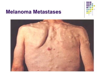
Cancer
Introduction A group of more than 200 diseases characterized by uncontrolled and unregulated growth of cells In NZ 14,808 new cases of cancer 1 in 3 men and 1 in 4 women will be affected by age 75yrs Most common cause of death in NZ (29% of deaths overall)
Cancer Most common cancers causing death in males are lung, prostate and colorectal Most common cancers causing death in females are breast, lung & colorectal Cancer rates for Maori & non Maori are similar but mortality rates are 51% (males) and 78%(females) higher for Maori
5-year survival rate is now 62% for those who are disease free, in remission, or under treatment 5-year survival rates do not include the number of people who are “cured” of cancer
Defect in Cellular Proliferation  (process by which cells divide & reproduce) Cancer cells are characterized by the loss of contact inhibition – have no regard for cellular boundaries Grow on top of one another and on top of or between normal cells Cancer cells divide indiscriminately and haphazardly – sometimes they produce more than 2 cells at the time of mitosis
Defect in Cell Proliferation In most situations, cancer cells multiply at the same rate as the normal cells from which they originate The difference is that proliferation of the cancer cells is indiscriminate and continuous Because of this there is continuous growth of a tumour mass
Defect in Cellular Differentiation Cellular differentiation (the development of specific & distinctive features in cells) is normally an orderly process Protooncogenes Normal cellular genes that are important regulators on normal cellular processes Mutations that alter their expression can activate them to act as oncogenes (tumour-inducing)
Neoplasms Characterised as: Benign neoplasms Malignant neoplasms Major difference between malignant & benign neoplasms is the ability of malignant tumour cells to invade and metastasize
Benign Neoplasms Well differentiated (specific & distinctive features of cells are well developed).  Cells resemble the cells of tissue of origin. Well-defined fibrous capsule Characterized by slow, progressive rate of growth. Tumours grow by expansion Metastases absent Recurrence unusual Do not usually cause death unless they interfere with vital functions because of location e.g.. Benign cranial tumour compressing brain structures
Malignant Neoplasms Tend to grow rapidly, spread widely, & kill regardless of original location Able to metastasize Because of rapid growth, they compress blood vessels & outgrow their blood supply causing ischaemia & tissue necrosis Rob normal tissues of essential nutrients Liberate enzymes & toxins that destroy normal & tumour tissue Infiltrative and expansive Frequent recurrence Moderate to marked vascularity Rarely encapsulated Cells abnormal; become more unlike parent cells
Effects on Body Function with Cancer Growth Altered function of involved tissue Bleeding & haemorrhage Ulceration, necrosis & infection of tumour area Obstruction of hollow organs or pathways Effusion in serous cavities Increased risk of vascular thrombosis Anaemia Bone destruction
Effects (cont.) Hypercalcaemia Pain Cachexia – weight loss & wasting of body fat & lean protein Inappropriate hormone production (e.g. ADH or ACTH secretion by cancers such as bronchogenic carcinaoma)
Cachexia
Advanced Breast Cancer
Cancer of Kidney
Ascites – Liver Cancer
Development of Cancer Carcinogens  – cancer-causing agents capable of producing cellular alterations Can be chemical, environmental, immunologic, viral, or spontaneous in origin Some genetic abnormalities increase the susceptibility of individuals to certain cancers Hereditary predisposition in approx 50 types of cancers e.g. breast cancer
Process of Cancer Development Fig. 15-3
Metastasis The spread of cancer  from the initial or primary site to a distant site Some metastasize early e.g. pre-menopausal breast cancer Others spread regionally and rarely metastasize e.g.. BCC of skin Certain cancers have an affinity  for a particular tissue or organ as a site of metastasis, others are unpredictable e.g. melanoma
Metastasis (cont.) Certain cancers (‘seed’), such as ovarian cancer, require a particular site for proliferation (‘soil’) Most frequent sites for metastases are lungs, brain, , bone, liver & adrenals
Metastasis Metastasis process begins with rapid growth of primary tumour Development of its own blood supply is critical for survival  Tumour angiogenesis  is formation of blood vessels within the tumour
Melanoma Metastases
Progression of Cancer Metatases
 
Role of Immune System Immune response is to reject or destroy cancer cells if perceived as non-self May be inadequate as cancer cells arise from normal human cells Some cancer cells have changes on their surface antigens Tumor-associated antigens (TAAs)
Role of Immune System Response to TAAs is termed immunologic surveillance Lymphocytes continually check cell surfaces and detect and destroy cells with abnormalities Involves cytotoxic T cell, natural killer cells, macrophages, and B lymphocytes
Role of Immune System Escape mechanisms by which cancer cells evade immune system Suppression of factors that stimulate T cells Weak surface antigens allow cancer cells to “sneak through” surveillance
Role of Immune System Development of tolerance of immune system Suppression of immune response to products secreted by cancer cells Induction of suppressor T cells  Blocking antibodies that bind TAAs
Classification of Cancer Anatomic Site Classification Identified by  tissue origin anatomic site behaviour of the tumour
Classification of Cancer Anatomic Site Classification Carcinomas originate from embryonal ectoderm and endoderm Sarcomas originate from embryonic mesoderm Lymphomas and leukaemias originate from hepatopoietic system
Classification of Cancer Histologic Analysis Classification Appearance of cells and degree of differentiation are evaluated Grade 1: Cells differ slightly from normal cells and are well differentiated Grade 2: Cells more abnormal and moderately differentiated
Classification of Cancer Grade 3: Cells very abnormal and poorly differentiated Grade 4: Cells immature and primitive and undifferentiated; cell of origin difficult to determine
Classification of Cancer Clinical Staging 0: cancer in situ 1: tumor limited to tissue of origin 2: limited local spread 3: extensive local and regional spread 4: metastasis
Classification of Cancer TNM Classification Tumor size Spread to lymph nodes Metastasis
TNM Classification System T (tumour) Tx Tumour unable to be adequately  assessed T0 No evidence of primary tumour Tis Carcinoma in situ T1-4 Progressive increase in tumour size or  involvement
TNM Classification System N (nodes ) Nx Regional lymph nodes cannot be  assessed N0 No evidence of regional node metastasis N1-3 Increasing involvement of regional lymph  nodes
TNM Classification System M (metastasis) Mx Not assessed M0 No distant metastasis M1 Distant metastasis present, specify  sites
Prevention and Detection of Cancer Reduce or avoid exposure to known or suspected carcinogens Eat balanced diet Exercise regularly Adequate rest Health examination on a regular basis
Prevention and Detection of Cancer Eliminate, reduce, or change perceptions of stressors and enhance ability to cope Enjoy consistent periods of relaxation and leisure Know 7 warning signs of cancer Self-examination Seek medical care if cancer is suspected
Early Warning Signs of Cancer Change in bowel or bladder habits A sore that does not heal Unusual bleeding or discharge Thickening or lump in breast or elsewhere Indigestion or difficulty in swallowing Obvious change in mole or wart Nagging cough or hoarseness

Cancer Ppt 2008

  • 1.
  • 2.
    Introduction A groupof more than 200 diseases characterized by uncontrolled and unregulated growth of cells In NZ 14,808 new cases of cancer 1 in 3 men and 1 in 4 women will be affected by age 75yrs Most common cause of death in NZ (29% of deaths overall)
  • 3.
    Cancer Most commoncancers causing death in males are lung, prostate and colorectal Most common cancers causing death in females are breast, lung & colorectal Cancer rates for Maori & non Maori are similar but mortality rates are 51% (males) and 78%(females) higher for Maori
  • 4.
    5-year survival rateis now 62% for those who are disease free, in remission, or under treatment 5-year survival rates do not include the number of people who are “cured” of cancer
  • 5.
    Defect in CellularProliferation (process by which cells divide & reproduce) Cancer cells are characterized by the loss of contact inhibition – have no regard for cellular boundaries Grow on top of one another and on top of or between normal cells Cancer cells divide indiscriminately and haphazardly – sometimes they produce more than 2 cells at the time of mitosis
  • 6.
    Defect in CellProliferation In most situations, cancer cells multiply at the same rate as the normal cells from which they originate The difference is that proliferation of the cancer cells is indiscriminate and continuous Because of this there is continuous growth of a tumour mass
  • 7.
    Defect in CellularDifferentiation Cellular differentiation (the development of specific & distinctive features in cells) is normally an orderly process Protooncogenes Normal cellular genes that are important regulators on normal cellular processes Mutations that alter their expression can activate them to act as oncogenes (tumour-inducing)
  • 8.
    Neoplasms Characterised as:Benign neoplasms Malignant neoplasms Major difference between malignant & benign neoplasms is the ability of malignant tumour cells to invade and metastasize
  • 9.
    Benign Neoplasms Welldifferentiated (specific & distinctive features of cells are well developed). Cells resemble the cells of tissue of origin. Well-defined fibrous capsule Characterized by slow, progressive rate of growth. Tumours grow by expansion Metastases absent Recurrence unusual Do not usually cause death unless they interfere with vital functions because of location e.g.. Benign cranial tumour compressing brain structures
  • 10.
    Malignant Neoplasms Tendto grow rapidly, spread widely, & kill regardless of original location Able to metastasize Because of rapid growth, they compress blood vessels & outgrow their blood supply causing ischaemia & tissue necrosis Rob normal tissues of essential nutrients Liberate enzymes & toxins that destroy normal & tumour tissue Infiltrative and expansive Frequent recurrence Moderate to marked vascularity Rarely encapsulated Cells abnormal; become more unlike parent cells
  • 11.
    Effects on BodyFunction with Cancer Growth Altered function of involved tissue Bleeding & haemorrhage Ulceration, necrosis & infection of tumour area Obstruction of hollow organs or pathways Effusion in serous cavities Increased risk of vascular thrombosis Anaemia Bone destruction
  • 12.
    Effects (cont.) HypercalcaemiaPain Cachexia – weight loss & wasting of body fat & lean protein Inappropriate hormone production (e.g. ADH or ACTH secretion by cancers such as bronchogenic carcinaoma)
  • 13.
  • 14.
  • 15.
  • 16.
  • 17.
    Development of CancerCarcinogens – cancer-causing agents capable of producing cellular alterations Can be chemical, environmental, immunologic, viral, or spontaneous in origin Some genetic abnormalities increase the susceptibility of individuals to certain cancers Hereditary predisposition in approx 50 types of cancers e.g. breast cancer
  • 18.
    Process of CancerDevelopment Fig. 15-3
  • 19.
    Metastasis The spreadof cancer from the initial or primary site to a distant site Some metastasize early e.g. pre-menopausal breast cancer Others spread regionally and rarely metastasize e.g.. BCC of skin Certain cancers have an affinity for a particular tissue or organ as a site of metastasis, others are unpredictable e.g. melanoma
  • 20.
    Metastasis (cont.) Certaincancers (‘seed’), such as ovarian cancer, require a particular site for proliferation (‘soil’) Most frequent sites for metastases are lungs, brain, , bone, liver & adrenals
  • 21.
    Metastasis Metastasis processbegins with rapid growth of primary tumour Development of its own blood supply is critical for survival Tumour angiogenesis is formation of blood vessels within the tumour
  • 22.
  • 23.
  • 24.
  • 25.
    Role of ImmuneSystem Immune response is to reject or destroy cancer cells if perceived as non-self May be inadequate as cancer cells arise from normal human cells Some cancer cells have changes on their surface antigens Tumor-associated antigens (TAAs)
  • 26.
    Role of ImmuneSystem Response to TAAs is termed immunologic surveillance Lymphocytes continually check cell surfaces and detect and destroy cells with abnormalities Involves cytotoxic T cell, natural killer cells, macrophages, and B lymphocytes
  • 27.
    Role of ImmuneSystem Escape mechanisms by which cancer cells evade immune system Suppression of factors that stimulate T cells Weak surface antigens allow cancer cells to “sneak through” surveillance
  • 28.
    Role of ImmuneSystem Development of tolerance of immune system Suppression of immune response to products secreted by cancer cells Induction of suppressor T cells Blocking antibodies that bind TAAs
  • 29.
    Classification of CancerAnatomic Site Classification Identified by tissue origin anatomic site behaviour of the tumour
  • 30.
    Classification of CancerAnatomic Site Classification Carcinomas originate from embryonal ectoderm and endoderm Sarcomas originate from embryonic mesoderm Lymphomas and leukaemias originate from hepatopoietic system
  • 31.
    Classification of CancerHistologic Analysis Classification Appearance of cells and degree of differentiation are evaluated Grade 1: Cells differ slightly from normal cells and are well differentiated Grade 2: Cells more abnormal and moderately differentiated
  • 32.
    Classification of CancerGrade 3: Cells very abnormal and poorly differentiated Grade 4: Cells immature and primitive and undifferentiated; cell of origin difficult to determine
  • 33.
    Classification of CancerClinical Staging 0: cancer in situ 1: tumor limited to tissue of origin 2: limited local spread 3: extensive local and regional spread 4: metastasis
  • 34.
    Classification of CancerTNM Classification Tumor size Spread to lymph nodes Metastasis
  • 35.
    TNM Classification SystemT (tumour) Tx Tumour unable to be adequately assessed T0 No evidence of primary tumour Tis Carcinoma in situ T1-4 Progressive increase in tumour size or involvement
  • 36.
    TNM Classification SystemN (nodes ) Nx Regional lymph nodes cannot be assessed N0 No evidence of regional node metastasis N1-3 Increasing involvement of regional lymph nodes
  • 37.
    TNM Classification SystemM (metastasis) Mx Not assessed M0 No distant metastasis M1 Distant metastasis present, specify sites
  • 38.
    Prevention and Detectionof Cancer Reduce or avoid exposure to known or suspected carcinogens Eat balanced diet Exercise regularly Adequate rest Health examination on a regular basis
  • 39.
    Prevention and Detectionof Cancer Eliminate, reduce, or change perceptions of stressors and enhance ability to cope Enjoy consistent periods of relaxation and leisure Know 7 warning signs of cancer Self-examination Seek medical care if cancer is suspected
  • 40.
    Early Warning Signsof Cancer Change in bowel or bladder habits A sore that does not heal Unusual bleeding or discharge Thickening or lump in breast or elsewhere Indigestion or difficulty in swallowing Obvious change in mole or wart Nagging cough or hoarseness