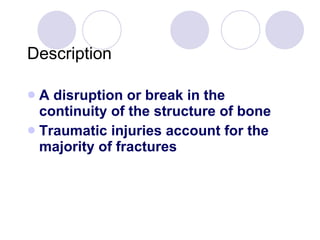
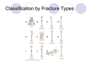
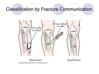
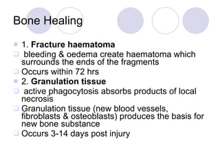
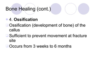
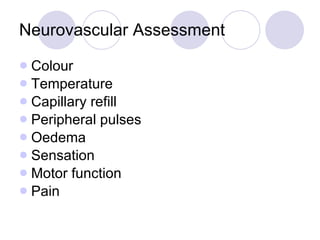
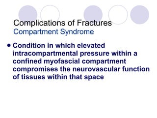
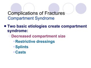
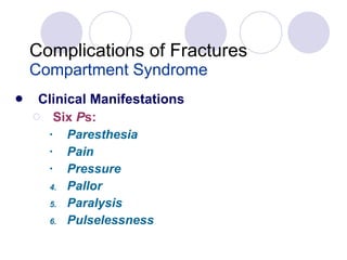
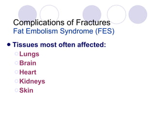
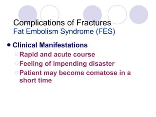
Fractures
Objectives Describe the sequence of fracture healing Differentiate between open and closed reduction, cast immobilization, and traction Describe neurovascular assessment of injured extremity Explain common complications associated with fracture injury and healing
Description A disruption or break in the continuity of the structure of bone Traumatic injuries account for the majority of fractures
Description Described and classified according to: Type Communication or noncommunication with external environment Anatomic location
Classification by Fracture Types
Classification by Fracture Communication
Classification by Fracture Location
Description Described and classified according to: Appearance, position, and alignment of the fragments Classic names Stable or unstable
Description Closed (simple) Open (compound)
Description Stable fractures Occur when a piece of the periosteum is intact across the fracture External or internal fixation has rendered the fragments stationary
Description Stable fractures Transverse Spiral Greenstick
Description Unstable fractures Comminuted Oblique
Clinical Manifestations Patient history indicates a mechanism of injury associated with: Immediate localized pain    Function Inability to bear weight or use affected part Guarding May not be accompanied by obvious bone deformity
Fracture Healing Reparative process of self-healing ( union ) occurs in the following stages: Fracture hematoma Granulation tissue Callus formation Consolidation Ossification Remodeling
Bone Healing 1.  Fracture haematoma bleeding & oedema create haematoma which surrounds the ends of the fragments Occurs within 72 hrs 2.  Granulation tissue active phagocytosis absorbs products of local necrosis Granulation tissue (new blood vessels, fibroblasts & osteoblasts) produces the basis for new bone substance Occurs 3-14 days post injury
Bone Healing (cont.) 3.  Callus formation As minerals are deposited, an unorganised network of bone is formed that is woven about the fracture parts Callus is composed of cartilage, osteoblasts, calcium & phosphorus Begins to appear by end of 2 nd  week
Bone Healing (cont.) 4.  Ossification Ossification (development of bone) of the callus Sufficient to prevent movement at fracture site Occurs from 3 weeks to 6 months
Bone Healing (cont.) 5.  Consolidation As callus develops, the distance between bone fragments diminishes & eventually closes 6. Remodelling Excess bone tissue is reabsorbed & union is completed
Bone Healing
Collaborative Care Overall goals of treatment : Anatomic realignment of bone fragments (reduction) Immobilization to maintain alignment Restoration of normal function
Fracture Reduction Closed reduction Nonsurgical, manual realignment Open reduction Correction of bone alignment through a surgical incision
Fracture Immobilization Casts Temporary circumferential immobilization device Common treatment following closed reduction
Fracture Immobilization External fixation Metallic device composed of pins that are inserted into the bone and attached to external rods
Fracture Immobilization Internal fixation Pins, plates, intermedullary rods, and screw Surgically inserted at the time of realignment
Traction Application of a pulling force to an injured part of the body while counter-traction pulls in the opposite direction
Fracture Reduction - Traction Skin traction (short-term) Skeletal traction (longer periods)
Purpose of Traction Prevent or reduce muscle spasm Immobilization Reduction Treat a pathologic condition
Neurovascular Assessment Colour Temperature Capillary refill Peripheral pulses Oedema Sensation Motor function Pain
Complications of Fractures Infection Open fractures and soft tissue injuries have    incidence Osteomyelitis can become chronic
Complications of Fractures Infection Open fractures require aggressive surgical debridement Post-op IV antibiotics for 3 to 7 days
Complications of Fractures Compartment Syndrome Condition in which elevated intracompartmental pressure within a confined myofascial compartment compromises the neurovascular function of tissues within that space
Complications of Fractures Compartment Syndrome Two basic etiologies create compartment syndrome: Decreased compartment size Restrictive dressings Splints Casts
Complications of Fractures Compartment Syndrome Two basic etiologies create compartment syndrome: Increased compartment content Bleeding Oedema
Complications of Fractures Compartment Syndrome Clinical Manifestations Six  P s: Paresthesia Pain   Pressure Pallor Paralysis Pulselessness
Complications of Fractures Venous Thrombosis Veins of the lower extremities and pelvis are highly susceptible to thrombus formation after fracture, especially hip fracture
Complications of Fractures Venous Thrombosis Precipitating factors: Venous stasis caused by incorrectly applied casts or traction Local pressure on a vein Immobility
Complications of Fractures Fat Embolism Syndrome (FES) Characterized by the presence of fat globules in tissues and organs after a traumatic skeletal injury
Complications of Fractures Fat Embolism Syndrome (FES) Fractures that most often cause FES: Long bones Ribs Tibia Pelvis
Complications of Fractures Fat Embolism Syndrome (FES) Tissues most often affected: Lungs Brain Heart  Kidneys  Skin
Complications of Fractures Fat Embolism Syndrome (FES) Clinical Manifestations Usually occur 24 to 48 hours after injury Interstitial pneumonitis  Produce symptoms of ARDS
Complications of Fractures Fat Embolism Syndrome (FES) Clinical Manifestations Symptoms of ARDS: Chest pain Tachypnea Cyanosis    PaO 2
Complications of Fractures Fat Embolism Syndrome (FES) Clinical Manifestations Symptoms of ARDS: Dyspnea Apprehension Tachycardia
Complications of Fractures Fat Embolism Syndrome (FES) Clinical Manifestations Rapid and acute course Feeling of impending disaster Patient may become comatose in a short time

4 Fractures 2010

  • 1.
  • 2.
    Objectives Describe thesequence of fracture healing Differentiate between open and closed reduction, cast immobilization, and traction Describe neurovascular assessment of injured extremity Explain common complications associated with fracture injury and healing
  • 3.
    Description A disruptionor break in the continuity of the structure of bone Traumatic injuries account for the majority of fractures
  • 4.
    Description Described andclassified according to: Type Communication or noncommunication with external environment Anatomic location
  • 5.
  • 6.
  • 7.
  • 8.
    Description Described andclassified according to: Appearance, position, and alignment of the fragments Classic names Stable or unstable
  • 9.
  • 10.
    Description Stable fracturesOccur when a piece of the periosteum is intact across the fracture External or internal fixation has rendered the fragments stationary
  • 11.
    Description Stable fracturesTransverse Spiral Greenstick
  • 12.
  • 13.
    Clinical Manifestations Patienthistory indicates a mechanism of injury associated with: Immediate localized pain  Function Inability to bear weight or use affected part Guarding May not be accompanied by obvious bone deformity
  • 14.
    Fracture Healing Reparativeprocess of self-healing ( union ) occurs in the following stages: Fracture hematoma Granulation tissue Callus formation Consolidation Ossification Remodeling
  • 15.
    Bone Healing 1. Fracture haematoma bleeding & oedema create haematoma which surrounds the ends of the fragments Occurs within 72 hrs 2. Granulation tissue active phagocytosis absorbs products of local necrosis Granulation tissue (new blood vessels, fibroblasts & osteoblasts) produces the basis for new bone substance Occurs 3-14 days post injury
  • 16.
    Bone Healing (cont.)3. Callus formation As minerals are deposited, an unorganised network of bone is formed that is woven about the fracture parts Callus is composed of cartilage, osteoblasts, calcium & phosphorus Begins to appear by end of 2 nd week
  • 17.
    Bone Healing (cont.)4. Ossification Ossification (development of bone) of the callus Sufficient to prevent movement at fracture site Occurs from 3 weeks to 6 months
  • 18.
    Bone Healing (cont.)5. Consolidation As callus develops, the distance between bone fragments diminishes & eventually closes 6. Remodelling Excess bone tissue is reabsorbed & union is completed
  • 19.
  • 20.
    Collaborative Care Overallgoals of treatment : Anatomic realignment of bone fragments (reduction) Immobilization to maintain alignment Restoration of normal function
  • 21.
    Fracture Reduction Closedreduction Nonsurgical, manual realignment Open reduction Correction of bone alignment through a surgical incision
  • 22.
    Fracture Immobilization CastsTemporary circumferential immobilization device Common treatment following closed reduction
  • 23.
    Fracture Immobilization Externalfixation Metallic device composed of pins that are inserted into the bone and attached to external rods
  • 24.
    Fracture Immobilization Internalfixation Pins, plates, intermedullary rods, and screw Surgically inserted at the time of realignment
  • 25.
    Traction Application ofa pulling force to an injured part of the body while counter-traction pulls in the opposite direction
  • 26.
    Fracture Reduction -Traction Skin traction (short-term) Skeletal traction (longer periods)
  • 27.
    Purpose of TractionPrevent or reduce muscle spasm Immobilization Reduction Treat a pathologic condition
  • 28.
    Neurovascular Assessment ColourTemperature Capillary refill Peripheral pulses Oedema Sensation Motor function Pain
  • 29.
    Complications of FracturesInfection Open fractures and soft tissue injuries have  incidence Osteomyelitis can become chronic
  • 30.
    Complications of FracturesInfection Open fractures require aggressive surgical debridement Post-op IV antibiotics for 3 to 7 days
  • 31.
    Complications of FracturesCompartment Syndrome Condition in which elevated intracompartmental pressure within a confined myofascial compartment compromises the neurovascular function of tissues within that space
  • 32.
    Complications of FracturesCompartment Syndrome Two basic etiologies create compartment syndrome: Decreased compartment size Restrictive dressings Splints Casts
  • 33.
    Complications of FracturesCompartment Syndrome Two basic etiologies create compartment syndrome: Increased compartment content Bleeding Oedema
  • 34.
    Complications of FracturesCompartment Syndrome Clinical Manifestations Six P s: Paresthesia Pain Pressure Pallor Paralysis Pulselessness
  • 35.
    Complications of FracturesVenous Thrombosis Veins of the lower extremities and pelvis are highly susceptible to thrombus formation after fracture, especially hip fracture
  • 36.
    Complications of FracturesVenous Thrombosis Precipitating factors: Venous stasis caused by incorrectly applied casts or traction Local pressure on a vein Immobility
  • 37.
    Complications of FracturesFat Embolism Syndrome (FES) Characterized by the presence of fat globules in tissues and organs after a traumatic skeletal injury
  • 38.
    Complications of FracturesFat Embolism Syndrome (FES) Fractures that most often cause FES: Long bones Ribs Tibia Pelvis
  • 39.
    Complications of FracturesFat Embolism Syndrome (FES) Tissues most often affected: Lungs Brain Heart Kidneys Skin
  • 40.
    Complications of FracturesFat Embolism Syndrome (FES) Clinical Manifestations Usually occur 24 to 48 hours after injury Interstitial pneumonitis Produce symptoms of ARDS
  • 41.
    Complications of FracturesFat Embolism Syndrome (FES) Clinical Manifestations Symptoms of ARDS: Chest pain Tachypnea Cyanosis  PaO 2
  • 42.
    Complications of FracturesFat Embolism Syndrome (FES) Clinical Manifestations Symptoms of ARDS: Dyspnea Apprehension Tachycardia
  • 43.
    Complications of FracturesFat Embolism Syndrome (FES) Clinical Manifestations Rapid and acute course Feeling of impending disaster Patient may become comatose in a short time