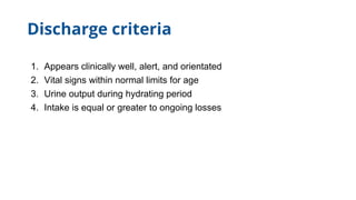
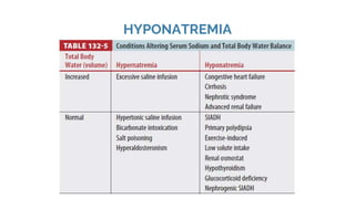
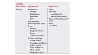
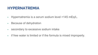
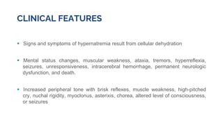
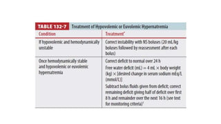
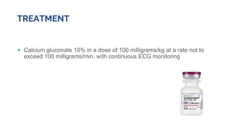
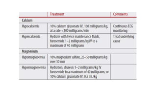
SlideShare a Scribd company logo
Fluid and Electrolyte Therapy in
Infants and Children
Dr Raseena vattamkandathil
MEM Resident
Why Dehydration..?
High percentage of total body
water
Decreased ability to control
water loss
Decreased ability to concentrate
the urine
TYPES OF DEHYDRATION
1. Isonatremic (isotonic) dehydration - MC
2. Hyponatremic (hypotonic) dehydration
3. Hypernatremic (hypertonic) dehydration
HISTORY
● Symptoms? what they are? when they started? where they started?
e.g., was the child in a hot environment?
● Whether fever is present?
● Tachypneic? and prior treatment.?
Breast feeding history
 Frequency of feeds
 Whether the mother feels she has good milk production
 Whether the infant is feeding or engaged in
nonnutrient sucking for comfort.
 If not breastfeeding, ask what type of fluid has been
Given- hypotonic fluids (e.g., water) increase the risk of
hyponatremia.
BOTTLE FED INFANTS
● Whether the formula is premixed or made from
powder
● hypernatremia or hyponatremia can result from inappropriately prepared
formula.
ASSESS OUTPUT
● Questions surrounding output aid in assessing whether replacement of losses has been
adequate
● Excess output results from vomiting or diarrhea (quantify the frequency and volume if
possible).
● Assess urine output by asking how often the child is urinating
● The number of wet diapers if the child is not yet toilet trained.
Inquire about volume status
● Ask about tear production
● The presence or absence of sweat
● Child’s general appearance
● Mental status: is the child increasingly irritable or lethargic?
● Has the parent noticed a change in the skin (cyanotic, pale, mottled)?
● Ask about signs or symptoms of infection
● Recent travel, sick contacts
● Underlying chronic disease, which may point to a specific cause of
dehydration.
● Children are at risk for accidental ingestion of toxins or plants, many of
which can cause vomiting and lead to electrolyte disturbances.
Most important, is a change in weight, because weight loss
is the gold standard for assessment of volume status
On examination..
● Tachycardia is an early sign of dehydration
● Tachypnea - metabolic acidosis
● Mental Status and the presence of lethargy or hypotonia - severe dehydration or
electrolyte abnormalities.
● Infants- fontanelle
● Presence or absence of tears when crying
● Mucous Membranes - cracked, dry lips or decreased saliva in the mouth
● Temperature, colour, and turgor of the skin
● Capillary refill time- less than 2 seconds when normal.
● Character of the pulses - diminished pulses in significant dehydration.
‘’Perform a bedside glucose test in any child presenting with altered
level of consciousness, and rapidly correct hypoglycaemia’’
NASOGASTRIC VS IV HYDRATION
● For children unable to tolerate oral rehydration, nasogastric hydration is effective,
even in vomiting patients.
● Nasogastric treatment is more cost effective than IV treatment.
“In a large study comparing nasogastric hydration versus IV hydration over 3 hours,
subjects in the nasogastric-treated group had fewer complications, achieved resolution of
ketonuria more often, and had greater reduction in specific gravity than IV-treated
subjects”
INITIAL TREATMENT OF DEHYDRATION
MODERATE AND SEVERE DEHYDRATION
● The child unable to tolerate oral/nasogastric rehydration therapy or with severe
dehydration requires prompt fluid resuscitation with large volumes of fluid over a short
period of time
● Give 20 mL/kg boluses over 5 to 10 minutes repetitively until hemodynamic
stabilization
● Up to 60 mL/kg or more may be required in the 1st hour, unless contraindicated
● Use an isotonic solution such as 0.9% saline or a lactated Ringer’s
solution during this resuscitation phase.
● After initial volume expansion, continue replacement with either normal
saline or 5% dextrose
The Formula for daily fluid requirements
● For the first 10 kg: 100 mL/kg/d (4 mL/kg/h)
● For the second 10 kg: 50 mL/kg/d (2 mL/kg/h)
● For each kg >20 kg: 20 mL/kg/d (1 mL/kg/h)
“Because hyponatremia is the most common intragenic complication of IV
fluid therapy, it is important that isotonic solutions be used as maintenance
fluid, such as normal saline with 5% dextrose”
Discharge criteria
1. Appears clinically well, alert, and orientated
2. Vital signs within normal limits for age
3. Urine output during hydrating period
4. Intake is equal or greater to ongoing losses
HYPONATREMIA
 First determine if a low sodium value is a true value by relating the
sodium value to the osmolarity.
 If hyponatremia occurs in a hyperosmolar state - osmotically active solute
in the plasma such as excess glucose or alcohol.
 If hyponatremia occurs in the presence of normal osmolarity-
hyperlipidemia or hyperproteinemia- correct the underlying disorder rather
than the serum sodium level.
 When hyponatremia occurs in a hypo-osmolar state - excess of free water
or loss of sodium.
CLINICAL FEATURES
 Signs and symptoms of hyponatremia depend on the serum sodium level and
the speed at which the sodium level falls.
 Symptoms primarily involve the CNS
 Neurologic symptoms: nausea, vomiting, headache, mental status changes,
altered consciousness, diminished reflexes, hypothermia, pseudobulbar
palsy, and seizures.
 Musculoskeletal symptoms: weakness, muscle cramps, and lethargy.
 Although patients may be only mildly symptomatic with sodium levels as low
as 120 mEq/L if the low level is chronic (>48 hours), symptoms usually occur
with an acute drop in serum sodium level below 120 mEq/L.
 Without appropriate treatment, complications include respiratory failure,
seizures, and death.
 Treatment depends on the stability of the patient and associated symptoms.
 Rapid correction can cause severe demyelination of brainstem neurons.
 If severe neurologic symptoms, such as confusion, altered level of consciousness, or
seizures, typically with sodium level <120 mmol/L - a rapid, controlled increase in sodium
level is required until neurologic symptoms resolve or a sodium level of 120 mmol/L is
achieved.
● For euvolemic hyponatremia, after correction of serum sodium level, begin water
restriction and treat the underlying disorder.
● For hypervolemic hyponatremia (edema), start sodium and water restriction and
administer diuretics if needed to treat the clinical condition
HYPERNATREMIA
 Hypernatremia is a serum sodium level >145 mEq/L.
 Because of dehydration
 secondary to excessive sodium intake
 if free water is limited or if the formula is mixed improperly.
● Serum sodium levels of >160 mEq/L require immediate attention due to
the potential for serious complications and permanent neurologic sequelae
● Patients who have a sodium level of <160 mEq/L and receive treatment
typically have symptoms that are relatively mild and self-limited.
CLINICAL FEATURES
 Signs and symptoms of hypernatremia result from cellular dehydration
 Mental status changes, muscular weakness, ataxia, tremors, hyperreflexia,
seizures, unresponsiveness, intracerebral hemorrhage, permanent neurologic
dysfunction, and death.
 Increased peripheral tone with brisk reflexes, muscle weakness, high-pitched
cry, nuchal rigidity, myoclonus, asterixis, chorea, altered level of consciousness,
or seizures
● Correct serum sodium gradually to avoid cerebral edema and associated
central pontine myelinolysis
● Closely monitor serum sodium levels every hour initially to ensure that the
level is reduced no faster than 1 mEq/L/h and no more than 15 mEq/L in
the first 24 hours.
● Monitor urine output given the risk of acute tubular necrosis
● Correct underlying causes.
● Hypervolemic hypernatremia may require dialysis if sodium levels cannot be
decreased without volume overload.
● Dialysis may also be required for hypernatremia of any type if the initial serum
sodium is >180 mmol/L.
Hypokalemia
● Most commonly occurs secondary to profuse vomiting and/or diarrhea.
● Therapy with loop or thiazide diuretics, mineralocorticoids, or laxatives and
diabetic ketoacidosis.
● In diabetic ketoacidosis, profound hypokalemia can result from osmotic diuresis,
although in the face of the hydrogen–potassium shift that accompanies acidemia,
serum levels may be normal or falsely elevated.
Uncommon causes of hypokalemia
 Renal tubular acidosis
 Bartter’s or Gitelman’s syndrome
 Cushing’s syndrome
 Familial hypokalemia-induced paralysis.
CLINICAL FEATURES
● In most cases, hypokalemia occurs slowly, and thus patients are asymptomatic.
● Clinical signs tend to reflect the rate of fall of serum potassium rather than the
absolute level.
● Severe potassium depletion can result in skeletal muscle weakness, ileus, and
cardiac conduction disturbances.
● A prominent ECG manifestation is the U wave.
HYPOCALCEMIA
▪ Serum calcium level <8 milligrams/dL (2 mmol/L) or
▪ Ionized calcium level <4.4 milligrams/dL (1.1 mmol/L);
CAUSES
Hypoparathyroidism
 Idiopathic
 DiGeorge’s syndrome
 after thyroid surgery
 associated with magnesium
deficiency.
End-organ resistance to
parathyroid hormone.
 Associated with vitamin D
deficiency.
 Dietary deficiency
 Chronic renal failure.
 Young infants fed cow’s milk, which is high in phosphate, can develop
severe hypocalcemia.
 hyperventilation: the decreased Pco2 results in an acute respiratory
alkalosis that rapidly decreases levels of ionized calcium.
Clinical features
 Muscle weakness, vomiting, and irritability.
 Infants may simply appear “jittery.”
 In severe cases, tetany, laryngospasm, carpopedal spasm, and seizures can
occur.
 Carpopedal spasm is especially common in children with hyperventilation
syndrome.
“The most characteristic ECG abnormality of hypocalcemia
is a prolonged QT interval”
INVESTIGATION
 Total serum and ionized calcium, phosphate
 Total protein and albumin
 Parathyroid hormone
 BUN, and creatinine levels.
 Urine calcium level should also be collected.
 In neonates, a chest radiograph should be done to look for a thymic shadow in infants and
young children. If the thymus is not present, consider DiGeorge’s syndrome.
TREATMENT
 Calcium gluconate 10% in a dose of 100 milligrams/kg at a rate not to
exceed 100 milligrams/min, with continuous ECG monitoring
HYPERCALCEMIA Etiology
 Malignancy involving the lymphoreticular system.
 Vitamin A or D intoxication
 Hyperparathyroid syndromes
 Hyperthyroidism
 Adrenal Insufficiency
 Pheochromocytoma
Clinical features
 Hypotonia
 Fatigue, irritability, anorexia
 Vomiting, and constipation.
 Affected children may be clinically dehydrated and complain of polyuria
and/or polydipsia.
 An ECG may reveal bradycardia and a shortened QT interval.
INVESTIGATIONS
 Total Serum and ionized calcium levels
 CBC
 Total protein and albumin
 Alkaline phosphatase levels.
 An evaluation of the vitamin D level may also be indicated, depending on
the patient’s medical history
 Acutely, patients with functioning kidneys can be treated with aggressive
IV hydration with or without furosemide, 1 to 2 milligrams/kg IV, to a
maximum of 40 milligrams.
 Then, treat the underlying cause
HYPOMAGNESEMIA ETIOLOGY
 GI loss : Diarrhea, malabsorption
short gut, and fistulas
 Hyperaldosteronism
 Iatrogenic causes of renal loss :
• Osmotic diuretics
• parenteral fluids
• Antibiotics
• Chemotherapeutics
 DM
 Disorders of the
parathyroid glands
“Hypercalcemia may cause magnesium loss as well as
hypophosphatemia”
CLINICAL FEATURES
 Hypocalcaemia like symptoms : muscle spasms, weakness, or even
atrophy
 CNS symptoms include ataxia, abnormal movements, nystagmus, and
seizures
 Cardiac changes - prolonged PR and QT intervals and may predispose to
arrhythmias such as torsades de pointes.
TREATMENT
 In symptomatic patients give IV magnesium sulfate, 25 to 50 milligrams/kg as
10% solution over 30 minutes, and repeat every 4 to 6 hours as needed.
 Include magnesium in parenteral or enteral nutritional liquids in chronically ill
children
HYPERMAGNESEMIA
 Hypermagnesemia is rare.
 Serum levels of >2.2 mEq/L are considered elevated.
 The most common cause is ingestion of exogenous magnesium, typically
found in antacids and laxatives.
 Patients with renal dysfunction are at increased risk.
CLINICAL FEATURES
 Hypotension
 loss of deep tendon reflexes
 respiratory failure.
 Cardiac manifestations include widening of the QRS, PR, and QT intervals.
 Treatment is removal of exogenous sources
 hydration accompanied by diuresis.
 Severe symptoms may be mitigated with IV calcium, 0.5 mL/kg delivered
as calcium gluconate.
 Dialysis is effective in patients with renal failure.
………….THANK YOU

More Related Content

Similar to Fluid and Electrolyte Therapy in paediatrics

nelson hyponatremia.pptx
nelson hyponatremia.pptxnelson hyponatremia.pptx
nelson hyponatremia.pptx
Shubhendra4
 
hyponatremiappt-170315180214 (1) (1).pptx
hyponatremiappt-170315180214 (1) (1).pptxhyponatremiappt-170315180214 (1) (1).pptx
hyponatremiappt-170315180214 (1) (1).pptx
AkhilGamingYt
 
Neonatal emergencies
Neonatal emergenciesNeonatal emergencies
Neonatal emergencies
Natangwe Tangi
 
Endocrine Emergencies.pptx
Endocrine Emergencies.pptxEndocrine Emergencies.pptx
Endocrine Emergencies.pptx
munriz
 
Sodium metabolism, hyponatraemia &amp; hypernatraemia
Sodium metabolism, hyponatraemia &amp; hypernatraemiaSodium metabolism, hyponatraemia &amp; hypernatraemia
Sodium metabolism, hyponatraemia &amp; hypernatraemia
AshikMajumder1
 
Endocrine emergencies
Endocrine emergenciesEndocrine emergencies
Endocrine emergenciesDr. Rubz
 
Endocrine emergencies
Endocrine emergenciesEndocrine emergencies
Endocrine emergencies
Mohd Hanafi
 
hyponatremiappt-170315180214.pptx
hyponatremiappt-170315180214.pptxhyponatremiappt-170315180214.pptx
hyponatremiappt-170315180214.pptx
SushantaPaudel
 
Approach to hyponatremia
Approach to hyponatremiaApproach to hyponatremia
Approach to hyponatremia
DrHammadArshi
 
Diabetic complications
Diabetic complicationsDiabetic complications
Diabetic complications
Lama K Banna
 
Hypernatremia
HypernatremiaHypernatremia
Hypernatremia
Khushi Devgan
 
fluids in children maintenance therapy and normal
fluids in children maintenance therapy and normalfluids in children maintenance therapy and normal
fluids in children maintenance therapy and normal
richardkikondo5
 
electrolyte।।।।।।।।।।।।।।।।।।।।।।।।।।।।।.pptx
electrolyte।।।।।।।।।।।।।।।।।।।।।।।।।।।।।.pptxelectrolyte।।।।।।।।।।।।।।।।।।।।।।।।।।।।।.pptx
electrolyte।।।।।।।।।।।।।।।।।।।।।।।।।।।।।.pptx
imrulsujon1
 
Approach to a patient with hyponatremia (2) (1)
Approach to a patient with hyponatremia (2) (1)Approach to a patient with hyponatremia (2) (1)
Approach to a patient with hyponatremia (2) (1)
Mohit Aggarwal
 
HHS.pptx
HHS.pptxHHS.pptx
HHS.pptx
HaziqMars1
 
Hyponatremia ppt .final
Hyponatremia ppt .finalHyponatremia ppt .final
Hyponatremia ppt .final
Arun Karmakar
 
Neurology of electrolyte imbalance
Neurology of electrolyte imbalanceNeurology of electrolyte imbalance
Neurology of electrolyte imbalanceNeurologyKota
 
Metabolic-Emergencies.pptx
Metabolic-Emergencies.pptxMetabolic-Emergencies.pptx
Metabolic-Emergencies.pptx
WengelRedkiss
 
Sodium imbalance
Sodium imbalanceSodium imbalance
Sodium imbalance
mauryaramgopal
 
hyponatremia -my prensentation
hyponatremia -my prensentationhyponatremia -my prensentation
hyponatremia -my prensentationSudhir K. Yadav
 

Similar to Fluid and Electrolyte Therapy in paediatrics (20)

nelson hyponatremia.pptx
nelson hyponatremia.pptxnelson hyponatremia.pptx
nelson hyponatremia.pptx
 
hyponatremiappt-170315180214 (1) (1).pptx
hyponatremiappt-170315180214 (1) (1).pptxhyponatremiappt-170315180214 (1) (1).pptx
hyponatremiappt-170315180214 (1) (1).pptx
 
Neonatal emergencies
Neonatal emergenciesNeonatal emergencies
Neonatal emergencies
 
Endocrine Emergencies.pptx
Endocrine Emergencies.pptxEndocrine Emergencies.pptx
Endocrine Emergencies.pptx
 
Sodium metabolism, hyponatraemia &amp; hypernatraemia
Sodium metabolism, hyponatraemia &amp; hypernatraemiaSodium metabolism, hyponatraemia &amp; hypernatraemia
Sodium metabolism, hyponatraemia &amp; hypernatraemia
 
Endocrine emergencies
Endocrine emergenciesEndocrine emergencies
Endocrine emergencies
 
Endocrine emergencies
Endocrine emergenciesEndocrine emergencies
Endocrine emergencies
 
hyponatremiappt-170315180214.pptx
hyponatremiappt-170315180214.pptxhyponatremiappt-170315180214.pptx
hyponatremiappt-170315180214.pptx
 
Approach to hyponatremia
Approach to hyponatremiaApproach to hyponatremia
Approach to hyponatremia
 
Diabetic complications
Diabetic complicationsDiabetic complications
Diabetic complications
 
Hypernatremia
HypernatremiaHypernatremia
Hypernatremia
 
fluids in children maintenance therapy and normal
fluids in children maintenance therapy and normalfluids in children maintenance therapy and normal
fluids in children maintenance therapy and normal
 
electrolyte।।।।।।।।।।।।।।।।।।।।।।।।।।।।।.pptx
electrolyte।।।।।।।।।।।।।।।।।।।।।।।।।।।।।.pptxelectrolyte।।।।।।।।।।।।।।।।।।।।।।।।।।।।।.pptx
electrolyte।।।।।।।।।।।।।।।।।।।।।।।।।।।।।.pptx
 
Approach to a patient with hyponatremia (2) (1)
Approach to a patient with hyponatremia (2) (1)Approach to a patient with hyponatremia (2) (1)
Approach to a patient with hyponatremia (2) (1)
 
HHS.pptx
HHS.pptxHHS.pptx
HHS.pptx
 
Hyponatremia ppt .final
Hyponatremia ppt .finalHyponatremia ppt .final
Hyponatremia ppt .final
 
Neurology of electrolyte imbalance
Neurology of electrolyte imbalanceNeurology of electrolyte imbalance
Neurology of electrolyte imbalance
 
Metabolic-Emergencies.pptx
Metabolic-Emergencies.pptxMetabolic-Emergencies.pptx
Metabolic-Emergencies.pptx
 
Sodium imbalance
Sodium imbalanceSodium imbalance
Sodium imbalance
 
hyponatremia -my prensentation
hyponatremia -my prensentationhyponatremia -my prensentation
hyponatremia -my prensentation
 

Recently uploaded

Surat @ℂall @Girls ꧁❤8527049040❤꧂@ℂall @Girls Service Vip Top Model Safe
Surat @ℂall @Girls ꧁❤8527049040❤꧂@ℂall @Girls Service Vip Top Model SafeSurat @ℂall @Girls ꧁❤8527049040❤꧂@ℂall @Girls Service Vip Top Model Safe
Surat @ℂall @Girls ꧁❤8527049040❤꧂@ℂall @Girls Service Vip Top Model Safe
Savita Shen $i11
 
Alcohol_Dr. Jeenal Mistry MD Pharmacology.pdf
Alcohol_Dr. Jeenal Mistry MD Pharmacology.pdfAlcohol_Dr. Jeenal Mistry MD Pharmacology.pdf
Alcohol_Dr. Jeenal Mistry MD Pharmacology.pdf
Dr Jeenal Mistry
 
Triangles of Neck and Clinical Correlation by Dr. RIG.pptx
Triangles of Neck and Clinical Correlation by Dr. RIG.pptxTriangles of Neck and Clinical Correlation by Dr. RIG.pptx
Triangles of Neck and Clinical Correlation by Dr. RIG.pptx
Dr. Rabia Inam Gandapore
 
How STIs Influence the Development of Pelvic Inflammatory Disease.pptx
How STIs Influence the Development of Pelvic Inflammatory Disease.pptxHow STIs Influence the Development of Pelvic Inflammatory Disease.pptx
How STIs Influence the Development of Pelvic Inflammatory Disease.pptx
FFragrant
 
basicmodesofventilation2022-220313203758.pdf
basicmodesofventilation2022-220313203758.pdfbasicmodesofventilation2022-220313203758.pdf
basicmodesofventilation2022-220313203758.pdf
aljamhori teaching hospital
 
KDIGO 2024 guidelines for diabetologists
KDIGO 2024 guidelines for diabetologistsKDIGO 2024 guidelines for diabetologists
KDIGO 2024 guidelines for diabetologists
د.محمود نجيب
 
Non-respiratory Functions of the Lungs.pdf
Non-respiratory Functions of the Lungs.pdfNon-respiratory Functions of the Lungs.pdf
Non-respiratory Functions of the Lungs.pdf
MedicoseAcademics
 
TEST BANK for Operations Management, 14th Edition by William J. Stevenson, Ve...
TEST BANK for Operations Management, 14th Edition by William J. Stevenson, Ve...TEST BANK for Operations Management, 14th Edition by William J. Stevenson, Ve...
TEST BANK for Operations Management, 14th Edition by William J. Stevenson, Ve...
kevinkariuki227
 
micro teaching on communication m.sc nursing.pdf
micro teaching on communication m.sc nursing.pdfmicro teaching on communication m.sc nursing.pdf
micro teaching on communication m.sc nursing.pdf
Anurag Sharma
 
Basavarajeeyam - Ayurvedic heritage book of Andhra pradesh
Basavarajeeyam - Ayurvedic heritage book of Andhra pradeshBasavarajeeyam - Ayurvedic heritage book of Andhra pradesh
Basavarajeeyam - Ayurvedic heritage book of Andhra pradesh
Dr. Madduru Muni Haritha
 
For Better Surat #ℂall #Girl Service ❤85270-49040❤ Surat #ℂall #Girls
For Better Surat #ℂall #Girl Service ❤85270-49040❤ Surat #ℂall #GirlsFor Better Surat #ℂall #Girl Service ❤85270-49040❤ Surat #ℂall #Girls
For Better Surat #ℂall #Girl Service ❤85270-49040❤ Surat #ℂall #Girls
Savita Shen $i11
 
Knee anatomy and clinical tests 2024.pdf
Knee anatomy and clinical tests 2024.pdfKnee anatomy and clinical tests 2024.pdf
Knee anatomy and clinical tests 2024.pdf
vimalpl1234
 
Are There Any Natural Remedies To Treat Syphilis.pdf
Are There Any Natural Remedies To Treat Syphilis.pdfAre There Any Natural Remedies To Treat Syphilis.pdf
Are There Any Natural Remedies To Treat Syphilis.pdf
Little Cross Family Clinic
 
Ocular injury ppt Upendra pal optometrist upums saifai etawah
Ocular injury  ppt  Upendra pal  optometrist upums saifai etawahOcular injury  ppt  Upendra pal  optometrist upums saifai etawah
Ocular injury ppt Upendra pal optometrist upums saifai etawah
pal078100
 
Novas diretrizes da OMS para os cuidados perinatais de mais qualidade
Novas diretrizes da OMS para os cuidados perinatais de mais qualidadeNovas diretrizes da OMS para os cuidados perinatais de mais qualidade
Novas diretrizes da OMS para os cuidados perinatais de mais qualidade
Prof. Marcus Renato de Carvalho
 
Ophthalmology Clinical Tests for OSCE exam
Ophthalmology Clinical Tests for OSCE examOphthalmology Clinical Tests for OSCE exam
Ophthalmology Clinical Tests for OSCE exam
KafrELShiekh University
 
Phone Us ❤85270-49040❤ #ℂall #gIRLS In Surat By Surat @ℂall @Girls Hotel With...
Phone Us ❤85270-49040❤ #ℂall #gIRLS In Surat By Surat @ℂall @Girls Hotel With...Phone Us ❤85270-49040❤ #ℂall #gIRLS In Surat By Surat @ℂall @Girls Hotel With...
Phone Us ❤85270-49040❤ #ℂall #gIRLS In Surat By Surat @ℂall @Girls Hotel With...
Savita Shen $i11
 
Pictures of Superficial & Deep Fascia.ppt.pdf
Pictures of Superficial & Deep Fascia.ppt.pdfPictures of Superficial & Deep Fascia.ppt.pdf
Pictures of Superficial & Deep Fascia.ppt.pdf
Dr. Rabia Inam Gandapore
 
Superficial & Deep Fascia of the NECK.pptx
Superficial & Deep Fascia of the NECK.pptxSuperficial & Deep Fascia of the NECK.pptx
Superficial & Deep Fascia of the NECK.pptx
Dr. Rabia Inam Gandapore
 
MANAGEMENT OF ATRIOVENTRICULAR CONDUCTION BLOCK.pdf
MANAGEMENT OF ATRIOVENTRICULAR CONDUCTION BLOCK.pdfMANAGEMENT OF ATRIOVENTRICULAR CONDUCTION BLOCK.pdf
MANAGEMENT OF ATRIOVENTRICULAR CONDUCTION BLOCK.pdf
Jim Jacob Roy
 

Recently uploaded (20)

Surat @ℂall @Girls ꧁❤8527049040❤꧂@ℂall @Girls Service Vip Top Model Safe
Surat @ℂall @Girls ꧁❤8527049040❤꧂@ℂall @Girls Service Vip Top Model SafeSurat @ℂall @Girls ꧁❤8527049040❤꧂@ℂall @Girls Service Vip Top Model Safe
Surat @ℂall @Girls ꧁❤8527049040❤꧂@ℂall @Girls Service Vip Top Model Safe
 
Alcohol_Dr. Jeenal Mistry MD Pharmacology.pdf
Alcohol_Dr. Jeenal Mistry MD Pharmacology.pdfAlcohol_Dr. Jeenal Mistry MD Pharmacology.pdf
Alcohol_Dr. Jeenal Mistry MD Pharmacology.pdf
 
Triangles of Neck and Clinical Correlation by Dr. RIG.pptx
Triangles of Neck and Clinical Correlation by Dr. RIG.pptxTriangles of Neck and Clinical Correlation by Dr. RIG.pptx
Triangles of Neck and Clinical Correlation by Dr. RIG.pptx
 
How STIs Influence the Development of Pelvic Inflammatory Disease.pptx
How STIs Influence the Development of Pelvic Inflammatory Disease.pptxHow STIs Influence the Development of Pelvic Inflammatory Disease.pptx
How STIs Influence the Development of Pelvic Inflammatory Disease.pptx
 
basicmodesofventilation2022-220313203758.pdf
basicmodesofventilation2022-220313203758.pdfbasicmodesofventilation2022-220313203758.pdf
basicmodesofventilation2022-220313203758.pdf
 
KDIGO 2024 guidelines for diabetologists
KDIGO 2024 guidelines for diabetologistsKDIGO 2024 guidelines for diabetologists
KDIGO 2024 guidelines for diabetologists
 
Non-respiratory Functions of the Lungs.pdf
Non-respiratory Functions of the Lungs.pdfNon-respiratory Functions of the Lungs.pdf
Non-respiratory Functions of the Lungs.pdf
 
TEST BANK for Operations Management, 14th Edition by William J. Stevenson, Ve...
TEST BANK for Operations Management, 14th Edition by William J. Stevenson, Ve...TEST BANK for Operations Management, 14th Edition by William J. Stevenson, Ve...
TEST BANK for Operations Management, 14th Edition by William J. Stevenson, Ve...
 
micro teaching on communication m.sc nursing.pdf
micro teaching on communication m.sc nursing.pdfmicro teaching on communication m.sc nursing.pdf
micro teaching on communication m.sc nursing.pdf
 
Basavarajeeyam - Ayurvedic heritage book of Andhra pradesh
Basavarajeeyam - Ayurvedic heritage book of Andhra pradeshBasavarajeeyam - Ayurvedic heritage book of Andhra pradesh
Basavarajeeyam - Ayurvedic heritage book of Andhra pradesh
 
For Better Surat #ℂall #Girl Service ❤85270-49040❤ Surat #ℂall #Girls
For Better Surat #ℂall #Girl Service ❤85270-49040❤ Surat #ℂall #GirlsFor Better Surat #ℂall #Girl Service ❤85270-49040❤ Surat #ℂall #Girls
For Better Surat #ℂall #Girl Service ❤85270-49040❤ Surat #ℂall #Girls
 
Knee anatomy and clinical tests 2024.pdf
Knee anatomy and clinical tests 2024.pdfKnee anatomy and clinical tests 2024.pdf
Knee anatomy and clinical tests 2024.pdf
 
Are There Any Natural Remedies To Treat Syphilis.pdf
Are There Any Natural Remedies To Treat Syphilis.pdfAre There Any Natural Remedies To Treat Syphilis.pdf
Are There Any Natural Remedies To Treat Syphilis.pdf
 
Ocular injury ppt Upendra pal optometrist upums saifai etawah
Ocular injury  ppt  Upendra pal  optometrist upums saifai etawahOcular injury  ppt  Upendra pal  optometrist upums saifai etawah
Ocular injury ppt Upendra pal optometrist upums saifai etawah
 
Novas diretrizes da OMS para os cuidados perinatais de mais qualidade
Novas diretrizes da OMS para os cuidados perinatais de mais qualidadeNovas diretrizes da OMS para os cuidados perinatais de mais qualidade
Novas diretrizes da OMS para os cuidados perinatais de mais qualidade
 
Ophthalmology Clinical Tests for OSCE exam
Ophthalmology Clinical Tests for OSCE examOphthalmology Clinical Tests for OSCE exam
Ophthalmology Clinical Tests for OSCE exam
 
Phone Us ❤85270-49040❤ #ℂall #gIRLS In Surat By Surat @ℂall @Girls Hotel With...
Phone Us ❤85270-49040❤ #ℂall #gIRLS In Surat By Surat @ℂall @Girls Hotel With...Phone Us ❤85270-49040❤ #ℂall #gIRLS In Surat By Surat @ℂall @Girls Hotel With...
Phone Us ❤85270-49040❤ #ℂall #gIRLS In Surat By Surat @ℂall @Girls Hotel With...
 
Pictures of Superficial & Deep Fascia.ppt.pdf
Pictures of Superficial & Deep Fascia.ppt.pdfPictures of Superficial & Deep Fascia.ppt.pdf
Pictures of Superficial & Deep Fascia.ppt.pdf
 
Superficial & Deep Fascia of the NECK.pptx
Superficial & Deep Fascia of the NECK.pptxSuperficial & Deep Fascia of the NECK.pptx
Superficial & Deep Fascia of the NECK.pptx
 
MANAGEMENT OF ATRIOVENTRICULAR CONDUCTION BLOCK.pdf
MANAGEMENT OF ATRIOVENTRICULAR CONDUCTION BLOCK.pdfMANAGEMENT OF ATRIOVENTRICULAR CONDUCTION BLOCK.pdf
MANAGEMENT OF ATRIOVENTRICULAR CONDUCTION BLOCK.pdf
 

Fluid and Electrolyte Therapy in paediatrics

  • 1. Fluid and Electrolyte Therapy in Infants and Children Dr Raseena vattamkandathil MEM Resident
  • 2. Why Dehydration..? High percentage of total body water Decreased ability to control water loss Decreased ability to concentrate the urine
  • 3. TYPES OF DEHYDRATION 1. Isonatremic (isotonic) dehydration - MC 2. Hyponatremic (hypotonic) dehydration 3. Hypernatremic (hypertonic) dehydration
  • 4. HISTORY ● Symptoms? what they are? when they started? where they started? e.g., was the child in a hot environment? ● Whether fever is present? ● Tachypneic? and prior treatment.?
  • 5. Breast feeding history  Frequency of feeds  Whether the mother feels she has good milk production  Whether the infant is feeding or engaged in nonnutrient sucking for comfort.  If not breastfeeding, ask what type of fluid has been Given- hypotonic fluids (e.g., water) increase the risk of hyponatremia.
  • 6. BOTTLE FED INFANTS ● Whether the formula is premixed or made from powder ● hypernatremia or hyponatremia can result from inappropriately prepared formula.
  • 7. ASSESS OUTPUT ● Questions surrounding output aid in assessing whether replacement of losses has been adequate ● Excess output results from vomiting or diarrhea (quantify the frequency and volume if possible). ● Assess urine output by asking how often the child is urinating ● The number of wet diapers if the child is not yet toilet trained.
  • 8. Inquire about volume status ● Ask about tear production ● The presence or absence of sweat ● Child’s general appearance ● Mental status: is the child increasingly irritable or lethargic? ● Has the parent noticed a change in the skin (cyanotic, pale, mottled)?
  • 9. ● Ask about signs or symptoms of infection ● Recent travel, sick contacts ● Underlying chronic disease, which may point to a specific cause of dehydration. ● Children are at risk for accidental ingestion of toxins or plants, many of which can cause vomiting and lead to electrolyte disturbances.
  • 10. Most important, is a change in weight, because weight loss is the gold standard for assessment of volume status
  • 11. On examination.. ● Tachycardia is an early sign of dehydration ● Tachypnea - metabolic acidosis ● Mental Status and the presence of lethargy or hypotonia - severe dehydration or electrolyte abnormalities. ● Infants- fontanelle ● Presence or absence of tears when crying ● Mucous Membranes - cracked, dry lips or decreased saliva in the mouth
  • 12. ● Temperature, colour, and turgor of the skin ● Capillary refill time- less than 2 seconds when normal. ● Character of the pulses - diminished pulses in significant dehydration.
  • 13. ‘’Perform a bedside glucose test in any child presenting with altered level of consciousness, and rapidly correct hypoglycaemia’’
  • 14. NASOGASTRIC VS IV HYDRATION ● For children unable to tolerate oral rehydration, nasogastric hydration is effective, even in vomiting patients. ● Nasogastric treatment is more cost effective than IV treatment.
  • 15. “In a large study comparing nasogastric hydration versus IV hydration over 3 hours, subjects in the nasogastric-treated group had fewer complications, achieved resolution of ketonuria more often, and had greater reduction in specific gravity than IV-treated subjects”
  • 16. INITIAL TREATMENT OF DEHYDRATION
  • 17. MODERATE AND SEVERE DEHYDRATION ● The child unable to tolerate oral/nasogastric rehydration therapy or with severe dehydration requires prompt fluid resuscitation with large volumes of fluid over a short period of time ● Give 20 mL/kg boluses over 5 to 10 minutes repetitively until hemodynamic stabilization ● Up to 60 mL/kg or more may be required in the 1st hour, unless contraindicated
  • 18. ● Use an isotonic solution such as 0.9% saline or a lactated Ringer’s solution during this resuscitation phase. ● After initial volume expansion, continue replacement with either normal saline or 5% dextrose
  • 19.
  • 20. The Formula for daily fluid requirements ● For the first 10 kg: 100 mL/kg/d (4 mL/kg/h) ● For the second 10 kg: 50 mL/kg/d (2 mL/kg/h) ● For each kg >20 kg: 20 mL/kg/d (1 mL/kg/h)
  • 21. “Because hyponatremia is the most common intragenic complication of IV fluid therapy, it is important that isotonic solutions be used as maintenance fluid, such as normal saline with 5% dextrose”
  • 22. Discharge criteria 1. Appears clinically well, alert, and orientated 2. Vital signs within normal limits for age 3. Urine output during hydrating period 4. Intake is equal or greater to ongoing losses
  • 24.
  • 25.  First determine if a low sodium value is a true value by relating the sodium value to the osmolarity.  If hyponatremia occurs in a hyperosmolar state - osmotically active solute in the plasma such as excess glucose or alcohol.  If hyponatremia occurs in the presence of normal osmolarity- hyperlipidemia or hyperproteinemia- correct the underlying disorder rather than the serum sodium level.  When hyponatremia occurs in a hypo-osmolar state - excess of free water or loss of sodium.
  • 26. CLINICAL FEATURES  Signs and symptoms of hyponatremia depend on the serum sodium level and the speed at which the sodium level falls.  Symptoms primarily involve the CNS  Neurologic symptoms: nausea, vomiting, headache, mental status changes, altered consciousness, diminished reflexes, hypothermia, pseudobulbar palsy, and seizures.  Musculoskeletal symptoms: weakness, muscle cramps, and lethargy.
  • 27.  Although patients may be only mildly symptomatic with sodium levels as low as 120 mEq/L if the low level is chronic (>48 hours), symptoms usually occur with an acute drop in serum sodium level below 120 mEq/L.  Without appropriate treatment, complications include respiratory failure, seizures, and death.
  • 28.
  • 29.  Treatment depends on the stability of the patient and associated symptoms.  Rapid correction can cause severe demyelination of brainstem neurons.  If severe neurologic symptoms, such as confusion, altered level of consciousness, or seizures, typically with sodium level <120 mmol/L - a rapid, controlled increase in sodium level is required until neurologic symptoms resolve or a sodium level of 120 mmol/L is achieved.
  • 30. ● For euvolemic hyponatremia, after correction of serum sodium level, begin water restriction and treat the underlying disorder. ● For hypervolemic hyponatremia (edema), start sodium and water restriction and administer diuretics if needed to treat the clinical condition
  • 31. HYPERNATREMIA  Hypernatremia is a serum sodium level >145 mEq/L.  Because of dehydration  secondary to excessive sodium intake  if free water is limited or if the formula is mixed improperly.
  • 32. ● Serum sodium levels of >160 mEq/L require immediate attention due to the potential for serious complications and permanent neurologic sequelae ● Patients who have a sodium level of <160 mEq/L and receive treatment typically have symptoms that are relatively mild and self-limited.
  • 33. CLINICAL FEATURES  Signs and symptoms of hypernatremia result from cellular dehydration  Mental status changes, muscular weakness, ataxia, tremors, hyperreflexia, seizures, unresponsiveness, intracerebral hemorrhage, permanent neurologic dysfunction, and death.  Increased peripheral tone with brisk reflexes, muscle weakness, high-pitched cry, nuchal rigidity, myoclonus, asterixis, chorea, altered level of consciousness, or seizures
  • 34. ● Correct serum sodium gradually to avoid cerebral edema and associated central pontine myelinolysis ● Closely monitor serum sodium levels every hour initially to ensure that the level is reduced no faster than 1 mEq/L/h and no more than 15 mEq/L in the first 24 hours.
  • 35.
  • 36. ● Monitor urine output given the risk of acute tubular necrosis ● Correct underlying causes. ● Hypervolemic hypernatremia may require dialysis if sodium levels cannot be decreased without volume overload. ● Dialysis may also be required for hypernatremia of any type if the initial serum sodium is >180 mmol/L.
  • 37. Hypokalemia ● Most commonly occurs secondary to profuse vomiting and/or diarrhea. ● Therapy with loop or thiazide diuretics, mineralocorticoids, or laxatives and diabetic ketoacidosis. ● In diabetic ketoacidosis, profound hypokalemia can result from osmotic diuresis, although in the face of the hydrogen–potassium shift that accompanies acidemia, serum levels may be normal or falsely elevated.
  • 38. Uncommon causes of hypokalemia  Renal tubular acidosis  Bartter’s or Gitelman’s syndrome  Cushing’s syndrome  Familial hypokalemia-induced paralysis.
  • 39. CLINICAL FEATURES ● In most cases, hypokalemia occurs slowly, and thus patients are asymptomatic. ● Clinical signs tend to reflect the rate of fall of serum potassium rather than the absolute level. ● Severe potassium depletion can result in skeletal muscle weakness, ileus, and cardiac conduction disturbances. ● A prominent ECG manifestation is the U wave.
  • 40.
  • 41. HYPOCALCEMIA ▪ Serum calcium level <8 milligrams/dL (2 mmol/L) or ▪ Ionized calcium level <4.4 milligrams/dL (1.1 mmol/L);
  • 42. CAUSES Hypoparathyroidism  Idiopathic  DiGeorge’s syndrome  after thyroid surgery  associated with magnesium deficiency. End-organ resistance to parathyroid hormone.  Associated with vitamin D deficiency.
  • 43.  Dietary deficiency  Chronic renal failure.  Young infants fed cow’s milk, which is high in phosphate, can develop severe hypocalcemia.  hyperventilation: the decreased Pco2 results in an acute respiratory alkalosis that rapidly decreases levels of ionized calcium.
  • 44. Clinical features  Muscle weakness, vomiting, and irritability.  Infants may simply appear “jittery.”  In severe cases, tetany, laryngospasm, carpopedal spasm, and seizures can occur.  Carpopedal spasm is especially common in children with hyperventilation syndrome.
  • 45.
  • 46. “The most characteristic ECG abnormality of hypocalcemia is a prolonged QT interval”
  • 47. INVESTIGATION  Total serum and ionized calcium, phosphate  Total protein and albumin  Parathyroid hormone  BUN, and creatinine levels.  Urine calcium level should also be collected.  In neonates, a chest radiograph should be done to look for a thymic shadow in infants and young children. If the thymus is not present, consider DiGeorge’s syndrome.
  • 48. TREATMENT  Calcium gluconate 10% in a dose of 100 milligrams/kg at a rate not to exceed 100 milligrams/min, with continuous ECG monitoring
  • 49. HYPERCALCEMIA Etiology  Malignancy involving the lymphoreticular system.  Vitamin A or D intoxication  Hyperparathyroid syndromes  Hyperthyroidism  Adrenal Insufficiency  Pheochromocytoma
  • 50. Clinical features  Hypotonia  Fatigue, irritability, anorexia  Vomiting, and constipation.  Affected children may be clinically dehydrated and complain of polyuria and/or polydipsia.  An ECG may reveal bradycardia and a shortened QT interval.
  • 51. INVESTIGATIONS  Total Serum and ionized calcium levels  CBC  Total protein and albumin  Alkaline phosphatase levels.  An evaluation of the vitamin D level may also be indicated, depending on the patient’s medical history
  • 52.  Acutely, patients with functioning kidneys can be treated with aggressive IV hydration with or without furosemide, 1 to 2 milligrams/kg IV, to a maximum of 40 milligrams.  Then, treat the underlying cause
  • 53. HYPOMAGNESEMIA ETIOLOGY  GI loss : Diarrhea, malabsorption short gut, and fistulas  Hyperaldosteronism  Iatrogenic causes of renal loss : • Osmotic diuretics • parenteral fluids • Antibiotics • Chemotherapeutics  DM  Disorders of the parathyroid glands
  • 54. “Hypercalcemia may cause magnesium loss as well as hypophosphatemia”
  • 55. CLINICAL FEATURES  Hypocalcaemia like symptoms : muscle spasms, weakness, or even atrophy  CNS symptoms include ataxia, abnormal movements, nystagmus, and seizures  Cardiac changes - prolonged PR and QT intervals and may predispose to arrhythmias such as torsades de pointes.
  • 56. TREATMENT  In symptomatic patients give IV magnesium sulfate, 25 to 50 milligrams/kg as 10% solution over 30 minutes, and repeat every 4 to 6 hours as needed.  Include magnesium in parenteral or enteral nutritional liquids in chronically ill children
  • 57. HYPERMAGNESEMIA  Hypermagnesemia is rare.  Serum levels of >2.2 mEq/L are considered elevated.  The most common cause is ingestion of exogenous magnesium, typically found in antacids and laxatives.  Patients with renal dysfunction are at increased risk.
  • 58. CLINICAL FEATURES  Hypotension  loss of deep tendon reflexes  respiratory failure.  Cardiac manifestations include widening of the QRS, PR, and QT intervals.
  • 59.  Treatment is removal of exogenous sources  hydration accompanied by diuresis.  Severe symptoms may be mitigated with IV calcium, 0.5 mL/kg delivered as calcium gluconate.  Dialysis is effective in patients with renal failure.
  • 60.