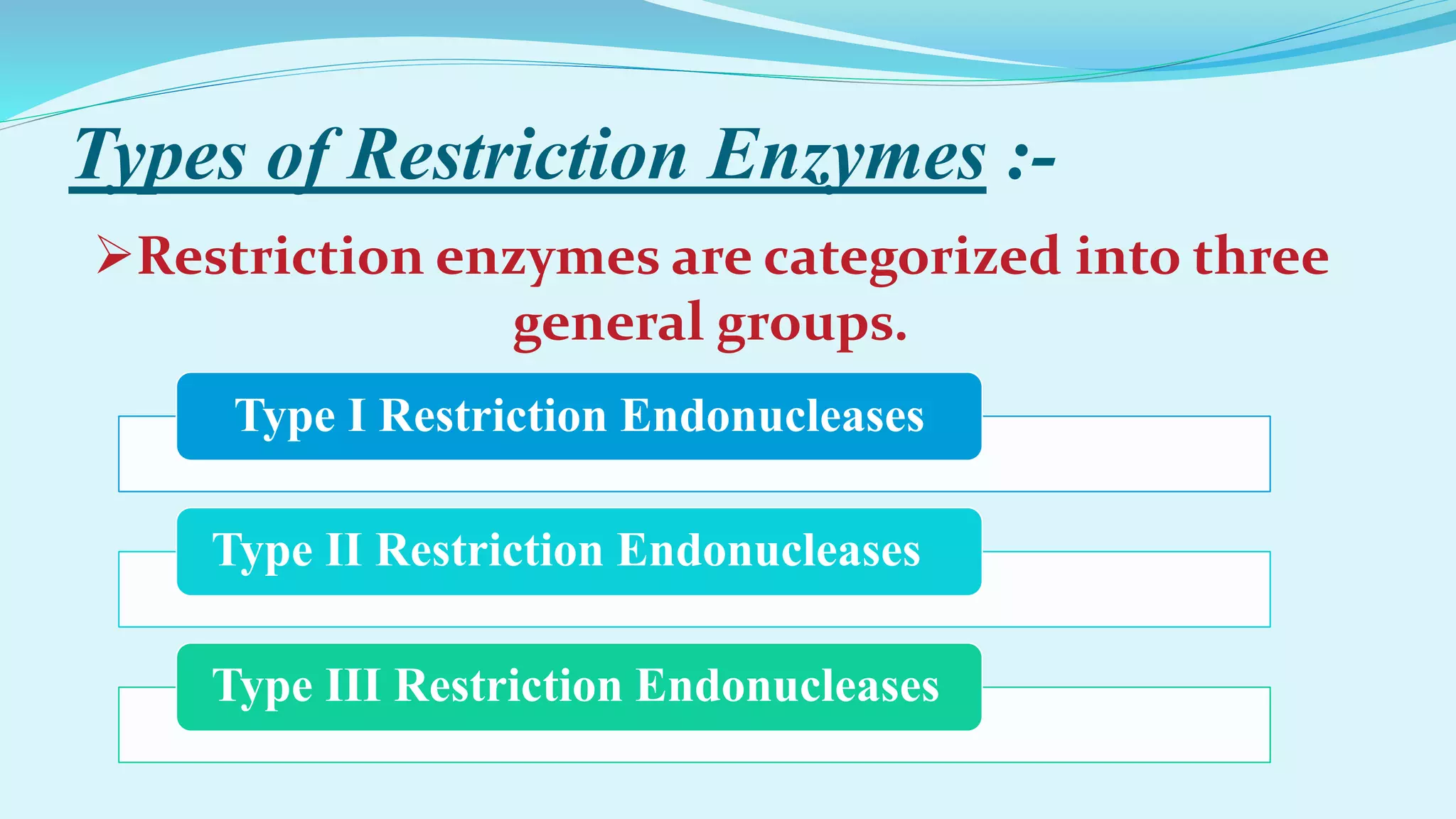
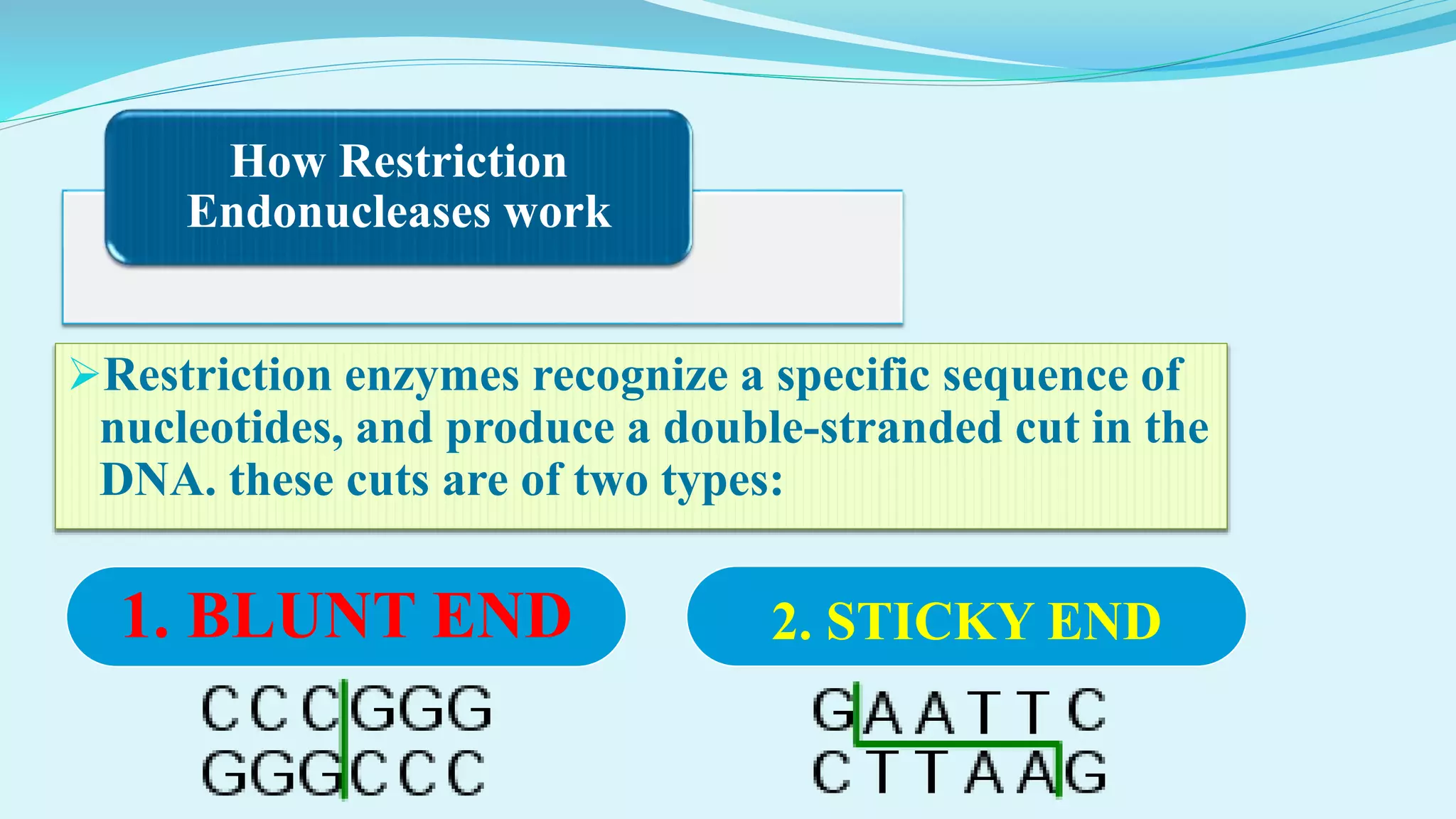
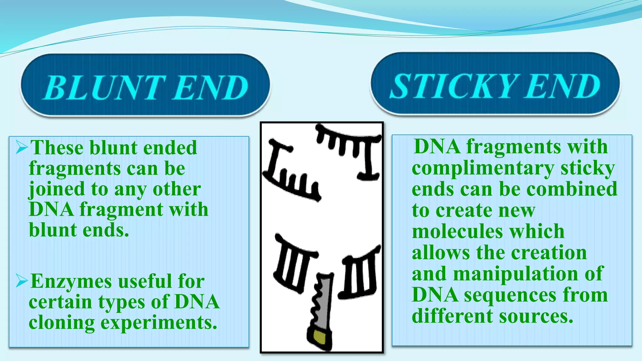
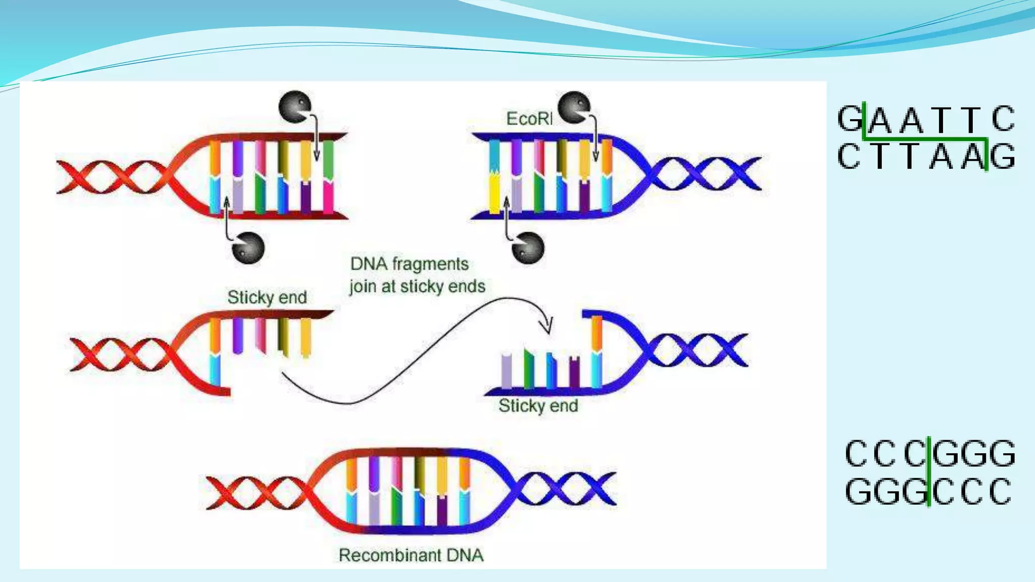
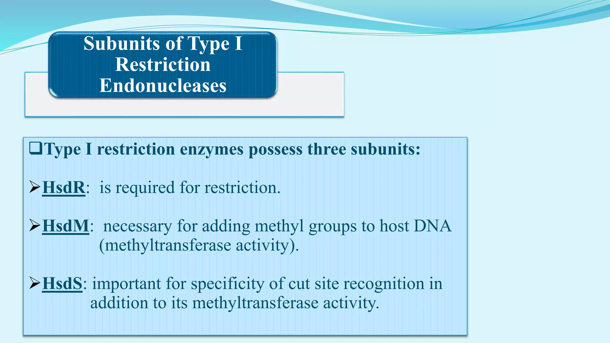
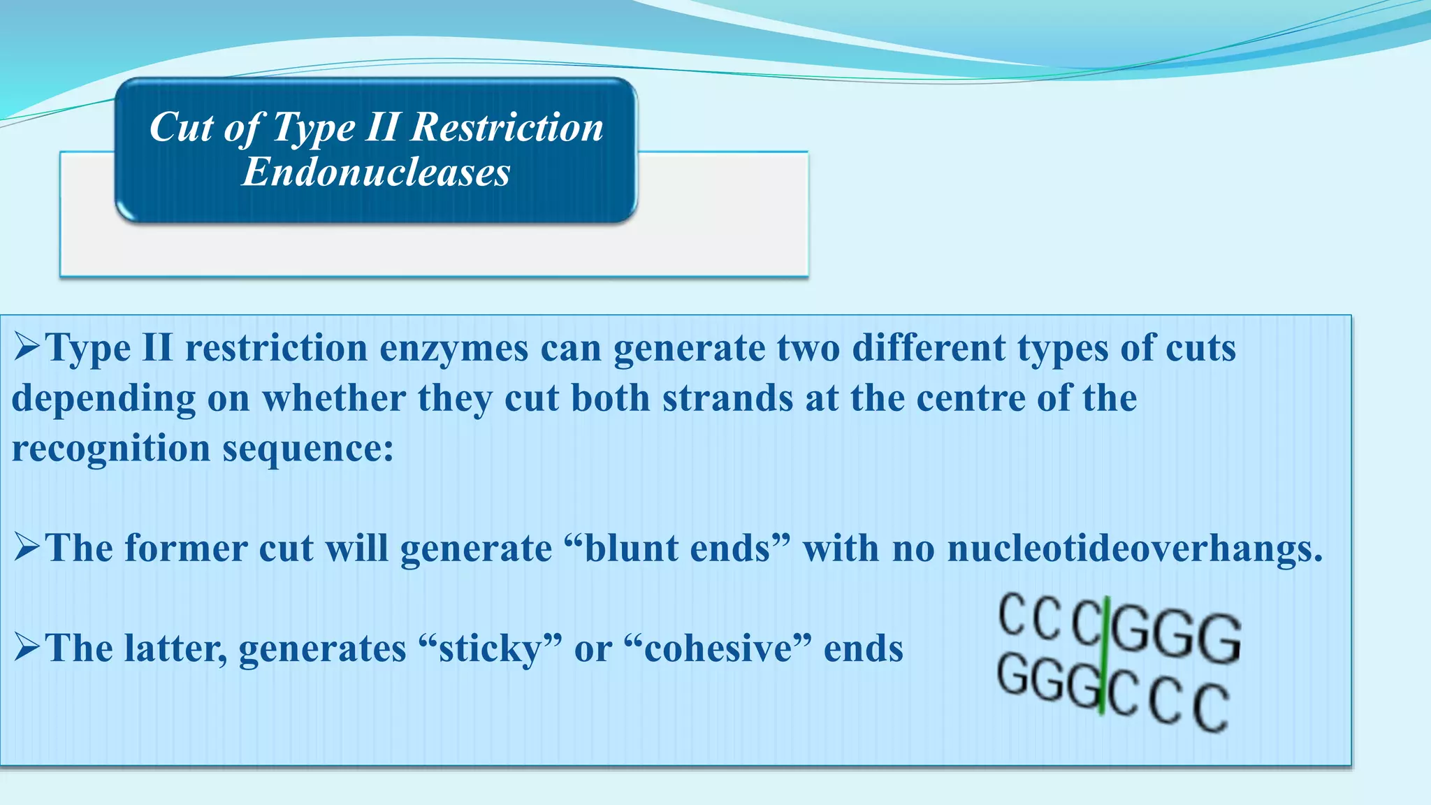
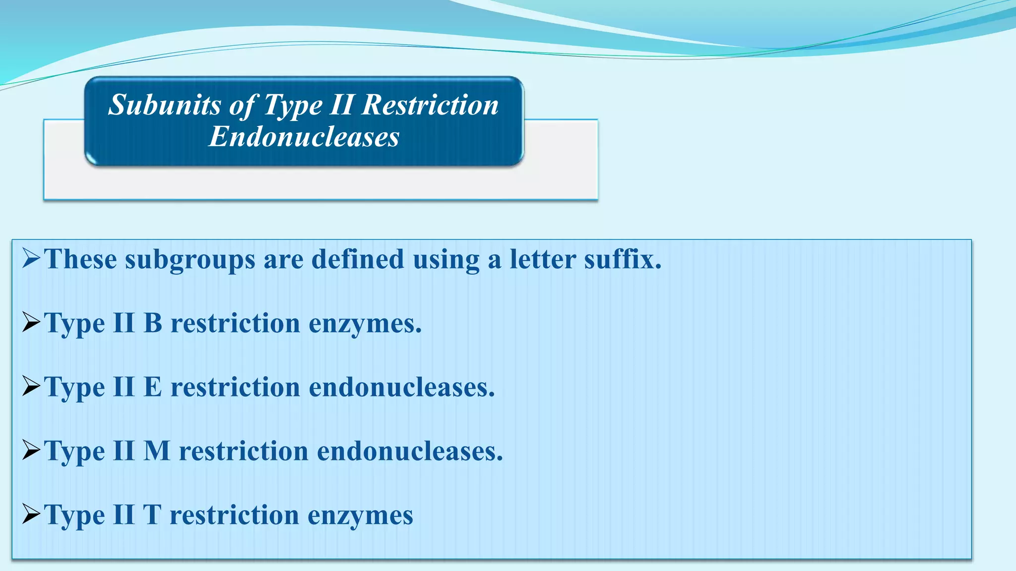
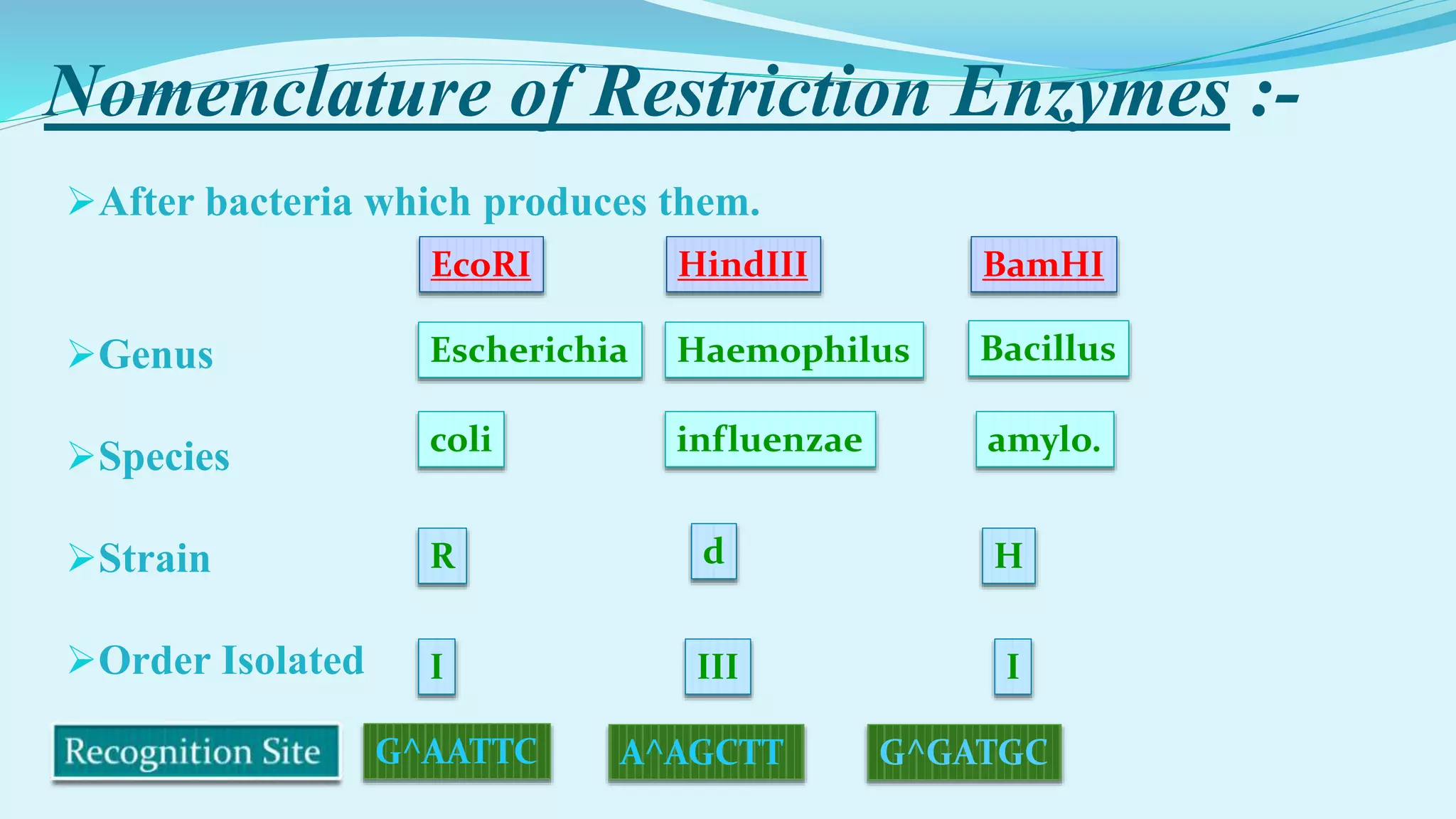
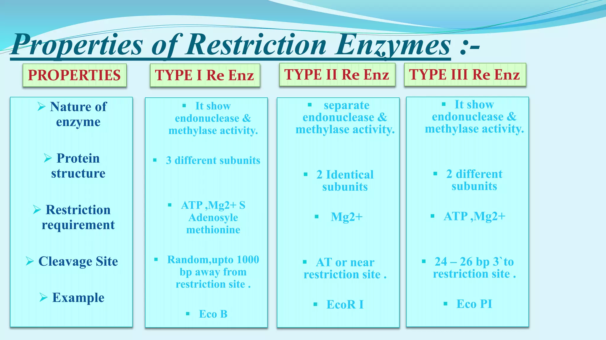
Presented by:-
MOHD ANAS
DEPT.-BIOSCIENCE
SHRI RAM COLLEGE,MUZAFFARNAGAR
Contents :-
Introduction
Discovery
How Restriction Enzymes work
Types of Restriction Enzymes
Subunits of Restriction Enzymes
Nomenclature of Restriction Enzymes
Recognition Sequences of restriction Enzymes
Properties of Restriction Enzymes
Applications of Restriction Enzymes
Introduction :-
 Restriction Endonucleases are enzymes that produce internal
cuts, called cleavage, In the DNA molecule.
 Restriction Endonuclease (Restriction Enzyme) is a bacterial
enzyme that cuts dsDNA into fragments after recognizing
specific nucleotide sequence known as recognition or
restriction site.
 Restriction Enzymes are beleved to be evolved by bacteria to
resist viral attack.
 Restriction Enzymes are also known as molecular scessor.
Discovery Of Restriction Enzyme :-
The term restriction enzyme originated from the studies of phage λ.
In 1970, Hamilton O. Smith, Thomas Kelly and Kent Wilcox isolated and characterized the first type II
restriction enzyme, HindII, from the bacterium Haemophilus influenzae
For their work in the discovery and characterization of restriction enzymes, the 1978 Nobel Prize for
Physiology or Medicine was awarded to Werner Arber, Daniel Nathans, and Hamilton O. Smith.
Types of Restriction Enzymes :-
Type I Restriction Endonucleases
Type II Restriction Endonucleases
Type III Restriction Endonucleases
Restriction enzymes are categorized into three
general groups.
Categorization of Restriction Enzymes
on the bases of :-
Their composition.
Enzyme co-factor requirement.
the nature of their target sequence.
position of their DNA cleavage site relative to
the target sequence.
How Restriction
Endonucleases work
Restriction enzymes recognize a specific sequence of
nucleotides, and produce a double-stranded cut in the
DNA. these cuts are of two types:
1. BLUNT END 2. STICKY END
These blunt ended
fragments can be
joined to any other
DNA fragment with
blunt ends.
Enzymes useful for
certain types of DNA
cloning experiments.
DNA fragments with
complimentary sticky
ends can be combined
to create new
molecules which
allows the creation
and manipulation of
DNA sequences from
different sources.
Type I Restriction
Endonucleases
 Type I restriction enzymes are complex endonucleases, and have recognition
sequence of 15 bp; they cleave the DNA about 1000 bp away from the 5` end of
the sequence “TCA” located within the recognition site. Eg:- EcoK-12,EcoB,etc.
 The type I Restriction Enzyme work with the help of many co factor like as S-
Adenosyl Methionine, Hydrolysed Adenosine Triphosphate (ATP) ad
Magnesium (Mg2+).
 The type I Restriction Enzyme methylates the DNA sequence at the site of
reognition.
 The type I Restriction Enzyme translocate the DNA sequence.
Subunits of Type I
Restriction
Endonucleases
Type I restriction enzymes possess three subunits:
HsdR: is required for restriction.
HsdM: necessary for adding methyl groups to host DNA
(methyltransferase activity).
HsdS: important for specificity of cut site recognition in
addition to its methyltransferase activity.
Type II Restriction
Endonucleases
The type II restriction enzymes are remarcably stable and
induce cleavage either, in most cases, within or outside their
recognition sequences,which are symmetrical.
The type I Restriction Enzyme work with the help of only
single co factor like as Magnesium (Mg2+).
Type II Restriction
Endonucleases
The Ist type II Enzyme to be isolated was Hind
II in 1970.
Only type II restriction endonucleases are used
for restriction mapping and gene cloning in
view of their cleavage only at specific site .
Cut of Type II Restriction
Endonucleases
Type II restriction enzymes can generate two different types of cuts
depending on whether they cut both strands at the centre of the
recognition sequence:
The former cut will generate “blunt ends” with no nucleotideoverhangs.
The latter, generates “sticky” or “cohesive” ends
Subunits of Type II Restriction
Endonucleases
These subgroups are defined using a letter suffix.
Type II B restriction enzymes.
Type II E restriction endonucleases.
Type II M restriction endonucleases.
Type II T restriction enzymes
Type III Restriction
Endonucleases
 Type III restriction enzymes are intermediate between type I and type II
endonucleases, they cleave DNA in the immediate vacinity of their recognition sites,
Eg;- EcoP1, EcoP15, Hind III etc.
They cut DNA upto 20-30 base pairs away from the recognition site.
These enzymes contain more than one subunit.(ATP & Mg2+)
And require AdoMet and ATP cofactors for their roles in DNA methylation and
restriction.
Nomenclature of Restriction Enzymes :-
After bacteria which produces them.
Genus
Species
Strain
Order Isolated
Escherichia
coli
R
I
EcoRI
Haemophilus
influenzae
d
III
HindIII
Bacillus
amylo.
H
I
BamHI
G^AATTC A^AGCTT G^GATGC
Recognition sequence of Res. Enz. :-
The recognition sequence of Type II
endonucleases form palindrome with
rotational symmetry.
Mostly Type II endonucleases have
recognition sites of 4,5 or 6 bp. Which
are predominantly GC rich.
Properties of Restriction Enzymes :-
TYPE I Re EnzPROPERTIES TYPE II Re Enz TYPE III Re Enz
 Nature of
enzyme
 Protein
structure
 Restriction
requirement
 Cleavage Site
 Example
 It show
endonuclease &
methylase activity.
 3 different subunits
 ATP ,Mg2+ S
Adenosyle
methionine
 Random,upto 1000
bp away from
restriction site .
 Eco B
 separate
endonuclease &
methylase activity.
 2 Identical
subunits
 Mg2+
 AT or near
restriction site .
 EcoR I
 It show
endonuclease &
methylase activity.
 2 different
subunits
 ATP ,Mg2+
 24 – 26 bp 3`to
restriction site .
 Eco PI
Applications of Restriction Enzymes :-
 They are used in gene cloning and protein expression experiments.
 Restriction enzymes are used in biotechnology to cut DNA into
smaller strands in order to study fragment length differences among
individuals (Restriction Fragment Length Polymorphism – RFLP).
 Each of these methods depends on the use of agarose gel
electrophoresis for separation of the DNA fragments.
Applications of Restriction Enzymes :-
Provides different ways of manipulating DNA such as the creation
of recombinant DNA, which has endless applications
Allows for the large scale production human insulin for diabetics
using E. coli, as well as for the Hepatitis B and HPV vaccines
Cloning DNA Molecules
Studying nucleotide sequence
Restriction endonucleases

Restriction endonucleases

  • 1.
  • 2.
    Contents :- Introduction Discovery How RestrictionEnzymes work Types of Restriction Enzymes Subunits of Restriction Enzymes Nomenclature of Restriction Enzymes Recognition Sequences of restriction Enzymes Properties of Restriction Enzymes Applications of Restriction Enzymes
  • 3.
    Introduction :-  RestrictionEndonucleases are enzymes that produce internal cuts, called cleavage, In the DNA molecule.  Restriction Endonuclease (Restriction Enzyme) is a bacterial enzyme that cuts dsDNA into fragments after recognizing specific nucleotide sequence known as recognition or restriction site.  Restriction Enzymes are beleved to be evolved by bacteria to resist viral attack.  Restriction Enzymes are also known as molecular scessor.
  • 4.
    Discovery Of RestrictionEnzyme :- The term restriction enzyme originated from the studies of phage λ. In 1970, Hamilton O. Smith, Thomas Kelly and Kent Wilcox isolated and characterized the first type II restriction enzyme, HindII, from the bacterium Haemophilus influenzae For their work in the discovery and characterization of restriction enzymes, the 1978 Nobel Prize for Physiology or Medicine was awarded to Werner Arber, Daniel Nathans, and Hamilton O. Smith.
  • 5.
    Types of RestrictionEnzymes :- Type I Restriction Endonucleases Type II Restriction Endonucleases Type III Restriction Endonucleases Restriction enzymes are categorized into three general groups.
  • 6.
    Categorization of RestrictionEnzymes on the bases of :- Their composition. Enzyme co-factor requirement. the nature of their target sequence. position of their DNA cleavage site relative to the target sequence.
  • 7.
    How Restriction Endonucleases work Restrictionenzymes recognize a specific sequence of nucleotides, and produce a double-stranded cut in the DNA. these cuts are of two types: 1. BLUNT END 2. STICKY END
  • 8.
    These blunt ended fragmentscan be joined to any other DNA fragment with blunt ends. Enzymes useful for certain types of DNA cloning experiments. DNA fragments with complimentary sticky ends can be combined to create new molecules which allows the creation and manipulation of DNA sequences from different sources.
  • 10.
    Type I Restriction Endonucleases Type I restriction enzymes are complex endonucleases, and have recognition sequence of 15 bp; they cleave the DNA about 1000 bp away from the 5` end of the sequence “TCA” located within the recognition site. Eg:- EcoK-12,EcoB,etc.  The type I Restriction Enzyme work with the help of many co factor like as S- Adenosyl Methionine, Hydrolysed Adenosine Triphosphate (ATP) ad Magnesium (Mg2+).  The type I Restriction Enzyme methylates the DNA sequence at the site of reognition.  The type I Restriction Enzyme translocate the DNA sequence.
  • 11.
    Subunits of TypeI Restriction Endonucleases Type I restriction enzymes possess three subunits: HsdR: is required for restriction. HsdM: necessary for adding methyl groups to host DNA (methyltransferase activity). HsdS: important for specificity of cut site recognition in addition to its methyltransferase activity.
  • 12.
    Type II Restriction Endonucleases Thetype II restriction enzymes are remarcably stable and induce cleavage either, in most cases, within or outside their recognition sequences,which are symmetrical. The type I Restriction Enzyme work with the help of only single co factor like as Magnesium (Mg2+).
  • 13.
    Type II Restriction Endonucleases TheIst type II Enzyme to be isolated was Hind II in 1970. Only type II restriction endonucleases are used for restriction mapping and gene cloning in view of their cleavage only at specific site .
  • 14.
    Cut of TypeII Restriction Endonucleases Type II restriction enzymes can generate two different types of cuts depending on whether they cut both strands at the centre of the recognition sequence: The former cut will generate “blunt ends” with no nucleotideoverhangs. The latter, generates “sticky” or “cohesive” ends
  • 15.
    Subunits of TypeII Restriction Endonucleases These subgroups are defined using a letter suffix. Type II B restriction enzymes. Type II E restriction endonucleases. Type II M restriction endonucleases. Type II T restriction enzymes
  • 16.
    Type III Restriction Endonucleases Type III restriction enzymes are intermediate between type I and type II endonucleases, they cleave DNA in the immediate vacinity of their recognition sites, Eg;- EcoP1, EcoP15, Hind III etc. They cut DNA upto 20-30 base pairs away from the recognition site. These enzymes contain more than one subunit.(ATP & Mg2+) And require AdoMet and ATP cofactors for their roles in DNA methylation and restriction.
  • 17.
    Nomenclature of RestrictionEnzymes :- After bacteria which produces them. Genus Species Strain Order Isolated Escherichia coli R I EcoRI Haemophilus influenzae d III HindIII Bacillus amylo. H I BamHI G^AATTC A^AGCTT G^GATGC
  • 18.
    Recognition sequence ofRes. Enz. :- The recognition sequence of Type II endonucleases form palindrome with rotational symmetry. Mostly Type II endonucleases have recognition sites of 4,5 or 6 bp. Which are predominantly GC rich.
  • 19.
    Properties of RestrictionEnzymes :- TYPE I Re EnzPROPERTIES TYPE II Re Enz TYPE III Re Enz  Nature of enzyme  Protein structure  Restriction requirement  Cleavage Site  Example  It show endonuclease & methylase activity.  3 different subunits  ATP ,Mg2+ S Adenosyle methionine  Random,upto 1000 bp away from restriction site .  Eco B  separate endonuclease & methylase activity.  2 Identical subunits  Mg2+  AT or near restriction site .  EcoR I  It show endonuclease & methylase activity.  2 different subunits  ATP ,Mg2+  24 – 26 bp 3`to restriction site .  Eco PI
  • 20.
    Applications of RestrictionEnzymes :-  They are used in gene cloning and protein expression experiments.  Restriction enzymes are used in biotechnology to cut DNA into smaller strands in order to study fragment length differences among individuals (Restriction Fragment Length Polymorphism – RFLP).  Each of these methods depends on the use of agarose gel electrophoresis for separation of the DNA fragments.
  • 21.
    Applications of RestrictionEnzymes :- Provides different ways of manipulating DNA such as the creation of recombinant DNA, which has endless applications Allows for the large scale production human insulin for diabetics using E. coli, as well as for the Hepatitis B and HPV vaccines Cloning DNA Molecules Studying nucleotide sequence