
Recommended
More Related Content
What's hot
What's hot (20)
Similar to Gingival recession part 2
Similar to Gingival recession part 2 (20)
More from ManishaSinha17
More from ManishaSinha17 (13)
Recently uploaded
Recently uploaded (20)
Gingival recession part 2
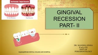
- 1. DR. MANISHA SINHA II nd yr PG DEPT OF PERIODONTOLOGY GINGIVAL RECESSION PART- II RAJARAJESWARI DENTAL COLLEGE AND HOSPITAL
- 2. Diagnosis HISTORY RADIOGRAPHIC ASSESSMENT Treatment Conclusion References
- 3. DIAGNOSIS
- 4. Chief Complaint, Specific Reason for the Visit or Referral
- 5. Dental History Orthodontic treatment Oral appliances A history of periodontitis, or necrotizing ulcerative gingivitis/periodontitis or mechanical/chemical trauma Periodontal treatment or surgical procedures Aesthetic dentistry/splinting/filling on anterior teeth Oral (hygiene) habits
- 6. Personal History Tobacco use (duration, daily consumption): Dietary habits: Recreational drugs (cocaine, meth, smokeless tobacco, bethel nut, etc.) either
- 7. Mucosa Special attention should be given to: Depth of vestibulum: adequate space for oral hygiene procedures. Frenula: possible frenum pull at place of insertion. Scar tissue might exert tension. Piercings: position of the intraoral disc in relation to the gingiva
- 8. Gingiva Thin scalloped: association with triangular-shaped crown, subtle cervical convexity, interproximal contacts close to incisal edge, narrow zone of KT, thin delicate gingiva, and relatively thin alveolar bone Thick scalloped: associated with slender teeth, thick fibrotic gingiva, narrow zone of KT, and a high gingival scallop Thick flat: associated with more square-shaped tooth crowns, pronounced cervical convexity, large interproximal contact located more apically, broad zone of KT, thick, fibrotic gingiva, and thick alveolar bone
- 12. • Aberrant frenal insertions: Ankyloglossia? Blanching? • Oral hygiene-induced or self-induced lesions –– Stilman’s clefts? Incomplete (red) or complete (white) lesions –– McCall’s festoons –– Gingival erosion
- 19. Radiographic Assessment Periapical Radiograph Bitewing Radiograph Cone Beam Computed Tomography Panoramic Radiograph
- 21. Cone Beam Computed Tomography
- 23. Miller,1994 •Root coverage to CEJ •Adequate band of attached gingiva •An esthetic tissue contour •Minimal post op pain
- 25. 1. Correction of tooth brushing technique 2. Removal of masochistic habits 3. Correction of malocclusion 4. Treating the dentinal sensitivity
- 26. Correction of tooth brushing technique
- 27. Removal of masochistic habits
- 29. Treating the dentinal sensitivity
- 30. 1. Pedicle soft tissue graft procedures : • Rotational flaps • Laterally positioned flap • Double papilla flap • Coronally positioned flap • Semilunar flap 2. Free soft tissue grafts • Nonsubmerged graft • One stage (free gingival graft) • Two stage (free gingival graft + coronally positioned flap) • Submerged grafts • Connective tissue graft + laterally positioned flap • Connective tissue graft + double papilla flap • Connective tissue graft + coronally positioned flap • (subepithelial connective tissue graft) • Envelope techniques ROOT COVERAGE TECHNIQUES
- 31. 3. Additive treatments • Root surface modification agents • Enamel matrix proteins • Guided tissue regeneration • Nonresorbable membrane barriers • Resorbable membrane barriers
- 32. Recipient Site 1. Gingival recession is limited to one tooth or extends to multiple teeth 2. Degree of gingival recession 3. Amount and thickness of existing keratinized gingiva in the area of recession 4. Whether the area of recession protrudes labially from the dental arch 5. The relation between the gingival recession area and smile line 6. Restorative/Prosthodonti c treatment after root coverage is necessary
- 33. • Thickness of the alveolar bone covering the donor tissue 2. Thickness of palatal soft tissue used as donor tissue Donor Site 1. Whether area adjacent to gingival recession can be used as a donor site • Amount of Keratinized gingiva • Thickness of keratinized gingiva • Size of adjacent interdental papilla
- 35. Presurgical preparation Mechanical modification of the root surface as well as root conditioning procedures have been used prior to the surgical root coverage techniques to achieve improved results (Miller, 1985). Root surface modification using agents such as citric acid or tetracycline hydrochloride has been advocated in an effort to promote the healing response following root surface coverage, although clinical studies have failed to show any improvement in root surface coverage when using such agents (Tolmie et al., 1991; Zucchelli et al., 2009; Pini-Prato et al., 1999)
- 36. Laterally Positioned Flaps Indications: 1. Sufficient width, length, and thickness of keratinised tissue exist adjacent to the area of gingival recession. 2. Coverage of the exposed root is limited to one or two teeth. 3. This method is most suitable for root coverage in gingival recession with narrow mesiodistal dimension.( mandibular anterior region) Introduced by Grupe and warren 1956
- 37. Contraindications: 1. Insufficient width and thickness of keratinised tissue in the adjacent donor site. 2. Extremely thin bone in the donor site or an osseous defect such as a dehiscence or fenestration. 3. Gingival recession area extremely protrusive. 4. Deep periodontal pocket and remarkable loss of interdental alveolar bone in the adjacent area. 5. Narrow oral vestibule. 6. Multiple teeth involved.
- 40. • Good vascularity • Ability to cover denuded root surface • One surgical site Recession at donor site,Guinard,1978 • Dehiscence or fenestration at donor site • Limited to 1 or 2 teeth Advantages Disadvantages
- 41. VARIANTS Staffileno,1964 and Pfeifer and Heller,1971 partial thickness flap to avoid dehiscence at donor site Grupe,1966 submarginal incision to avoid recession
- 43. Oblique rotated pedicle flap Dhalberg,1969
- 44. ADVANTAGES : 1. Good tissue blend 2. Usually one surgical site 3. Pedicle to be moved over donor site without tension and releasing incision 4. Usually complete root coverage DISADVANTAGE : Possible recession at the donor site
- 45. Transpositional Flaps Bahat et al" modified the oblique rotated flap. This is called the transpositional flap. Advantages 1. Predictability in areas of narrow root exposure 2. Possible to avoid gingival recession at the donor site Disadvantages 1. Sufficient length and width of the interdental papilla adjacent to the gingival recession area necessary 2. Not suitable for multiple tooth root coverage
- 47. Disadvantages 1. Technically demanding. 2. Limited application. The technique is generally used for multiple interdental papilla grafting, not for root coverage. The objective is to increase the width of the attached gingiva. Indication 1. Sufficient width and length of the interdental papilla on both sides of the area of gingival recession. Advantages 1. The amount of donor tissue is small because interdental papilla adjacent to the gingival recession area is displaced. Therefore, the procedure can be achieved with less tension to the pedicle flap. 2. While interdental bone is exposed if a full-thickness pedicle flap including interdental papilla is used, there is little damage to the alveolar bone because interdental alveolar bone is thick. Double Papilla Flap
- 50. Coronally advanced flap Indications 1. Esthetic coverage of exposed roots 2. For tooth sensitivity owing to gingival recession
- 52. Advantages • Treatment of multiple area of root exposure • No need for involvement of adjacent teeth • High degree of success Disadvantages • Need of 2 surgical procedure if zone of KG is less
- 53. Semilunar coronally advanced flap Introduced by Tarnow in 1986
- 54. Advantages • No vestibular shortening • No need for sutures Disadvantages • Inability to treat large area of recession • Requires FGG if underlying Dehiscence or fenestration is present
- 55. Free autogenous gingival graft This method was not indicated for root coverage of deep and wide gingival recession areas because of insufficient blood supply to graft. However, a series of procedures modified by Miller and Holbrook and Oschenbein demonstrated successful root coverage using free autogenous gingival graft. The common techniques are: 1. Through planing of the root surface to reduce the thickness of cementum , thereby reducing the mesiodistal dimension of the root surface. 2. Creating close adaptation of the coronal margin of the recipient site and the graft with a butt joint. 3. Using a graft of 1.5-2.0 mm thickness. 4. Stretching the graft to regenerate vascularity.
- 56. Indications: All cases where root coverage is necessary except where a graft of sufficient thickness (1.5-2.0 mm) cannot be harvested. (palatal tissue). Disadvantages: 1. Method with poorest ability to provide blood supply to the graft for root coverage. 2. Because a large and thick graft is necessary, a deep and large wound is exposed on the palatal mucosa( donor tissue). Various problems may occur, such as difficulty achieving hemostasis and patient pain and discomfort due to slow healing( healing by secondary intention). 3. Scarring occurs with wound healing, therefore, esthetic results may be inferior to other methods. 4. Surgery is required in two areas.
- 57. Preparation of Recipient Site.
- 58. . Make a partial-thickness incision. Complete recipient site preparation. • Prepare recipient site 3-5 mm or more apical to the most apical part of the exposed root.
- 59. Obtaining Graft Tissue. Donor tissue, although obtainable from various sites—the edentulous ridge, the tuberosity area, gingivectomy tissue— is most often secured from palatal tissue. The area of choice is the gingival zone distal to the anterior ruga on the posterior portion of the palate . This has the widest gingival zone with the least amount of submucosa . The submucosal tissue is fatty anteriorly and glandular posteriorly.
- 61. a. Tinfoil is placed over the recipient site to determine the size. The tinfoil is then placed over the palatal area. b. An incision is made to the palatal tissue with a depth of about 2 mm. The incision should be about 1 mm larger than the outline of the tinfoil to accommodate graft shrinkage. c. Small tissue pliers are used to lift the graft's edge. The graft is separated along the outline and a uniform-thickness graft (about 2 mm) is harvested.
- 62. d. After harvest, the donor site is immediately sutured and an absorbable hemostat gauze is placed. e. The harvested graft is placed onto a gauze soaked with physiologic saline solution. The thickness of the adipose tissue and glandular tissue is checked. f. The graft thickness necessary for root coverage is 1.5-2.5 mm.
- 63. Holbrook and Ochsenbein (1983) noted that when grafts are used for root coverage and underlying anatomic osseous factors must be taken into account, the teeth with the most prominent roots generally exhibit the least amount of bone over them, the most dehiscences and fenestrations, the greatest gingival scalloping, the thinnest type of periodontium, the most esthetic form, and the most mucogingival problems. They pointed out that prominent, bulging roots produce deep interproximal valleys , which require close adaptation of the grafts. These interradicular concavities necessitate graft stabilization to promote intimate graft contact and prevent dead space and hematoma formation. Suture of graft Suture technique of Holbrook and Ochsenbein
- 64. After making the ligature, pass the needle through the body of the graft and pull it out from the bottom without cutting the thread. Engage the periosteum 2-3 mm from the mesial edge of the flap. Leave a slack in the suture. Last, make a ligature and stretch to eliminate the sag. Stretching prevents primary shrinkage of the graft (primary contraction) and regenerates graft vascularity. Horizontal suture
- 65. Circumferential suture Insert the needle in the periosteum of the recipient site slightly apical to the bottom edge of the graft. Carry the suture around the cervical area and tie it to the tail on the lingual aspect. The thread presses the graft at the border of the exposed root (dotted line).
- 66. Insert the needle in the periosteum at the bottom of the interdental concavity area. Circle the needle around the tooth, suture the graft diagonally, make a sling, and make a ligature on the lingual aspect. Perform the same procedure in the other Interdental area. Interdental concavity suture
- 67. Insert the needle from outside of the distal aspect of the papilla flap and then insert the needle from the inside of the mesial aspect of the papilla flap. Join the distal aspect of the papilla flap and the bevel of the mesial papilla flap. Make an interrupted suture to overlap each connective tissue surface. If the flap edge is not straight after suture of the bilateral papilla, make a releasing incision of the periosteum to relieve flap tension. Place the flap edge on the enamel coronal to the CEJ and stabilize the overlapped bilateral papilla with a sling suture. Next, make a horizontal suture onto the flap mesiodistally for close adaptation of the flap to the root surface. Suturing in double papilla flap
- 68. Connective Tissue Grafts Inroduced by Langer and Langer in 1985 • The techniques utilizing a subepithelial soft tissue graft, i.e. the connective tissue, involve the placement of the graft directly over the exposed root and the mobilization of a mucosal flap coronally or laterally for coverage of the graft (Langer & Langer 1985; Nelson 1987; Harris 1992; Bruno 1994; Zucchelli et al. 2003). • An alternative technique is to place the base of the connective tissue graft within an “envelope” prepared by an undermining partial-thickness incision from the soft tissue margin, i.e. part of the graft will rest on the root surface coronal to the soft tissue margin (Raetzke 1985; Allen 1994). • For the treatment of multiple adjacent recessions, a multi-envelope recipient bed (“tunnel”) may be prepared (Zabalegui et al. 1999).
- 69. Indications Root coverage necessary in the gingival recession area. Contraindications Inadequate thickness of donor tissue. The necessary thickness of the connective tissue graft for root coverage is 1.5-2.0 mm, and the thickness of the palatal flap should be 1.5-2.0 mm after graft harvest to prevent necrosis. Therefore, at least 3-mm thickness is necessary in the palatal soft tissue of the donor site.
- 70. Connective tissue graft combined with a coronally advanced flap a. Make a trapezoidal partial-thickness flap with two vertical incisions mesiodistally. Vertical incision should be at least 0.5 mm from the gingival margin of the adjacent teeth. b. In the interdental papilla, make a horizontal i ncision at the CEJ or slightly coronal to it.
- 71. c. Prepare a partial-thickness flap. The interdental papillae should be de-epithelialized to allow for maximum coronal positioning of the tissue fl ap over the exposed root surface at suturing d. Reflect the partial-thickness flap. Make a releasing incision of the periosteum at the base of the flap for easy coronal migration.
- 72. Harvesting of graft Subepithelial connective tissue graft of masticatory mucosa is harvested on the palatal aspect of the maxillary premolars/first molar (or from the retromolar pad) by the use of a “trap door” approach. Before incisions are placed, the available thickness of the mucosa is estimated by the use of the tip of the syringe. . Primary incision. Make a horizontal incision with a partial-thickness flap 3-5 mm apical to the gingival margin in the palate (preparation of primary flap). . Secondary incision. Make a secondary incision 1-2 mm coronal to the primary horizontal incision line. This incision, which is perpendicular to the surface of the gingiva, should extend to the bone.
- 73. Make a vertical incision mesiodistally approximating the width and length of the necessary graft. Prepare a primary partial-thickness flap (1.5-mm thick) toward the center of the palate, parallel to the palatal gingiva. Expose the underlying connective tissue.
- 74. For the secondary incision, the blade contacts the bone. Use a small periosteal elevator or Kirkland 'S/6 knife to reflect the connective tissue graft, bringing it toward the center of the palate. Extend the base of the primary incision to the bone. Separate the connective tissue graft from the bone.
- 75. After harvesting of the connective tissue graft, the bone surface is exposed.
- 76. I. Suture the primary flap. Close the wound with an interrupted suture and a cross horizontal sling suture.
- 77. In subepithelial connective tissue grafts, place the thin border of the epithelium, left on the marginal area of graft, coronal to the CEJ. Make an interrupted suture in the interdental papilla with resorbable suture material and then stabilize the graft. Displace the flap coronally, covering the graft as much as possible, and suture.
- 78. Free connective tissue graft combined with a coronally advanced fl ap procedure – single recession (a) Deep gingival recession at a cuspid with minimal height of keratinized tissue apical to the root exposure. (b) The graft has been sutured to leave an area between the cemento-enamel junction and the graft available for the marginal keratinized tissue of the flap. (c) The flap has been advanced coronally and sutured. (d) Clinical healing at 1 year.
- 79. Advantages 1. High predictability. 2. The graft receives abundant blood supply from both the inside of the flap and the periosteum- connective tissue. 3. Wound closed at palatal donor site after harvest of connective tissue graft. Therefore, hemostasis is easy and healing is rapid. There is also less discomfort and pain during healing. 4. The graft fits the surrounding tissue on the recipient site, therefore, results are esthetically pleasing. 5. Applicable for gingival recession on multiple teeth. Disadvantages 1. Technically demanding. 2. Because a thick graft is used, the grafted tissue is thick. Gingivoplasty may be necessary postoperatively to obtain better morphology.
- 80. Modified Technique of Langer and Langer Do not use a vertical incision when preparing a recipient site to: • Ensure excellent blood supply to flap. • Alleviate postoperative discomfort. • Avoid scarring. Make a partial-thickness horizontal incision perpendicular to the interdental papilla of the recipient site. Close adaptation to donor tissue is obtained with a butt joint.
- 81. a. Make a partial-thickness horizontal incision on the CEJ or slightly coronally and perpendicular to the interdental papilla. b. Connect each horizontal incision with a sulcular incision. Extend the incision mesiodistally and prepare a large flap for proper access. After reflecting a partial-thickness flap, prepare a periosteum-connective tissue recipient site. Extend the partial-thickness incision apically for coronal migration of the flap.
- 82. d. Cover the exposed root with a connective tissue graft and suture. Cover the graft completely with the flap and suture.
- 83. Connective Tissue Graft Using an Envelope Flap Raetzke introduced a connective tissue graft using an envelope technique. A partial-thickness envelope flap is prepared on the soft tissue adjacent to the gingival recession area from the gingival sulcus. No horizontal or vertical incision is made.
- 84. Prepare the envelope flap. With the use of the “envelope” technique the recipient site is prepared by fi rst eliminating the sulcular epithelium by an internal beveled incision an “envelope” is prepared apically and laterally to the recession by split incisions. The depth of the preparation should be 3–5 mm in all directions. In an apical direction, the preparation of the site should extend beyond the mucogingival junction to facilitate the placement of the connective tissue graft and to allow for coronal advancement of the mucosal fl ap at time of suturing.
- 85. • A foil template may be used for the harvest of an appropriately sized connective tissue graft. The graft, , is inserted into the prepared “envelope” and positioned to cover the exposed root surface. • Sutures are placed to secure graft in position . A crossed sling suture may be placed to advance the mucosal fl ap coronally. Pressure is applied for 5 minutes to adapt the graft closely to the root surface and covering soft tissue.
- 86. The advantages of this technique are: simplicity, minimal surgical invasiveness, and good esthetics because the interdental papilla is preserved. One limitation of the envelope flap is that it cannot be displaced coronally. The envelope flap is also not applicable in areas of extensive gingival recession because there is a limit to the size of the graft that can be placed in the envelope flap.
- 87. Pouch and Tunnel Technique In case multiple adjacent recessions are to be treated, “envelopes” are prepared for each tooth. However, the lateral split incisions are extended so that the multi-envelopes are connected mesially and distally to form a mucosal tunnel. Care should be taken to avoid detachment of the papillae. • The graft is gently positioned inside the tunnel and its mesial and distal extremities are fixed with two interrupted sutures. Sling sutures may be placed to advance the mucosal fl ap coronally over the exposed portions of the connective tissue graft. Pressure is applied for 5 minutes to closely adapt the graft to the root surface and covering soft tissue. Application of a periodontal dressing is often not required. Introduced by Zabalegui, 1999
- 88. Subpedicle Connective Tissue Grafts a. Double papilla flap design in recipient site. b. Prepare the partial-thickness pedicle flap, which includes mesiodistal interdental papilla.
- 89. c. Prepare the recipient site, which consists of periosteum-connective tissue. d. Suture and stabilize the connective tissue graft. e. Connect each papilla flap to make a double papilla flap. f. Cover the connective tissue graft on the root surface with the double papilla flap. Make a sling suture.
- 90. CAUSES OF FAILURE OF CONNECTIVE TISSUE GRAFTS • Insufficient height of interdental bone and soft tissue. • Horizontal incision placed apical to the CEJ. • Reflection of all interdental papilla. • Inadequate root planing. • Insufficient blood supply from surrounding tissue due to inadequate recipient site preparation. • Connective tissue graft too small. • Connective tissue graft too thick. • Connective tissue graft inadequate for root coverage and coronal placement. • Insufficient coronal migration of flap covering the graft.
- 91. Advantages 1. High predictability. 2. The graft receives abundant blood supply from both the inside of the flap and the periosteum- connective tissue. 3. Wound closed at palatal donor site after harvest of connective tissue graft. Therefore, hemostasis is easy and healing is rapid. There is also less discomfort and pain during healing. 4. The graft fits the surrounding tissue on the recipient site, therefore, results are esthetically pleasing. 5. Applicable for gingival recession on multiple teeth. Disadvantages 1. Technically demanding. 2. Because a thick graft is used, the grafted tissue is thick. Gingivoplasty may be necessary postoperatively to obtain better morphology.
- 92. Healing of pedicle soft tissue grafts 1. The adaptation stage (from 0–4 days). The laterally repositioned flap is separated from the exposed root surface by a thin fibrin layer. The epithelium covering the transplanted tissue flap starts to proliferate and reaches contact with the tooth surface at the coronal edge of the flap after a few days.
- 93. The proliferation stage (from 4–21 days). In the early phase of this stage the fibrin layer between the root surface and the flap is invaded by connective tissue proliferating from the subsurface of the flap. After 6–10 days a layer of fibroblasts is seen in apposition to the root surface. These cells are believed to differentiate into cementoblasts at a later stage of healing. At the end of the proliferation stage, thin collagen fibers are formed adjacent to the root surface, but a fibrous union between the connective tissue and the root has not been observed. From the coronal edge of the wound, epithelium proliferates apically along the root surface.
- 94. The attachment stage (from 27–28 days). During this stage of healing thin collagen fibers become inserted in a layer of new cementum formed at the root surface in the apical portion of the recession. The maturation stage. This last stage of healing is characterized by continuous formation of collagen fibers. After 2–3 months bundles of collagen fibers insert into the cementum layer on the curetted root surface in the apical portion of the recession.
- 95. HEALING FOLLOWING FREE SOFT TISSUE GRAFTS Healing of free soft tissue grafts placed entirely on a connective tissue recipient bed has been studied in monkeys and can be divided into the following three phases. (Oliver et al.1988) 0 – 3 day (Initial phase): During these fi rst days of healing a thin layer of exudate is present between the graft and the recipient bed. During this period the grafted tissue survives with an avascular “plasmatic circulation” from the recipient bed. Therefore, it is essential for the survival of the graft that a close contact is established to the underlying recipient bed at the time of operation. A thick layer of exudate or a blood clot may hamper the “plasmatic circulation” and result in rejection of the graft. The epithelium of the free graft degenerates early in the initial healing phase, and subsequently it becomes desquamated.
- 96. 2-11 day (revascularization phase) After 4-5 days of healing, anastomoses are established between the blood vessels of the recipient bed and those in the grafted tissue. At the same time, a fibrous union is established between the graft and the underlying connective tissue bed . If a free graft is placed over the denuded root surface, apical migration of epithelium along the tooth-facing surface of the graft may take place at this stage of healing.
- 97. 11-42 days (tissue maturation phase): After approximately 14 days the vascular system of the graft appears normal. Also the epithelium gradually matures with the formation of a keratin layer during this stage of healing. Another healing phenomenon frequently observed following the free graft procedures is “Creeping Attachment” i.e. coronal migration of the soft tissue margin. This occurs as a consequence of tissue maturation during a period of about 1 year post treatment.
- 98. Guided Tissue Regeneration for Gingival Recession The use of a barrier membrane, according to the principles of guided tissue regeneration, in conjunction with pedicle soft tissue graft procedures was introduced as a treatment modality for root coverage by Pini Prato et al. (1992). In order create space for tissue formation between the facial root surface and the membrane Pini Prato et al. (1992) suggested that extensive root planing should be carried out to produce concave root morphology. Specially designed membranes for the treatment of recession type defects are available, such as nonabsorbable titanium-reinforced expanded polytetrafluoroethylene (e-PTFE) membranes.
- 99. Guided Tissue Regeneration for Gingival Recession
- 100. a. Prepare a full-thickness trapezoidal flap • Horizontal incision to the mesiodistal interdental papilla at the level of the CEJ. • Two vertical incisions. b. Connect the horizontal and vertical incisions with a sulcular incision.
- 101. c. Prepare a full-thickness flap 3-4 mm apical to the crest of the osseous dehiscence. Apically, prepare a partial thickness flap. d. Remove the epithelial tissue in the interdental papilla area.
- 102. e. Perform root planing and preparation of root concavity. If a titanium-reinforced membrane is used, the root profile may not need to be changed to establish the required space between the root and the membrane f. Bend the membrane in a tentlike fashion with suture thread. The titanium frame is bent to create space under the membrane. g. Stabilize the membrane with a sling suture.
- 103. h. Displace the flap coronally and cover the membrane completely. The use of non-biodegradable membrane barriers requires a second surgery for membrane removal, usually after 5–6 weeks. A partial thickness trapezoidal fl ap is raised to expose the membrane. Following its removal, the flap is repositioned at the level of the CEJ to completely cover the newly formed tissue. Mechanical plaque control is reinstituted 4 weeks after membrane removal
- 104. Pedicle soft tissue graft procedures combined with enamel matrix proteins Abbas et al. (2003) described a surgical procedure for periodontal regenerative therapy of recession defects utilizing enamel matrix derivative bioactive material (Emdogain®) Following preparation of the coronally advanced flap, the exposed root surface is conditioned with PrefGelTM (24% EDTA-gel, pH 6.7; Straumann Biologics, Switzerland) for 2 minutes to remove the smear layer. After thorough rinsing with sterile saline, the enamel matrix protein gel (Emdogain®Straumann Biologics, Switzerland) is applied to the exposed root surface. The pedicle graft is advanced coronally and secured at a level slightly coronal to the CEJ by suturing the flap to the de- epithelialized papilla regions.
- 105. Acellular dermal grafts Silverstein and callan,1997 AlloDerm is donated human soft tissue that is processed to remove dermal cells, leaving behind a regenerative collagen matrix. It provides a matrix consisting of collagen, elastin, blood vessels. Silverstein and callan,1997
- 106. After scaling and root planning, the root surfaces are conditioned. A partial thickness flap creating a pouch is formed using a no. 15 blade. The AlloDerm is rehydrated in two consecutive 10- to 15- min sterile saline baths (depending on size and thickness of the piece used). The graft is inserted into the pouch with the connective tissue against the recipient bed. The papillae are de-epithelialized, and the graft is immobilized with resorbable sutures at the level of the cemento-enamel junction .
- 107. The buccal flap is then sutured over the AlloDerm to cover the graft as much as possible. It is important to not leave any AlloDerm exposed.
- 108. PRP Griffin, 2004 suggested use of platelet concentrate carried by collagen sponge as graft substitute Yen and Jankovic,2007 used PRP with CTG and found accelerated wound healing and attachment formation. Advantages • Decreases pain and bleeding as less invasive • Increases tissue thickness • Decreases infection and graft sloughing • Decreases healing time, mature tissue within 1 week • Promotes vascularization • Accelerates wound healing
- 109. PRF Choukroun’s PRF, is a second-generation platelet concentrate. PRF consists of an intimate assembly of cytokines, glycamic chains, and structural glycoproteins enmeshed within a slowly polymerized fibrin network. These biochemical components have well-known synergetic effects on healing processes.
- 110. Prior to surgery IV blood is collected in 10 ml vials without anticoagulant & centrifuged at 2700 rpm for 10 min
- 112. Factors influencing the degree of root coverage Patient-related factors. Poor oral hygiene is a factor that will negatively influence the success of root coverage procedures (Caffesse et al. ) Further, the predominant causative factor in the development of gingival recession is toothbrushing trauma, and hence this factor has to be corrected to secure an optimal outcome of any root coverage procedure. Treatment outcome in terms of root coverage is usually less favorable in smokers than in nonsmokers (Trombelli & Scabbia 1997; Zucchelli et al. 1998; Martins et al. 2004; Erley et al. 2006; Silva et al. 2006)
- 113. Site related factors. Among site-specific factors, the level of interdental periodontal support may be of greatest significance for the outcome of root coverage procedures. From a biological point of view complete root coverage is achievable in class I–II recession defects , while when loss of connective tissue attachment also involves proximal tooth sites (class III–IV recession defects), only partial facial root coverage is obtainable (Miller 1985b) An additional factor shown to influence the degree of attainable root coverage is the dimensions of the recession defect. Less favorable treatment outcome has been reported at sites with wide (>3 mm) and deep (≥5 mm) recessions (Holbrook & Ochsenbein 1983; Pini Prato et al. 1992; Trombelli et al. 1995).
- 114. Technique-related factors. Several technique-related factors may influence the treatment outcome of a pedicle graft procedure. In a systematic review including data from 15 studies (Hwang & Wang 2006) a positive correlation was demonstrated between the thickness of the tissue flap and recession reduction. For complete root coverage the critical threshold thickness was found to be about 1 mm. However, whether a full- or split-thickness pedicle graft is used for root coverage may not influence the treatment outcome (Espinel & Caffesse 1981).
- 115. Elimination of flap tension is considered an important factor for the outcome of the coronally advanced flap procedure. Pini Prato et al. (2000a) measured the tension in coronally advanced flaps to compare the amount of root coverage in sites with and without residual flap tension. At sites that had residual tension (mean 6.5 g) the root coverage amounted to 78% 3 months post-surgically and 18% of the treated sites showed complete root coverage. Sites without tension demonstrated mean root coverage of 87% and complete root coverage in 45% of the cases. Pini Prato et al. (2005) demonstrated that for 100% predictability of complete root coverage in the treatment of Miller class I recessions with a coronally advanced flap procedure the flap margin has to be positioned at least 2 mm coronal to the CEJ.
- 117. CONCLUSION The management of gingival recession and its sequelae is based on a thorough assessment of the etiological factors and the degree of involvement of the tissues. The initial part of the management of the patient with gingival recession should be preventive and any pain should be managed and disease should be treated. The degree of gingival recession should be monitored for signs of further progression. When esthetics is the priority and periodontal health is good then surgical root coverage is a potentially useful therapy. Careful case selection and surgical management are critical if a successful outcome is to be achieved.
- 118. Carranza’s Clinical periodontology – 9th & 10th ed Clinical Periodontology and Implant Dentistry – Jan Lindhe 5th ed Periodontal surgery, Sato Atlas of cosmetic and reconstructive periodontal surgery- cohen Chambrone L, Sukekava F, Araújo MG, Pustiglioni FE, Chambrone LA, Lima LA. Root coverage procedures for the treatment of localised recession-type defects (Review). The Cochrane Library 2009, Issue 2 Umberto Pagliaro, Michele Nieri, Debora Franceschi,Carlo Clauser,and Giovanpaolo Pini-Prato. Evidence-Based Mucogingival Therapy. Part 1: A Critical Review of the Literature on Root Coverage Procedures. J Periodontol May 2003
- 119. The etiology and Prevalence of gingival recession – Moawia M.Kassab, Rober E. Cohen – JADA Feb 2003 The use of free gingival grafts for aesthetic purposes Paulom. Camargo, Philip R.Melnick & E. Barrie Kenney : Periodontology 2000, Vol. 27, 2001, Decision-making in aesthetics: root coverage revisited - Philippe bouchard, jacquesmalet & alain borghetti - Periodontology 2000, Vol. 27, 2001 Surgical management of gingival recession: A clinical update Hamdan, The Saudi Dental Journal (2009) 21, 83–94 Gingival Recession: Clinical Examination 4 and Diagnostics Corinna Bruckmann and Gernot Wimmer
- 120. THANK YOU
Editor's Notes
- When regression of the gingival margin is noticed, a structured diagnostic process of information gathering should be initiated. As gingival recessions might have several aetiologies, it is of utmost importance for the practitioner to be able to compile anamnestic, clinical, and radiologic signs and symptoms, as well as laboratory information. This process allows for differential diagnoses of possible underlying reasons and the decision-making in respect to future treatment options or necessities.
- Comprehensive exploration is desirable as past (dental) treatment may be the reason of today’s problems. Comprehensive exploration is desirable as past (dental) treatment may be the reason of today’s problemsOrthodontic treatment in the past may be the reason for present recessions. • Oral appliances (removable partial dentures/denture clasps, occlusal splints, removable orthodontics, anti-snoring mouthpieces, etc.) may impinge on periodontal tissues. • A history of periodontitis, or necrotizing ulcerative gingivitis/periodontitis (NUG/NUP), or (mechanical/chemical) trauma may explain loss of soft and/or hard tissue attachment, especially interdentally [7]. • Periodontal treatment or surgical procedures may have caused soft tissue recessions. • Aesthetic dentistry/splinting/filling on anterior teeth may have been used to mask tooth drifting/pathological migration/recessions. • Oral (hygiene) habits –– Oral hygiene aids, toothpastes and mouth rinses, frequency/duration of use –– Nail biting /pen chewing/factitious lesions
- Several systemic diseases and conditions are associated with oral signs and symptoms [2], and many drugs are known to modify gingivitis/periodontitis [3]. Diabetes mellitus is an important risk factor for periodontal inflammation if poorly controlled. Last but not least, age, hormonal changes (e.g. puberty, pregnancy, menopause), and stress (at work, financial, domestic, etc.) influence oral tissues. Make sure that reported diseases and medications do correspond. Regular alcohol use may have a negative impact on either periodontal tissues and/or adherence to treatment. Of particular importance for the evaluation of gingival recessions are the following: Tobacco use (duration, daily consumption): Very important for the diagnostic process (less overt bleeding), risk for recession, and healing response [4]. • Dietary habits: Increased risk for caries on denuded root surfaces? Erosive potential of diet (hypersensitivity, abrasion) [5]? • Recreational drugs (cocaine, meth, smokeless tobacco, bethel nut, etc.) either have direct local influence on oral tissues, are a risk factor for caries (by diminishing saliva flow), or induce negligent behaviour
- Inspect for adequate lubrification, pigmentation, any lesions, or growths. Aphthous lesions are often seen secondary to medication (e.g. non-steroidal anti-inflammatory drugs), stress, or Behçet syndrome.
- Categorize according to visibility of periodontal probe after insertion into the facial sulcus.. Note that the biotype may differ between the lower and upper jaw within the same patient
- (a) Thin-scalloped biotype, periodontal probe shining through delicate free gingiva, PPD 1 mm; notelocation of papilla tips due to natural diastemas and recessions mostly at teeth with buccal position; (b) thick-scalloped biotype; © thick-flat biotype with broad band of keratinized tissue, thick, fibrotic gingiva
- (a) Rolling test: softly push the adjacent mucosa coronally with a periodontal probe to identify width of the blanching attached gingiva/tissue. (b) Staining test with Lugol’s iodine: glycogen containing mucosa stains brownish in contrast to orthokeratinized gingiva
- (a) Irregular gingival scallop due to developmental enamel indentation #11, loss of central papilla height (class II (Nordland and Tarnow), PPI 3 (Cardaropoli et al.)); (b) inconsistent height of gingival margin (incomplete eruption of #32, #42, recession in #31) Cardaropoli et al. [29]: Papilla Presence Index (PPI) 1–4
- (a) Buccal position of #31 and #41 and gingival recession, very thin zone of KT, frenum pull, blanching, missing contact point, low interdental central papilla (PPI 4 (Cardaropoli et al.); (b) irregular frenum, frenum pull at #13 with blanching; (c) irregular frenum, lingual position of #41, lingual recessions, persistent lingual frenum; (d) irregular frenula, buccal recessions #22–25, cervical abrasions #23, frenum pull and blanching in #23 and #24, possible plaque niche # 24 distal of frenum insertion
- (a) Red Stilman’s cleft at distobuccal root of #27 (note buccal malposition); (b) buccal malposition of #45, loss of buccal soft (red Stilman’s cleft) and hard tissue, due to overzealous toothbrushing (however, note insufficient plaque control interdentally); (c) generalized buccal recession and abrasions, white Stilman’s clefts #12 and #34; (d) gingival erosion due to self-inflicted trauma (brushing and flossing) #33, red Stilman’s cleft #32
- (a) Healthy (in #12 and #11 reduced) periodontium (note circumferential recession, loss of interdental papilla due to past periodontal disease/treatment), incomplete eruption of #13, papilla height class III between #12 and #11 (Nordland and Tarnow), (b) assessment of width, and (c) height of recession making use of a periodontal probe or (d) a caliper (note extremely thin blanching buccal tissue in #31)
- Tooth form [39] –– Square: associated with thick-flat tissue, large interproximal contact located more apically, a broad zone of KT, thick, fibrotic gingiva, and a comparatively thick alveolar bone. (a) Buccal position/rotation of #31, root proximity #31/32, interdental and buccal recessions up to 5 mm in 5th sextant, PPD up to 5 mm, CAL up to 10 mm (#31); (b) periapical radiograph of #31/21 with bone loss of more than 2/3 of the root length
- (a) Multiple misalignments of front teeth in all three planes; (b) same case as 4.6b–d: buccal malposition of #31 (Miller class I recession), #41 (Miller class II recession), minimal zone of keratinized attached gingiva, marginal gingivitis in #41. Tooth (mal)position in the arch in three planes: rotated, tilted, displaced, and incompletely erupted (Figs. 4.1a, 4.4a, c, 4.5b, 4.7a, and 4.8a, b) –– Vertical (apical-coronal): cervical portion apical or coronal of the FGM of adjacent teeth (Fig. 4.8a) –– Sagittal (buccal-lingual): variability of gingival thickness and underlying bone plate (Fig. 4.8b) –– Horizontal: crowding, rotation
- Clasps, bands, etc. • Non-passive orthodontic retainers • Piercings. (a) Tongue piercing; (b) lingual gingival recession at the opposed tooth #41
- Periapical Radiograph • Root morphology and crown-to-root ratio • Periodontal ligament (PDL) space: –– Widening of the PDL: sign of occlusal trauma or periapical pathology –– Bone hyperdensity of lamina dura: sign of functional adaptation to occlusal forces –– Loss of PDL: sign of ankylosis • Root proximity: possible risk factor for periodontal disease (Fig. 4.7b), might have influence on treatment options [42] • Furcation involvement: separation coefficient, length of root trunk Bitewing Radiograph Due to the perpendicular visualization of the teeth, it is ideal for reliable assessment of the alveolar crestal bone [43] and diagnosing caries/restorations. • Distance of CEJ to interdental bone crest –– 2 mm: crestal bone loss? “Fuzziness” on the mesial/distal aspect of the interdental septa indicating loss of mineral content? CEJ discrepancies of adjacent teeth: horizontal or vertical type of bone loss? Interradicular radiolucencies might indicate possible furcation involvement. –– < 2 mm: incomplete eruption? • Distance of interproximal alveolar crest to contact point: influence on presence (≤5 mm) or absence (>5 mm) of interdental papilla [44] • Calculus/caries/overhanging or open margins/resorptions?
- (a) Thick periodontal biotype, buccal position of #31, clinical signs of inflamed gingiva, 1.5 mm buccal recession, loss of interdental papilla, PPD 5 mm on mesial aspect, high frenum insertion #41; (b) CBCT of area of interest #31: note demineralized interdental bone Overcoming the limitations of two-dimensional radiographs CBCT is the only method that allows for an analysis of the buccal and lingual/palatal surfaces [45, 46] and an improved visualization of the morphology of a periodontal defect, especially in the evaluation of dehiscencies, fenestrations (Fig. 4.11a, b), interradicular bone (Fig. 4.12), and furcation defects [47]. A novel approach using a lip/tongue retractor allows for visualization and measurement of the periodontal dimensions, gingival thickness, and the dentogingival attachment
- The management of gingival recession and its sequelae is based on a thorough assessment of the etiological factors and the degree of tissue involvement. The initial part of the management of the patient with gingival recession should be directed towards correcting the etiological factors. The degree of gingival recession has to be monitored for signs of further progression. Surgical root coverage is indicated when esthetics is the prime concern and periodontal health is good.
- Treatment of gingival recession using low level laser.
- The techniques used for root coverage are based on tissue displacement whether by translation (pedicle flap procedures) or by grafting (free gingival or connective tissue graft procedures), and use of resorbable and non-resorbable membranes according to the principles of guided tissue regeneration (GTR) (Wennstrom, 1996). Several modifications to the conventional techniques have been developed in an attempt to obtain optimal root coverage and a better esthetic integration. Surgical procedures may be broadly divided into two different types: Pedicle soft tissue graft procedures. These types of graft remain attached at their base and involve the positioning of soft tissue over the recession defect; they retain their own blood, supply during their transfer to a new location. Examples include: Rotational flap procedures, including laterally positioned flap, double papilla flap. Flap advancement procedures, including coronally repositioned flap. Free soft tissue graft procedures. Soft tissues are transferred from an area distant to the recession to cover the defect. These techniques are used where there is inadequate donor tissue close to the recipient site or where the aim of treatment is to increase tissue thickness. Free gingival graft. Subepithelial connective tissue graft.
- Root surfaces are mechanically prepared prior to any mucogingival procedure to allow biological attachment of the grafted tissue to it. The root surface is thoroughly debrided with ultrasonic or hand instruments and irrigated with sterile saline. Mechanical modification of the root surface as well as root conditioning procedures have been used prior to the surgical root coverage techniques to achieve improved results (Miller, 1985). Root surface modification using agents such as citric acid or tetracycline hydrochloride has been advocated in an effort to promote the healing response following root surface coverage, although clinical studies have failed to show any improvement in root surface coverage when using such agents (Tolmie et al., 1991; Zucchelli et al., 2009; Pini-Prato et al., 1999).
- Root surfaces are mechanically prepared prior to any mucogingival procedure to allow biological attachment of the grafted tissue to it. The root surface is thoroughly debrided with ultrasonic or hand instruments and irrigated with sterile saline. Mechanical modification of the root surface as well as root conditioning procedures have been used prior to the surgical root coverage techniques to achieve improved results (Miller, 1985). Root surface modification using agents such as citric acid or tetracycline hydrochloride has been advocated in an effort to promote the healing response following root surface coverage, although clinical studies have failed to show any improvement in root surface coverage when using such agents (Tolmie et al., 1991; Zucchelli et al., 2009; Pini-Prato et al., 1999).
- tetracycline HCl solution at concentration of 10% by active burnishing technique for 3 minutes. Root surfaces are mechanically prepared prior to any mucogingival procedure to allow biological attachment of the grafted tissue to it. The root surface is thoroughly debrided with ultrasonic or hand instruments and irrigated with sterile saline.
- b. Make a V-shaped incision in the peripheral gingiva in the gingival recession area while preserving sufficient interdental papilla on the distal aspect of 10. c. A wide external bevel incision on the mesial aspect and an internal bevel incision on the distal aspect create close adaptation of the flap. Remove the V-shaped gingiva and make a bevel for flap adaptation. e. Make an internal bevel incision toward the alveolar bone crest from the free gingival margin of the donor site. Prepare a vertical incision one and one-half teeth from the recipient site.
- . f. Prepare a full-thickness pedicle flap. g. If the flap is strained after displacement to the recipi ent site, make a releasing incision of the periosteum or cut back the incision at the base of the flap. h. Cover the exposed root surface completely with the pedicle flap and suture the flap coronal to the CEJ. i. To minimize postoperative gingival recession at the donor site, place a free autogenous gingival graft.
- To prevent donor site recession, Grupe (1966) modified this to a submarginal incision on the donor site (Figure 6-25B). Staffileno (1964) solved this problem by using a partial-thickness flap to protect the donor site from recession. Corn (1964b) further modified this by adding a cutback incision to release tension (Figure 6-25C) Historical outline of the laterally positioned flap design.. C, Cutback or releasing incision for tension release. D, Rotated pedicle flap permitting placement without the need for a cutback incision. E, Use of more than one tooth to permit periosteal placement over exposed root with bone exposure (stimulated or nonstimulated).
- Advantage Prevent recession at donor site This type of incision can be used for all procedures provided that an adequate width (25 mm) of keratinized gingiva is present at the donor site. This will permit leaving a small collar of tissue about the neck of the teeth to prevent recession at the donor site, thus facilitating use of a full-thickness pedicle flap if desired.
- A, V-shaped incision and pedicle flap outlined. B, Incisions completed and V-shaped incision removed. Note the obliquely angled donor flap. C, Pedicle flap rotated over the tooth. Dahlberg (1969) designed incisions for pedicle flaps based on a center of rotation about an axis at the base of the vertical donor incision. This permitted the pedicle to be moved over the donor site without tension and without the need for releasing incisions.
- a. Make two vertical incisions including sufficient interdental papilla. b. Prepare the pedicle flap using a partial-thickness incision. Resect the epithelium of the mesial interdental papilla and prepare the recipient site. c. Suture the pedicle flap on the mesial interdental papilla area of the recipient site. d. Using partial thickness, extend the pedicle flap preparation apically beyond the MGJ so that it may be displaced to the exposed root surface. e. Completion of suture. Make a periosteal suture on the mesial aspect.
- Cohen and Ross" introduced the method in which bilateral interdental papilla is used as donor tissue for localized root coverage. In this technique, there is less chance of flap necrosis and suture is easy because interdental papilla is thicker and wider than labial gingiva on the root surface. Therefore, double papilla flaps are useful in cases where there is no gingiva on sites adjacent to areas of gingival recession or where there are periodontal pockets on the labial surfaces of the adjacent tooth. Laterally positioned flap surgery is not indicated in these cases
- Make a V-shaped incision with a bevel on the mesial i nterdental papilla surface b. Remove the V-shaped tissue. The flap design includes a hori . zontal incision to the mesiodistal interdental papilla on the coronal side and two vertical incisions. c. Prepare a full-thickness pedicle flap including sufficient interdental papilla on the mesial and distal sides. d. Make a partial-thickness flap on the apical part of the flap for easy flap migration. d. Make a partial-thickness flap on the apical part of the flap for easy flap migration.
- f. Suture each flap and make a double papilla flap. g. Cover the exposed root with the double papilla flap. Stabilize the flap coronal to the CEJ with a sling suture.
- The coronally advanced fl ap procedure is initiated with the placement of two apically divergent vertical releasing incisions, extending from a point coronal to the CEJ at the mesial and distal line axis of the tooth and apically into the lining mucosa . A split-thickness fl ap is prepared by sharp dissection mesial and distal to the recession and connected with an intracrevicular incision. Apical to the receded soft tissue margin on the facial aspect of the tooth, a full-thickness fl ap is elevated to maintain maximal thickness of the tissue fl ap to be used for root coverage . Vertical incisions to prepare recipient site. H, Split-thickness flap reflected. I, Connective tissue sutured over denuded root surface. J, Split-thickness flap sutured over donor connective tissue.
- Coronally positioned pedicle flap, diagrammatic view. A, Incisions outlined preoperatively. Note that the incisions do not go to the tips of the papillae. B, A full-thickness flap is reflected, exposing the underlying bone (B). The epithelium overlying the remaining portion of the papillae (P) is removed. C, The flap is sutured coronally for root coverage. Clinical view: A', Before. B', Full-thickness flap reflected. C', Epithelium over the remaining papillae removed and the flap sutured coronally. D', Two years later; compare with A'.
- A semilunar incision is placed apical to the recession and at a distance from the soft tissue margin, which should be approximately 3 mm greater than the depth of the recession. The outline of the incision should be parallel to the curvature of the gingival margin (Fig. 44-39a). The incision is extended into the papilla region on each side of the tooth, but care should be taken to maintain a broad base of anchorage to secure a collateral blood supply to the pedicle graft. • A split-thickness dissection of the facially located tissue is then made by an intracrevicular incision extending apically to the level of the semilunar incision (Fig. 44-39b). The mid-facial soft tissue graft is coronally repositioned to the level of the CEJ (Fig. 44-39c) and stabilized by light pressure for 5 minutes. • No suturing is needed but a light-cured dressing may be applied for wound protection.
- Free gingival grafts are used to create a widened zone of attached gingiva. They were initially described by Bjorn' in 1963 and have been extensively investigated since that time
- However, in areas of extensive gingival recession, there is the problem of blood supply to the graft. In such cases, connective tissue grafts are suitable. Because a disharmonious result at the receipent site is possible, the author limits root coverage using free autogenous gingival grafts to the mandibular anterior teeth and premolars.
- Site selection and thickness of donor tissue will vary according to the individual operator’s preference and the intended purpose and function of the graft tissue.
- FIGURE 6-15. Continued. M represents various zones of palate from which donor material may be selected. N, Cross-section enlargement of the posterior gingival zone from which material is usually selected. O, Outlining of the graft from a previously sized tinfoil template. P, Partial-thickness dissection of the graft. Q, Use of tissue pliers to reflect graft tissue as it is being dissected. R, Graft removed and palate sutured for hemostasis. S, Use of a sharp scalpel blade to remove any fat or glandular tissue and to reduce underlying tissue irregularities. T, Graft smoothed. U, Initial suture placed in the graft. V, Stabilization of the graft during the suturing phase. W, Graft placed below recession and sutured in position. Note the apical suturing or mucosal flap, which is optional. X, Coronal positioning for root coverage.
- d. After harvest, the donor site is immediately sutured and an absorbable hemostat gauze is placed. e. The harvested graft is placed onto a gauze soaked with physiologic saline solution. The thickness of the adipose tissue and glandular tissue is checked. f. The graft thickness necessary for root coverage is 1.5-2.5 mm.
- Slackmeaning not taut or held tightly in position; loose.
- A horizontal suture is made on the recipient site in the mesial interdental concavity area under the graft. This is done to prevent dead space in the interdental concavity area and for better adaptation of the graft to this area. b. The cervical area is surrounded and a sling made. A ligature is then made on the lingual aspect. The same suture is made on the distal aspect. Insert the needle in the periosteum at the bottom of the interdental concavity area. Circle the needle around the tooth, suture the graft diagonally, make a sling, and make a ligature on the lingual aspect. Perform the same procedure in the other Interdental area.
- Compared to the epithelialized graft, the connective tissue graft is preferable due to a less invasive palatal wound and an improved esthetic result.
- g.
- Supply to the flap after reflection depends primarily on the bilateral surrounding gingival tissue (laterally) and oral mucosa (apically) in the surgical area, because the blood supply from the interalveolar septum and periodontal membrane is terminated. The Langer and Langer technique requires a vertical incision to the flap, therefore reducing blood supply markedly. The envelope flap (modified flap), which is reflected without the vertical incision, is superior to the full flap with a vertical incision in this respect. Bruno modified the Langer and Langer technique to include a horizontal partial-thickness incision extending mesiodistally to the recipient site. Blood supply to the graft is increased because the grafted connective tissue is covered by this flap. Also, the lack of a vertical incision alleviates postoperative discomfort and facilitates healing, and no scar will develop in the vertical incision area, an esthetic advantage (Table 6-16). The horizontal incision aids close adaptation of the interdental papilla area and graft with a butt-joint effect. The blood supply from the interdental papilla area is enhanced by this close adaptation.
- a. Perform root planing of the exposed root and use a finishing bur to recontour it. b. (Fig. 44-48a). Secondly,
- According to Wilderman and Wentz (1965), the apical proliferation of epithelium may stop within the coronal half of the defect although further downgrowth of epithelium was also frequently observed.
- The establishment of collateral circulation from adjacent vascular borders of the bed allows the healing phenomenon of “bridging” (Sullivan & Atkins 1968a).
- Revascularization phase (from 2–11 days). After 4–5 days of healing, anastomoses are established between the blood vessels of the recipient bed and those in the grafted tissue. Thus, the circulation of blood is re-established in the pre-existing blood vessels of the graft. The subsequent time period is characterized by capillary proliferation, which gradually results in a dense network of blood vessels in the graft. At the same time a fi brous union is established between the graft and the underlying connective tissue bed. The re-epithelialization of the graft occurs mainly by proliferation of epithelium from the adjacent tissues. If a free graft is placed over the denuded root surface, apical migration of epithelium along the toothfacing surface of the graft may take place at this stage of healing
- Tissue maturation phase (from 11–42 days). During this period the number of blood vessels in the transplant becomes gradually reduced, and after approximately 14 days the vascular system of the graft appears normal. Also, the epithelium gradually matures with the formation of a keratin layer during this stage of healing.
- In addition, a variety of bioabsorbable membranes are commercially available, but many of these may not be rigid enough for maintaining required space during healing. Advantages: Gain of new attachment • Donor site not necessary • Predictable root coverage Disadvantages : Technically demanding • Costly Indications: deep and wide with more than 5mm attachment loss, particularly maxillary canine.
- e
- The surgical technique utilized is the coronally advanced fl ap as described above. The interdental papillae should be de-epithelialized to allow for maximum coronal positioning of the tissue fl ap over the exposed root surface at suturing.
- The buccal flap is then sutured over the AlloDerm to cover the graft as much as possible. It is important to not leave any AlloDerm exposed. The buccal flap is sutured over the AlloDerm by using a sling suture to provide the graft with maximum coverage.
- . Numerous therapeutic solutions for recession defects have been proposed in the periodontal literature and modified with time according to the evolution of clinical knowledge.