SlideShare a Scribd company logo
1 of 87
GINGIVAL
ENLARGEMENT
DR MANISHA SINHA
II YEAR PG
RAJARAJESWARI DENTAL COLLEGE AND HOSPITAL
CONTENTS
 INTRODUCTION
 CLASSIFICATIONS
 GRADING
 INFLAMMATORY GINGIVAL
ENLARGEMENT
 DRUG-INDUCED ENLARGEMENT
 IDIOPATHIC ENLARGEMENT
 ENLARGEMENTS ASS. WITH SYSTEMIC DISEASES
 NEOPLASTIC ENLARGEMENT
 FALSE ENLARGEMENT
 TREATMENT OF GINGIVAL ENLARGEMENT
 CONCLUSION
INTRODUCTION
 Gingival enlargement is the proliferation and intensification of the gingiva which is a prevailing
character of the diseased gingival tissues.
 Gingival enlargement and gingival overgrowth are terms used interchangeably with hyperplasia,
hypertrophy, and fibrosis.
 Hyperplasia is an increase in the number of cells in tissues that results in increased tissue volume.
Hypertrophy refers to increased tissue size and volume resulting from increased cell size.
 Fibrosis refers to a pathologic process in which disrupted wound healing is associated with
defective cell proliferation, cell-to-cell interactions, cell-to-matrix interactions, and matrix
deposition and with an impaired immune system response.
CLASSIFICATION
I. Inflammatory enlargement
A. Chronic
B. Acute
II. Drug-induced enlargement
III. Enlargements associated with systemic diseases or
conditions
A. Conditioned enlargement
1. Pregnancy
2. Puberty
3. Vitamin C deficiency
4. Plasma cell gingivitis
5. Nonspecific conditioned enlargement (granuloma
pyogenicum)
B. Systemic diseases causing gingival enlargement
1. Leukemia
2. Granulomatous disease (Wegener’s granulomatosis,
sarcoidosis)
IV. Neoplastic enlargement (gingival tumors)
A. Benign tumors
B. Malignant tumors
V. False enlargement
Using the criteria of location & distribution, gingival
enlargement is designated as follows:
LOCALISED GENERALISED MARGINAL
PAPILLARY
DIFFUSE DISCRETE
The degree of gingival enlargement can be scored as follows:
ANGELOPOULOS
AND GOAZ (1972)
None.
Not more than 1/3rd of the clinical
crown covered.
Any part of the middle
third of the crown covered.
Greater than
2/3rd of the
crown covered.
1
0
2
3
0 = No encroachment of the interdental
papilla onto the tooth surface
1 = Mild encroachment of the interdental
papilla, producing a blunted appearance to
papilla tip
2 = Moderate encroachment, involving lateral
spread of papilla across buccal tooth surface
of less than one quarter of tooth width.
3 = Marked encroachment of papilla, i.e. more
than 1/4th tooth width. Loss of normal
papilla form
Seymour et al 1985
Miller and Damm (1992)
Horizontal gingival overgrowth index
is described as:
Grade 0: < 1mm
Grade 1: 1 to 2 mm
Grade 2: >2mm
Vertical gingival overgrowth index
is described as:
Grade 0: Normal gingival, no alteration
Grade 1: Minimal overgrowth, ≤ 2mm, gingiva covering the
cervical third or less of the anatomic crown.
Grade 2: Moderate overgrowth: 2 to 4 mm, gingival
covering the middle third of the anatomic crown.
Grade 3: Severe overgrowth: ≥4mm, nodular
growth, gingival covering more than two thirds
of the dental crown.
Bokenkamp A and Bohnhorst B (1994)
 Grade 0: No signs of gingival overgrowth
 Grade I: Gingival hyperplasia confined to interdental papilla .
 Grade II: Hyperplasia of interdental papilla and marginal
gingiva
 Grade III: Gingival hyperplasia covering at least three-quarters
of tooth crowns
Eva and Ingles (1999)
Grade 0: No overgrowth,
firm adaptation of the
attached gingiva to the
underlying alveolar bone
Grade 1: Early
overgrowth, as evidenced
by an increase in density
of the gingiva with
marked stippling and
granular appearance. The
tip of the papilla is
rounded and the probing
depth is less than or equal
to 3mm.
Grade 2: Moderate
overgrowth, manifested
by an increase in the size
of the papilla and/ or
rolled gingival margins.
The contour of the margin
is still concave or
straight. The probing
depth is equal to or less
than 6mm and the papilla
is somewhat retractable.
Grade 3: Marked
overgrowth, represented
by encroachment of the
gingiva onto the clinical
crown. Contour of the
margin is convex rather
than concave. The probing
depth is greater than 6mm
and the papilla is clearly
retractable.
Grade 4: Severe
overgrowth, characterized
by a profound thickening
of the gingiva. A large
percentage of the clinical
crown is covered. The
papilla is retractable, the
probing depth is greater
than 6 mm and the
buccolingual dimension is
approximately 3 mm
Miranda and Brunet index (2001)
 described an index in which horizontal measurement of the enlargement is possible.
This index is also termed as nodullary papilla index.
 In this index the measurement is carried out with the help of a periodontal probe
from the enamel surface of the interdental contact point to the outer papillary area.
The scores of this index is as
mentioned below:
Score 0: Papilla thickness < 1 mm
Score 1: Papilla thickness 1- 2 mm
Score 2: Papilla thickness > 2 mm
INFLAMMATORY
ENLARGEMENT
 Gingival enlargement may result from chronic or acute
inflammatory changes; chronic changes are much
more common.
 In addition, inflammatory enlargements usually are a
secondary complication to any of the other types of
enlargement, creating a combined gingival
enlargement.
 In these cases it is important to understand the double
etiology and treat them adequately.
A) Acute Inflammatory Enlargement
 Acute inflammatory enlargement of the gingiva usually is caused by a mechanical,
chemical, or physical irritation and can be resolved by removal of the irritant.
 Mouth breathing, impacted food items, and poor oral hygiene are usually responsible for
acute inflammatory reactions in gingival tissues.
 Acute lesions are usually localized to marginal or papillary gingiva.
 Acute inflammatory enlargement can lead to the formation of gingival abscess. Gingival
abscess is a localized, painful, and rapidly expanding lesion.
GINGIVAL ABSCESS
 In its early stages, it appears as a red swelling with a smooth, shiny surface.
The lesion usually becomes fluctuant and pointed, with a surface orifice and
a purulent exudate in 24 to 48 hours
 Generally limited to marginal gingiva or interdental papilla.
 Etiology- caused by a mechanical, chemical, or physical irritation and can be
resolved by removal of the irritant. Mouth breathing, impacted food items,
and poor oral hygiene
Histopathology:-
A purulent focus in the connective tissue, surrounded by a diffuse infiltration of
polymorphonuclear leukocytes, edematous tissue, and vascular engorgement.
The surface epithelium has varying degrees of intra- and extracellular edema,
invasion by leukocytes, and sometimes ulceration.
The lesion is confined to the gingiva, and it should be distinguished from
periodontal abscess.
PERIODONTAL (LATERAL) ABSCESS
 It is a localized purulent inflammation in the periodontal tissues.
 Also known as Lateral or Parietal abscess.
 It involves the supporting periodontal tissues causing enlargement of gingiva.
 Periodontal abscess formation may occur in the following ways:
--Infection from
a periodontal
pocket deeply
into the
supporting
periodontal
tissues.
-- localization of
the suppurative
inflammatory
process
Lateral
extension of
inflammation
from the inner
surface of a
periodontal
pocket into the
connective tissue
of the pocket
wall.
pocket --
tortuous course
around the root,
a periodontal
abscess may
form in the cul -
de- sac.
Incomplete
removal of
calculus during
treatment of a
periodontal
pocket.
the absence of
periodontal
disease after
trauma to the
tooth or
perforation of
the lateral wall
of the root in
endodontic
therapy
B) CHRONIC INFLAMMATORY ENLARGEMENT
Etiology:--Prolonged exposure to dental plaque.
 Factors that favour plaque accumulation and retention include:-
Poor oral
hygiene.
Irritation by
anatomic
abnormalities
Improper
restorative and
orthodontic
appliances
Mouth
breathing
CLINICAL FEATURES
 Originates as Slight ballooning of the interdental papilla and/or the marginal gingiva.
 Produces a life preserver-shaped bulge around the involved teeth in early stages.
 May be localized or generalized and progresses slowly & painlessly, unless it is
complicated by acute infection or trauma.
 Occasionally, occurs as a discrete sessile or pedunculated mass resembling a
tumor.
 May be interproximal or on the marginal or attached gingiva.
 Lesions are slow-growing masses and usually painless.
 They may undergo spontaneous reduction in size,
followed by exacerbation and continued enlargement.
 Painful ulceration
sometimes occurs in the fold between the mass and the adjacent
gingiva
Gingival Changes Associated with Mouth
Breathing:-
o Gingiva appears red and edematous with a
diffuse surface shininess of the exposed area.
o Maxillary anterior region -common site
o Harmful effect is attributed to irritation from
surface dehydration.
Histopathology
 Show the exudative and proliferative features of chronic inflammation.
 Lesions that are clinically deep red or bluish red are soft and friable with a smooth, shiny surface, and they
bleed easily.
 They also have a preponderance of inflammatory cells and fluid, with vascular engorgement, new capillary
formation, and associated degenerative changes.
 Lesions that are relatively firm, resilient, and pink have a greater fibrotic component with an abundance of
fibroblasts and collagen fibers.
DRUG
INDUCED
ENLARGEMENT
 Predilection for anterior gingiva
 Higher prevalence in children
 Onset within 3 months
 Change in gingival contour leading to modifi
cation of gingival size
 Enlargement fi rst observed at the interdental
papilla
 Change in gingival color
 Increased gingival exudate
 Bleeding upon provocation
 Found in gingiva with or without bone loss but is not
associated with attachment loss
 Pronounced inflammatory response of gingiva in
relation to the plaque present
 Reductions in dental plaque can limit the severity of
lesion
 Must be using phenytoin, cyclosporine A or certain
calcium channel blockers; the plasma concentrations to
induce the lesion have not been clearly defi ned in
humans
Characteristics of drug-influenced gingival enlargement (Mariotti 1999)
Clinical Features
 Growth starts as a painless, bead-like enlargement of the interdental papilla
and extends to the facial and lingual gingival margins.
Case of phenytoin induced gingival enlargement
speech, mastication, tooth eruption,
and aesthetic problems.
ANTICONVULSANTS,
IMMUNOSUPPRESSANTS, AND CALCIUM
CHANNEL BLOCKERS
 As the condition progresses, the marginal and papillary enlargements unite
and may develop into a massive tissue fold covering a considerable portion
of the crowns; they may interfere with occlusion.
 Spontaneous disappearance occurs within a few months after
discontinuation of the drug.
 When UNCOMPLICATED BY INFLAMMATION- lesion is
mulberry shaped, firm,
Pale pink, and resilient
minutely lobulated surface
and no tendency to bleed.
 The presence of the enlargement makes plaque control difficult resulting in a
secondary inflammatory process that complicates the gingival overgrowth caused
by the drug.
 The resultant enlargement then becomes a combination of the increase in size caused
by the drug and the complicating inflammation caused by bacteria.
 Secondary inflammatory changes add to the size of the lesion caused by the drug
and produce a red or bluish red discoloration, obliterate the lobulated surface
demarcations, and increase bleeding tendency.
 Some investigators believe that inflammation is a prerequisite for development of
the enlargement, which therefore could be prevented by plaque removal and
fastidious oral hygiene (Ciancio and Yaffe J periodontol 1972)
Hassell et al (1982) have
hypothesized that in noninflamed
gingiva, fibroblasts are less active
or even quiescent and do not
respond to circulating phenytoin,
whereas fibroblasts within
inflamed tissue are in an active
state as a result of the
inflammatory mediators and the
endogenous growth factors
present.
Barclay S et al
(J Clin Periodontol 1992)
evaluated the incidence and
severity of nifedipine-induced
gingival overgrowth and
concluded that nifedipine therapy
results in significant gingival
changes, an effect which may be
mediated by the drug's action on
calcium transport.
Thomason JM et al
( J Clin Periodontol 1993)
studied the prevalence and
severity of cyclosporin and
nifedipine induced gingival
overgrowth and concluded that
patients taking cyclosporin or
cyclosporin and nifedipine
experience gingival overgrowth
and that the severity of the
overgrowth is greater in
patients taking the combined
therapy.
Histopathology
 Pronounced hyperplasia of the
connective tissue and epithelium.
 Acanthosis of the epithelium, and
elongated rete pegs extend deep into
the connective tissue, which exhibits
densely arranged collagen bundles
with an increase in the number of
fibroblasts and new blood vessels.
 An abundance of amorphous ground
substance as well as marked plasma
cell; infiltration has also been
reported (Mariani et al 1993).
Cyclosporine
enlargements
-- a more highly
vascularized
connective tissue with
foci of chronic
inflammatory cells,
particularly plasma
cells.
“mature” phenytoin
enlargements
fibroblast/collagen
ratio equal to that of
normal gingiva
from normal
individuals,
suggesting that at
some point in the
development of the
lesion, fibroblastic
proliferation must
have been
abnormally high
Recurring phenytoin
enlargements
appear as granulation
tissue composed of
numerous young
capillaries and fibroblasts
and irregularly arranged
collagen fibrils with
occasional lymphocytes
ANTICONVULSANTS
Phenytoin (Dilantin)
Ethotoin (Peganone)
and mephenytoin
(Mesantoin).
Succinimides
(ethosuximide
[Zarontin],
Methsuximide
[Celontin])
Valproic acid
[Depakene]).
Prevalence
Gingival overgrowth becomes clinically noticeable within 2 to 3 months after initial
administration of phenytoin and reaches its maximal severity at 12 to 18 months. (Livingston S
1990)
 occurs in 50% of patients receiving the drug, (Taicher S et al1991)
 Range is 3% to 84.5%. (Goaz PW,1972; Glickman 1941)
 It occurs more often in younger patients .
 Its occurrence and severity are not necessarily related to the dosage after a threshold level has
been exceeded.
Pathogenesis:-
Direct stimulating
action on gingival
fibroblasts or mast-cells
with secondary
fibroblastic involvement.
(Shafer et al 1960)
Existence of differential
proportions of fibroblast
subsets in each
individual which exhibit
a fibrogenic response to
these medications
(JISP 2013)
Fibroblasts with
abnormal susceptibility
to drug.
fibroblasts from
overgrown gingiva in
phenytoin treated
patients are
characterized by elevated
levels of proteins, esp.
Collagen
(J Periodontol 2004)
Genetic predisposition is a suspected factor .
Hassell et al (1994) hypothesized that gingival enlargement may result from the genetically determined
ability or inability of the host to deal effectively with prolonged administration of phenytoin.
Other proposed mechanisms include:
 the production of inactive fibroblastic collagenase causing a decrease in collagen turnover.
Phenytoin may induce a decrease in collagen degradation as a result of the production of an
inactive fibroblastic collagenase. (Hassell 1982)
 phenytoin-induced folic acid deficiency that can cause degenerative changes in the sulcular
epithelium and exacerbate the inflammatory response
 phenytoin-induced increase in the synthesis of testosterone metabolites by gingival fibroblasts
with resultant overgrowth
IMMUNOSUPPRESSANTS
potent immunosuppressive
agent used to prevent
organ transplant rejection
and to treat several
diseases of autoimmune
origin.
MOA-not known
selectively and reversibly
inhibit helperT cells,
which play a role in
cellular and humoral
immune responses.
Cyclosporin A
(Sandimmune, Neoral) is
administered
intravenously or by mouth
--dosages greater than 500
mg/day have been reported
to induce gingival
overgrowth.(Daley TD
1986)
Occurrence varies from
25% to 70% (Romito GA
2004)
CYCLOSPORINE
 Gingival enlargement is greater in patients who are medicated with both
cyclosporine and calcium channel blockers. (Taylor J 1987)
 The microscopic finding of many plasma cells plus the presence of an abundant
amorphous extracellular substance has suggested that the enlargement is a
hypersensitivity response to the cyclosporine.(Mariani G et al 1993)
 In addition to gingival enlargement, cyclosporine induces other major side effects
such as –nephrotoxicity,hypertension, hypertrichosis
 Another immunosuppressive drug, TACROLIMUS, has been used effectively and is
also nephrotoxic, but it results in much less severe hypertension, hypertrichosis, and
gingival overgrowth.(Bader G 1988)
CALCIUM CHANNEL BLOCKERS
They inhibit calcium ion influx across the cell membrane of heart and smooth muscle
cells, blocking intracellular mobilization of calcium. This induces direct dilation of
the coronary arteries and arterioles, improving oxygen supply to the heart muscle; it
also reduces hypertension by dilating the peripheral vasculature.
Drugs developed for the treatment of cardiovascular conditions such as
hypertension, angina pectoris, coronary artery spasms, and cardiac arrythmias.
• DIHYDROPYRIDINE
DERIVATIVES
(amlodipine [Lotrel,
Norvasc], felodipine
[Plendil], nicardipine
[Cardene], nifedipine
[Adalat, Procardia])
• BENZOTHIAZINE
DERIVATIVES(diltiazem
[Cardizem, Dilacor XR,
Tiazac])
• PHENYLALKYLAMINE
DERIVATIVES (verapamil
[Calan, Isoptin, Verelan,
Covera HS]).
 Nifedipine, one of the most often used, induces gingival enlargement in 20% of patients.
(Lucas RM 1985)
 It has been postulated that the drug may stimulate cell proliferation and
synthesis indirectly by one of three mechanisms:-
1. By giving rise to
the formation of a
more potent
Metabolic
byproduct.
2. By stimulating the
production of either
interleukin 2 by T cells or
metabolites of
testosterone by gingival
fibroblasts that in turn
would promote cellular
proliferation and
synthesis
(Nishikawa S 1991)
3.By causing a calcium
dependent inhibitory
effect on T cells that would
increase gingival
susceptibility to bacterial
infection through
immunosuppression.
(Seymour RA 1991)
IDIOPATHIC
GINGIVAL
ENLARGEMENT
 Idiopathic gingival enlargement is a rare condition of undetermined cause.
Etiology
 The cause is unknown and thus designated as idiopathic.
 Some cases- Hereditary basis but the genetic mechanisms are not well understood.
 Mode of inheritance is found to be autosomal recessive in some cases and autosomal dominant in
others. (Cocker et al 1974)
 Recently a locus for autosomal dominant HGF has been mapped to a region on chromosome 2 (Hart
et al. 1998, Xiao et al. 2000)
 In some families the gingival enlargement may be linked to impairment of physical development.
(Collan Y et al 1978)
Gingivomatosis,
elephantiasis, idiopathic
fibromatosis, hereditary
gingival hyperplasia, and
congenital familial
fibromatosis.
 HGF may be an isolated disease entity or part of a syndrome associated with other
clinical manifestations, such as hypertrichosis, mental retardation ,epilepsy, hearing
loss , growth retardation and abnormalities of extremities.
 Studies have suggested that an important pathogenic mechanism may be enhanced
production of transforming growth factor (TGF-beta 1) reducing the proteolytic
activities of HGF fibroblasts, which again favour the accumulation of extracellular
matrix (Coletta et al. 1999).
Clinical Features
Affects the attached gingiva, as well as the
gingival margin and interdental papillae
Enlarged gingiva is pink, firm, and almost
leathery in consistency
characteristic minutely pebbled surface
 Severe cases- teeth are almost completely covered & enlargement
projects into the oral vestibule.
 The jaws appear distorted because of the bulbous enlargement of the
gingiva.
ENLARGEMENTS
ASSOCIATED WITH
SYSTEMIC DISEASES
 Many systemic diseases can develop oral manifestations that may include gingival
enlargement.
 These diseases and conditions can affect the periodontium by two different
mechanisms, as follows:-
1. Magnification of an existing
inflammation initiated by
dental plaque.
This group of diseases
(Conditioned Enlargements)
includes:
 hormonal conditions (e.g.,
pregnancy and puberty)
 nutritional diseases such as
vitamin C deficiency
 some cases in which the
systemic influence is not
identified (nonspecific
conditioned enlargement).
2.Manifestation of the
systemic disease
independently of the
inflammatory
status of the gingiva.
This group involves Systemic
Diseases Causing Gingival
Enlargement and
Neoplastic Enlargement
(Gingival Tumors)
Bacterial plaque is necessary for the initiation of this type of enlargement. However, plaque
is not the sole determinant of the nature of the clinical features.
The three types of conditioned gingival enlargement are:
 Hormonal (pregnancy, puberty),
 Nutritional (associated with vitamin C deficiency),
 Allergic.
Gingival Overgrowth Associated With
Systemic Conditions
 Pregnancy gingival enlargement may be marginal and generalized or may occur as single or
multiple tumor-like masses .
 During pregnancy, there is an increase in levels of both progesterone and estrogen, which by
the end of the third trimester reach levels 10 and 30 times the levels during the menstrual
cycle, respectively. (Amar S 1994)
 These hormonal changes induce changes in vascular permeability leading to gingival edema
and an increased inflammatory response to dental plaque.
 The subgingival microbiota may also undergo changes, including an increase in Prevotella
intermedia, Prevotella melaninogenica, and Porphyromonas gingivalis (Kornman KS 1980)
1.Enlargement in Pregnancy
A 55 fold increase in the proportion of P intermedia has been demonstrated in pregnant females as
compared to non pregnant controls, implying a role for gestational hormones in causing a change in
microbial ecology in the gingival pocket. (Jensen et al 1981)
MARGINAL GINGIVAL ENLARGEMENT
 Marginal gingival enlargement during
pregnancy results from the aggravation of
previous inflammation,
 Incidence reported as 10%and 70%.
 The enlargement is usually generalized
and tends to be more prominent
interproximally than on the facial and
lingual surfaces.
 The enlarged gingiva is bright red or
magenta, soft, and friable and has a
smooth, shiny surface.
 Bleeding occurs spontaneously or on slight
provocation.
TUMORLIKE GINGIVAL ENLARGEMENT.
 The so-called pregnancy tumor is not a
neoplasm; it is an inflammatory response
to bacterial plaque and is modified by the
patient’s condition.
 Usually appears after the third month of
pregnancy but may occur earlier.
 The reported incidence is 1.8%to 5%.
 Appears as a discrete, mushroomlike,
flattened spherical mass that protrudes
from the gingival margin or more often
from the interproximal space and is
attached by a sessile or pedunculated base.
Histopathology
 A central mass of connective tissue, with numerous diffusely arranged, newly formed, and
engorged capillaries lined by cuboid endothelial cells and a moderately fibrous stroma.
 The stratified squamous epithelium is thickened, with prominent rete pegs and some degree of
intracellular and extracellular edema.
 It is marginal and interdental
 characterized by prominent bulbous interproximal papillae
 Often, only the facial gingivae are enlarged, and the lingual surfaces are relatively
unaltered.
 It is the degree of enlargement and its tendency to recur in the presence of relatively
scant plaque deposits that distinguish pubertal gingival enlargement from
uncomplicated chronic inflammatory gingival enlargement.
 After puberty the enlargement undergoes spontaneous reduction but does not
disappear completely until plaque and calculus are removed.
2.Enlargement in Puberty
 A longitudinal study of subgingival microbiota of children between ages 11 and 14
and their association with clinical parameters has implicated Capnocytophaga
species in the initiation of pubertal gingivitis.
(Mombelli A, Lang NP ,1990)
 Other studies have reported that hormonal changes coincide with an increase in the
proportion of Prevotella intermedia and Prevotella nigrescens.
(Fujii H, 1994)
The microscopic appearance of gingival enlargement in puberty is chronic inflammation with
prominent edema and associated degenerative changes
 Acute vitamin C deficiency itself does not cause gingival inflammation, but it
does cause hemorrhage, collagen degeneration, and edema of the gingival
connective tissue.
 These changes modify the response of the gingiva to plaque to the extent that the
normal defensive delimiting reaction is inhibited, and the extent of the
inflammation is exaggerated, resulting in the massive gingival enlargement seen
in scurvy.
3.Enlargement in Vitamin C Deficiency
marginal; the gingiva is bluish red, soft, and friable and has a
smooth, shiny surface.
Hemorrhage, occurring either spontaneously or on slight
provocation
surface necrosis with pseudomembrane formation
 Mild marginal gingival enlargement
 Gingiva appears red, friable, and sometimes granular
 Lesion is located in the oral aspect of the attached gingiva
and therefore differs from plaque-induced gingivitis.
4. Plasma Cell Gingivitis
 An associated cheilitis and glossitis have been reported.
 Plasma cell gingivitis is thought to be allergic in origin, possibly related to components of chewing
gum, dentifrices, or various diet components.
 Rare instances-marked inflammatory gingival enlargements with a predominance of plasma cells can
appear; these are associated with rapidly progressive periodontitis.
 more correctly called telangiectatic granuloma…
 frequently ulcerated and the appearance of the fibrin-coated ulcer may
resemble purulence.
 may occur in all areas of the oral mucosa, but is most frequently found
on the marginal gingiva (Makek & Sailer 1985).
 may develop rapidly and the size varies considerably.
 reddish or bluish, sometimes lobulated, and may be sessile or
pedunculated.
 Bleeding from the ulcerated lesion is common, but typically it is not
painful.
 Teeth may become separated due to interdental growth of the lesion.
5.Nonspecific Conditioned Enlargement (Pyogenic
Granuloma):
Gingival
Overgrowth
Associated With
Systemic Diseases
Leukemic Infiltration of the Periodontium
 Leukemic gingival enlargement can be diffuse or marginal and localized or generalized.
 It can appear as a diffuse enlargement of the gingival mucosa, an oversized extension of
the marginal gingiva or a discrete tumor-like interproximal mass.
 In patients with leukemic enlargement, the gingiva is bluish red, and it has a shiny surface.
The consistency is moderately firm, but there is a tendency toward friability and
hemorrhage that occur spontaneously or with slight irritation.
 True leukemic enlargement often occurs with acute leukemia, but it may also be seen with
subacute leukemia. It seldom occurs with chronic leukemia.
 Clinically, the gingiva appears bluish red and cyanotic, with a rounding and
tenseness of the gingival margin and has a shiny surface.
 The consistency is moderately firm, but there is a tendency towards friability and
hemorrhage, occurring either spontaneously or on slight irritation.
Enlargement may be diffuse or marginal, localized or generalized.
 Appear as a diffuse enlargement of the gingival mucosa, an oversized extension of
the marginal gingiva, or a discrete tumor like inter-proximal mass.
 Acute painful necrotizing ulcerative inflammatory involvement may occur in the
crevice formed at the junction of the enlarged gingiva and the contiguous tooth
surfaces.
Histopathology
 Areas of connective tissue infiltrated with a dense mass of immature and
proliferating leukocytes
 Engorged capillaries, oedematous and degenerated connective tissue, and
epithelium with various degrees of leukocytic infiltration and edema are found.
 Isolated surface areas of acute necrotizing inflammation with a pseudomembranous
meshwork of fibrin, necrotic epithelial cells, polymorphonuclear neutrophils
(PMNs), and bacteria are often seen.
2.Granulomatous Diseases
WEGENER'S GRANULOMATOSIS:
 Rare disease characterized by acute granulomatous necrotizing lesions of the respiratory tract,
including nasal and oral defects.
 immunologically mediated tissue injury. (Cotran RS 1989)
 The initial manifestations of Wegener’s granulomatosis may involve the orofacial region and
include:
oral mucosal ulceration
gingival enlargement
abnormal tooth mobility
exfoliation of teeth
delayed healing response
 The granulomatous papillary enlargement is reddish purple and bleeds easily on
stimulation.
Histopathology
Chronic inflammation occurs with scattered giant cells and foci of acute inflammation and
microabscesses covered by a thin acanthotic epithelium.
SARCOIDOSIS:
 granulomatous disease
 unknown etiology.
 can involve almost any organ, including the gingiva, in which a red, smooth,
painless enlargement may appear.
Histopathology
 Sarcoid granulomas consist of discrete, noncaseating whorls of epithelioid cells and
multinucleated, foreign body–type giant cells with peripheral mononuclear cells.
NEOPLASTIC
ENLARGEMENT
(GINGIVAL TUMORS)
arise from the
gingival
connective tissue
or from the
periodontal
ligament
slow-growing,
spherical tumors
that tend to be
firm and nodular
but may be soft
and vascular.
usually
pedunculated
--giant cell
fibroma
--peripheral
ossifying
fibroma
FIBROMA
Histopathology
Bundles of Well-formed collagen fibers with scattering
of fibrocytes and a variable vascularity.
A) Benign Tumors of the Gingiva
 Appear as Solitary, wartlike or "cauliflower"-like protuberances and may be small
and discrete or broad, hard elevations with minutely irregular surfaces.
Histopathology
 Finger like projections of stratified squamous epithelium often hyperkeratotic, with
a central core of fibrovascular tissue
PAPILLOMA
 Arise interdentally or from the gingival margin
 sessile or pedunculated
 smooth, regularly outlined masses to irregularly
shaped, multilobulated protuberances
 painless, vary in size
 firm or spongy,
 pink to deep red or purplish blue.
 Local irritation or trauma appears to be important
for these lesions to occur.
 It has growth potential and may cause separation of
teeth due to pressure exerted by the growth
PERIPHERAL GIANT CELL
GRANULOMA
Histopathology
Numerous foci of multinuclear giant cells and
hemosiderin particles in CT stroma.
Overlying epithelium is usually hyperplastic, with
ulceration at the base.
Bone formation occasionally occurs within the
lesion
 Leukoplakia of the gingiva varies in appearance from a grayish white,
flattened, scaly lesion to a thick, irregularly shaped, keratinous plaque.
LEUKOPLAKIA
Histopathology
•Leukoplakia exhibits hyperkeratosis and acanthosis.
•Premalignant and malignant cases have a variable
degree of atypical epithelial changes that may be mild,
moderate, or severe, depending on the extent of
involvement of the epithelial layers.
•Inflammatory involvement of the underlying connective
tissue is a common associated finding.
 involves the marginal & attached gingiva.
 occur in the mandibular canine and premolar areas.
 painless, but with expansion.
GINGIVAL CYST
Histopathology:-
 A gingival cyst cavity is lined by a thin, flattened epithelium with or without localized areas of
thickening.
 Less frequently, the following types of epithelium can be found: unkeratinized stratified
squamous epithelium, keratinized stratified squamous epithelium, and parakeratinized
epithelium with palisading basal cells.
B) Malignant Tumors of the Gingiva
Carcinoma:-
 Squamous cell carcinoma is the most common malignant tumor of the gingiva.
 It may be exophytic, presenting as an irregular outgrowth, or ulcerative, which appear as
flat, erosive lesions.
 It is often symptom free,
 locally invasive.
Malignant Melanoma
Sarcoma Metastasis
Rare oral tumor that tends to
occur in the hard palate and
maxillary gingiva of older
persons.
• Usually darkly pigmented
and is often preceded by
localized pigmentation.
• May be flat or nodular and
is characterized by rapid
growth and early metastasis.
• Arises from melanoblasts in
the gingiva, cheek, or palate.
• Infiltration into the
underlying bone and
metastasis to cervical
and axillary lymph nodes are
common
Fibrosarcoma,
lymphosarcoma, and
reticulum cell sarcoma
of the gingiva are rare
• Kaposi’s sarcoma
often
occurs in the oral
cavity of patients with
Acquired
immunodeficiency
syndrome (AIDS),
particularly in the
palate and the gingiva
Tumor metastasis to the
gingiva occurs
infrequently.
• Such metastasis has
been reported with
various tumors,
including
adenocarcinoma of the
colon, lung carcinoma,
melanoma, renal cell
carcinoma,
hypernephroma,
chondrosarcoma, and
testicular tumor.
FALSE
ENLARGEMENT
Underlying osseous lesions
Enlargement of bone subjacent to the gingival area occurs most often in tori and
exostoses but it can also occur in Paget’s disease, fibrous dysplasia, cherubism,central
giant cell granuloma, ameloblastoma, osteoma, and osteosarcoma.
The gingival tissue can appear normal or may have unrelated inflammatory changes.
Underlying Dental Tissues
During the various stages of eruption, particularly of the primary dentition, the labial gingiva
may show a bulbous marginal distortion caused by superimposition of the bulk of the gingiva on
the normal prominence of the enamel in the gingival half of the crown.
This enlargement has been termed developmental enlargement
and often persist until the junctional epithelium has migrated from
the enamel to the cementoenamel
MANAGEMENT OF GINGIVAL
ENLARGEMENTS
Treatment of chronic inflammatory enlargement
Treated by SCALING AND ROOT PLANING, (provided the size of the enlargement
does not interfere with complete removal of deposits from the involved tooth surfaces).
When chronic inflammatory gingival enlargements include a significant fibrotic
component that does not undergo shrinkage after scaling and root planing or are of such
size that they obscure deposits on the tooth surfaces and interfere with access to them.
SURGICAL REMOVAL is the treatment of choice
GINGIVECTOMY FLAP OPERATION.
Used when the enlarged gingiva
remains soft and friable even after
scaling and root planing
Indicated if the gingivectomy incision removes all of the
attached, keratinized gingiva, which will create a
mucogingival problem
Tumorlike inflammatory enlargements are treated by gingivectomy.
Treatment Of Drug induced gingival enlargement
First, consideration should be given to the possibility of DISCONTINUING THE DRUG
OR CHANGING THE MEDICATION. These possibilities should be examined with the
patient’s physician. Simple discontinuation of the offending drug is usually not practical,
but its SUBSTITUTION WITH ANOTHER MEDICATION might be an option.
Second, the clinician should EMPHASIZE PLAQUE CONTROL as the first step in the
treatment of drug-induced gingival enlargement
Third, in some patients, gingival enlargement persists after careful consideration of the
previous approaches. These patients may require SURGERY, either PERIODONTAL
FLAP OR GINGIVECTOMY.
DRUG SUBSTITUTES
PHENYTOIN
CARBAMAZEPINE AND
VALPROIC ACID,
NIFEDIPINE
DILTIAZEM OR
VERAPAMIL
CYCLOSPORINE
TACROLIMUS
 Also, consideration may be given to the use of another class of ANTIHYPERTENSIVE
medications rather than calcium channel blockers, none of which is known to induce gingival
enlargement.
 Clinical trials have also shown that the substitution of cyclosporine by tacrolimus results in a
significant decrease in the severity of gingival enlargement when compared to patients who are
kept on cyclosporine therapy.
Treatment of Leukemic gingival enlargement
The patient’s bleeding and clotting times and platelet count should be
checked, and the hematologist should be consulted before periodontal
treatment is instituted.
After acute symptoms subside, attention is directed to correction of the
gingival enlargement.
The rationale for therapy is to remove the local irritating factors to
control the inflammatory component of the enlargement.
The lesion is treated by scaling and root planing performed in stages
with topical and local anesthesia and instructing the patient in oral
hygiene for plaque control.
This portion of the therapy may include, at least initially, the
daily use of chlorhexidine mouthwashes. Oral hygiene
procedures are extremely important in these patients and
should be performed by the nurse if necessary.
Progressively deeper scaling is carried out at subsequent
visits. Treatments are confined to a small area of the mouth to
facilitate the control of bleeding.
ANTIBIOTICS are administered systemically the evening
before and for 48 hours after each treatment to reduce the risk
of infection.
Treatment of gingival enlargement in pregnancy
 Treatment requires elimination of all local irritants responsible for precipitating the gingival
changes in pregnancy.
 Marginal and interdental gingival inflammation and enlargement are treated by scaling and
root planing.
 Treatment of tumorlike gingival enlargements consists of surgical excision and scaling and
planing of the tooth surface. The enlargement will recur unless all of the irritants are removed
Timing of Treatment and Indications:-
 Gingival lesions in pregnancy should be treated as soon as they are detected, although not
necessarily by surgical means. Scaling and rootplaning procedures and adequate oral hygiene
measures may reduce the size of the enlargement.
 Gingival enlargements will shrink after pregnancy but may not completely disappear. After
pregnancy, the entire mouth should be reevaluated, a full set of radiographs taken, and the
necessary treatment undertaken.
 Lesions should be removed surgically during pregnancy only if they interfere with
mastication or produce an esthetic disfigurement that the patient wants removed.
Treatment of gingival enlargement in puberty
 Gingival enlargement in puberty is treated by performing scaling and curettage, removing all
sources of irritation, and controlling plaque.
 Surgical removal may be required in severe cases.
CONCLUSION
Inspite of a myriad of etiology, gingival enlargements can often be diagnosed
by a careful history, by location or by the clinical presentation. Presence of local
irritants (plaque and calculus) could be primary or associated cause of gingival
enlargements. Hence, plaque control is an essential aspect of management in all
the patients. An excisional/incisional biopsy and/or hematologic/histologic
examination may be needed occasionally to correctly diagnose the uncommon
cases of gingival enlargement.
REFERENCES:-
 Newman MG , Takei HH , Klokkevold PR , Carranza FA . Carranza’s Clinical Periodontology, 10th edition
 Newman MG , Takei HH , Klokkevold PR , Carranza FA . Carranza’s Clinical Periodontology, 13th edition
 Atlas of cosmetic and reconstructive periodontal surgery, Cohen
 Marshall R , Bartold M A clinical review of drug induced gingival overgrowth Australian dental journal 1999 ;44:4
219- 232
 Seymour RA, Ellis JS, Thomason JM: Risk factors for druginduced gingival overgrowth.J Clin Periodontol 2000; 27:
217-223.
 Seymour RA , Thomasan JM Pathogenesis of Drug Induced Gingival Overgrowth- J Clin Periodontol 1996;23:165-
175
 Strawberry –like gingival tumor as first sign of Wegener’s Granulomatosis. J Periodontol 2008; 79: 1297-1303
 Seymour RA and Heasman PA: Drugs and the periodoniium. J Clin Periodontol 1988: 15: 1-16
Gingival  enlargement

More Related Content

What's hot

Gingival recession aries
Gingival recession ariesGingival recession aries
Gingival recession ariesANIL KUMAR
 
Endodontics periodontal lesions
Endodontics periodontal lesionsEndodontics periodontal lesions
Endodontics periodontal lesionsArshe Gs
 
Gingival recession part 2
Gingival recession part 2Gingival recession part 2
Gingival recession part 2ManishaSinha17
 
2017 classification of periodontal and periimplant diseases
2017 classification of periodontal and periimplant diseases2017 classification of periodontal and periimplant diseases
2017 classification of periodontal and periimplant diseasesDr. Bibina George
 
Papilla Preservation Techniques.pptx
Papilla Preservation Techniques.pptxPapilla Preservation Techniques.pptx
Papilla Preservation Techniques.pptxRinisha Sinha
 
Periodontal medicine
Periodontal medicinePeriodontal medicine
Periodontal medicineMoola Reddy
 
Phase 1 periodontal therapy
Phase 1 periodontal therapyPhase 1 periodontal therapy
Phase 1 periodontal therapyDr.Shraddha Kode
 
4.furcation involvement and its treatment
4.furcation involvement and its treatment4.furcation involvement and its treatment
4.furcation involvement and its treatmentpunitnaidu07
 
Furcation involvement
Furcation involvementFurcation involvement
Furcation involvementneeti shinde
 
Mucogingival Surgery
Mucogingival SurgeryMucogingival Surgery
Mucogingival SurgeryVidya Vishnu
 
PERIODONTAL MEDICINE AN OVERVIEWPERIODONTAL MEDICINE AN OVERVIEW
PERIODONTAL MEDICINE  AN OVERVIEWPERIODONTAL MEDICINE  AN OVERVIEWPERIODONTAL MEDICINE  AN OVERVIEWPERIODONTAL MEDICINE  AN OVERVIEW
PERIODONTAL MEDICINE AN OVERVIEWPERIODONTAL MEDICINE AN OVERVIEWSupriyoGhosh15
 
furcation involvement
furcation involvementfurcation involvement
furcation involvementJignesh Patel
 

What's hot (20)

Gingival recession aries
Gingival recession ariesGingival recession aries
Gingival recession aries
 
Gingival Recession
Gingival RecessionGingival Recession
Gingival Recession
 
Endodontics periodontal lesions
Endodontics periodontal lesionsEndodontics periodontal lesions
Endodontics periodontal lesions
 
Gingival recession part 2
Gingival recession part 2Gingival recession part 2
Gingival recession part 2
 
2017 classification of periodontal and periimplant diseases
2017 classification of periodontal and periimplant diseases2017 classification of periodontal and periimplant diseases
2017 classification of periodontal and periimplant diseases
 
bone loss patterns
   bone loss patterns    bone loss patterns
bone loss patterns
 
Papilla Preservation Techniques.pptx
Papilla Preservation Techniques.pptxPapilla Preservation Techniques.pptx
Papilla Preservation Techniques.pptx
 
Periodontal medicine
Periodontal medicinePeriodontal medicine
Periodontal medicine
 
Pericoronitis
Pericoronitis  Pericoronitis
Pericoronitis
 
Periodontal pathogenesis
Periodontal pathogenesisPeriodontal pathogenesis
Periodontal pathogenesis
 
Gingival enlargement
Gingival enlargementGingival enlargement
Gingival enlargement
 
Phase 1 periodontal therapy
Phase 1 periodontal therapyPhase 1 periodontal therapy
Phase 1 periodontal therapy
 
4.furcation involvement and its treatment
4.furcation involvement and its treatment4.furcation involvement and its treatment
4.furcation involvement and its treatment
 
Furcation involvement
Furcation involvementFurcation involvement
Furcation involvement
 
Mucogingival Surgery
Mucogingival SurgeryMucogingival Surgery
Mucogingival Surgery
 
ROOT COVERAGE PROCEDURES
ROOT COVERAGE PROCEDURESROOT COVERAGE PROCEDURES
ROOT COVERAGE PROCEDURES
 
PERIODONTAL MEDICINE AN OVERVIEWPERIODONTAL MEDICINE AN OVERVIEW
PERIODONTAL MEDICINE  AN OVERVIEWPERIODONTAL MEDICINE  AN OVERVIEWPERIODONTAL MEDICINE  AN OVERVIEWPERIODONTAL MEDICINE  AN OVERVIEW
PERIODONTAL MEDICINE AN OVERVIEWPERIODONTAL MEDICINE AN OVERVIEW
 
furcation involvement
furcation involvementfurcation involvement
furcation involvement
 
HIV AND PERIODONTAL DISEASE
HIV AND PERIODONTAL DISEASEHIV AND PERIODONTAL DISEASE
HIV AND PERIODONTAL DISEASE
 
New classification of periodontal disease
New classification of periodontal diseaseNew classification of periodontal disease
New classification of periodontal disease
 

Similar to Gingival enlargement

Gingival enlargment and its treatment
Gingival enlargment and its treatmentGingival enlargment and its treatment
Gingival enlargment and its treatmentNavneet Randhawa
 
Gingival enlargement
Gingival enlargementGingival enlargement
Gingival enlargementyaskodental
 
Gingivalenlargment HOD SIR.pptx
Gingivalenlargment HOD SIR.pptxGingivalenlargment HOD SIR.pptx
Gingivalenlargment HOD SIR.pptxDentalYoutube
 
GINGIVAL_ENLARGEMENT[1].pptx
GINGIVAL_ENLARGEMENT[1].pptxGINGIVAL_ENLARGEMENT[1].pptx
GINGIVAL_ENLARGEMENT[1].pptxveena621629
 
Gingivalenlargment 180503181005-converted
Gingivalenlargment 180503181005-convertedGingivalenlargment 180503181005-converted
Gingivalenlargment 180503181005-convertedDr. Mansi Gandhi
 
Gingival_enlargement_ppt.pptx
Gingival_enlargement_ppt.pptxGingival_enlargement_ppt.pptx
Gingival_enlargement_ppt.pptxmalti19
 
Gingival enlrgment
Gingival enlrgmentGingival enlrgment
Gingival enlrgmentImran Bhatt
 
gingival enlargements / dental implant courses
gingival enlargements / dental implant coursesgingival enlargements / dental implant courses
gingival enlargements / dental implant coursesIndian dental academy
 
Gingival Enlargement.ppt
Gingival Enlargement.pptGingival Enlargement.ppt
Gingival Enlargement.pptpayampayamy1
 
Gingival Enlargement.ppt
Gingival Enlargement.pptGingival Enlargement.ppt
Gingival Enlargement.pptpayampayamy1
 
GINGIVAL ENLARGEMENT: PART-1 AND PART-2
GINGIVAL ENLARGEMENT: PART-1 AND PART-2GINGIVAL ENLARGEMENT: PART-1 AND PART-2
GINGIVAL ENLARGEMENT: PART-1 AND PART-2Dr. Shashi Kiran
 
Gingival enlargement
Gingival enlargementGingival enlargement
Gingival enlargementzebazareen
 
Gingival enlargement and recession
Gingival enlargement and recessionGingival enlargement and recession
Gingival enlargement and recessionDr. Ashif Iqbal
 
INFLAMMATORY GINGIVAL ENLARGEMENT.pptx
INFLAMMATORY GINGIVAL ENLARGEMENT.pptxINFLAMMATORY GINGIVAL ENLARGEMENT.pptx
INFLAMMATORY GINGIVAL ENLARGEMENT.pptxSaiLakshmi128
 
3.gingival enlargement.ppt
3.gingival enlargement.ppt3.gingival enlargement.ppt
3.gingival enlargement.pptDrNavyadidla
 
Gngival enlargement
Gngival enlargement Gngival enlargement
Gngival enlargement Parth Thakkar
 
DRUG INDUCED GINGIVAL ENLARGMENT (DIGO)
DRUG INDUCED GINGIVAL ENLARGMENT (DIGO)DRUG INDUCED GINGIVAL ENLARGMENT (DIGO)
DRUG INDUCED GINGIVAL ENLARGMENT (DIGO)Aishwarya Hajare
 
Gingival inflammation & clinical features of gingivitis
Gingival inflammation & clinical features of gingivitisGingival inflammation & clinical features of gingivitis
Gingival inflammation & clinical features of gingivitisKiranpreet Kaur Grewal
 
Diseases of periodontium by sidharth surana
Diseases of periodontium by sidharth suranaDiseases of periodontium by sidharth surana
Diseases of periodontium by sidharth suranaSidharthSurana1
 
Denture induced lesions- Aarti Dubey
Denture induced lesions- Aarti DubeyDenture induced lesions- Aarti Dubey
Denture induced lesions- Aarti Dubeyaartidubey1987
 

Similar to Gingival enlargement (20)

Gingival enlargment and its treatment
Gingival enlargment and its treatmentGingival enlargment and its treatment
Gingival enlargment and its treatment
 
Gingival enlargement
Gingival enlargementGingival enlargement
Gingival enlargement
 
Gingivalenlargment HOD SIR.pptx
Gingivalenlargment HOD SIR.pptxGingivalenlargment HOD SIR.pptx
Gingivalenlargment HOD SIR.pptx
 
GINGIVAL_ENLARGEMENT[1].pptx
GINGIVAL_ENLARGEMENT[1].pptxGINGIVAL_ENLARGEMENT[1].pptx
GINGIVAL_ENLARGEMENT[1].pptx
 
Gingivalenlargment 180503181005-converted
Gingivalenlargment 180503181005-convertedGingivalenlargment 180503181005-converted
Gingivalenlargment 180503181005-converted
 
Gingival_enlargement_ppt.pptx
Gingival_enlargement_ppt.pptxGingival_enlargement_ppt.pptx
Gingival_enlargement_ppt.pptx
 
Gingival enlrgment
Gingival enlrgmentGingival enlrgment
Gingival enlrgment
 
gingival enlargements / dental implant courses
gingival enlargements / dental implant coursesgingival enlargements / dental implant courses
gingival enlargements / dental implant courses
 
Gingival Enlargement.ppt
Gingival Enlargement.pptGingival Enlargement.ppt
Gingival Enlargement.ppt
 
Gingival Enlargement.ppt
Gingival Enlargement.pptGingival Enlargement.ppt
Gingival Enlargement.ppt
 
GINGIVAL ENLARGEMENT: PART-1 AND PART-2
GINGIVAL ENLARGEMENT: PART-1 AND PART-2GINGIVAL ENLARGEMENT: PART-1 AND PART-2
GINGIVAL ENLARGEMENT: PART-1 AND PART-2
 
Gingival enlargement
Gingival enlargementGingival enlargement
Gingival enlargement
 
Gingival enlargement and recession
Gingival enlargement and recessionGingival enlargement and recession
Gingival enlargement and recession
 
INFLAMMATORY GINGIVAL ENLARGEMENT.pptx
INFLAMMATORY GINGIVAL ENLARGEMENT.pptxINFLAMMATORY GINGIVAL ENLARGEMENT.pptx
INFLAMMATORY GINGIVAL ENLARGEMENT.pptx
 
3.gingival enlargement.ppt
3.gingival enlargement.ppt3.gingival enlargement.ppt
3.gingival enlargement.ppt
 
Gngival enlargement
Gngival enlargement Gngival enlargement
Gngival enlargement
 
DRUG INDUCED GINGIVAL ENLARGMENT (DIGO)
DRUG INDUCED GINGIVAL ENLARGMENT (DIGO)DRUG INDUCED GINGIVAL ENLARGMENT (DIGO)
DRUG INDUCED GINGIVAL ENLARGMENT (DIGO)
 
Gingival inflammation & clinical features of gingivitis
Gingival inflammation & clinical features of gingivitisGingival inflammation & clinical features of gingivitis
Gingival inflammation & clinical features of gingivitis
 
Diseases of periodontium by sidharth surana
Diseases of periodontium by sidharth suranaDiseases of periodontium by sidharth surana
Diseases of periodontium by sidharth surana
 
Denture induced lesions- Aarti Dubey
Denture induced lesions- Aarti DubeyDenture induced lesions- Aarti Dubey
Denture induced lesions- Aarti Dubey
 

More from ManishaSinha17

Contraversies in periodontics
Contraversies in periodontics Contraversies in periodontics
Contraversies in periodontics ManishaSinha17
 
Supportive Periodontal Therapy Part 2
Supportive Periodontal Therapy Part 2Supportive Periodontal Therapy Part 2
Supportive Periodontal Therapy Part 2ManishaSinha17
 
Supportive Periodontal Therapy Part 1
Supportive Periodontal Therapy Part 1Supportive Periodontal Therapy Part 1
Supportive Periodontal Therapy Part 1ManishaSinha17
 
Occlusal evaluation and therapy
Occlusal evaluation and therapyOcclusal evaluation and therapy
Occlusal evaluation and therapyManishaSinha17
 
INFLUENCE OF ENDOCRINE SYSTEM ON PERIODONTIUM
INFLUENCE OF ENDOCRINE SYSTEM ON PERIODONTIUMINFLUENCE OF ENDOCRINE SYSTEM ON PERIODONTIUM
INFLUENCE OF ENDOCRINE SYSTEM ON PERIODONTIUM ManishaSinha17
 
oral malodour/ halitosis
oral malodour/ halitosisoral malodour/ halitosis
oral malodour/ halitosisManishaSinha17
 
Defence mechanism of gingiva
Defence mechanism of gingivaDefence mechanism of gingiva
Defence mechanism of gingivaManishaSinha17
 
Smoking and peridontium part 2
Smoking and peridontium part 2Smoking and peridontium part 2
Smoking and peridontium part 2ManishaSinha17
 
Smoking and peridontium
Smoking and peridontiumSmoking and peridontium
Smoking and peridontiumManishaSinha17
 
Aging in periodontium
Aging in periodontium Aging in periodontium
Aging in periodontium ManishaSinha17
 

More from ManishaSinha17 (12)

Contraversies in periodontics
Contraversies in periodontics Contraversies in periodontics
Contraversies in periodontics
 
Supportive Periodontal Therapy Part 2
Supportive Periodontal Therapy Part 2Supportive Periodontal Therapy Part 2
Supportive Periodontal Therapy Part 2
 
Supportive Periodontal Therapy Part 1
Supportive Periodontal Therapy Part 1Supportive Periodontal Therapy Part 1
Supportive Periodontal Therapy Part 1
 
Occlusal evaluation and therapy
Occlusal evaluation and therapyOcclusal evaluation and therapy
Occlusal evaluation and therapy
 
INFLUENCE OF ENDOCRINE SYSTEM ON PERIODONTIUM
INFLUENCE OF ENDOCRINE SYSTEM ON PERIODONTIUMINFLUENCE OF ENDOCRINE SYSTEM ON PERIODONTIUM
INFLUENCE OF ENDOCRINE SYSTEM ON PERIODONTIUM
 
oral malodour/ halitosis
oral malodour/ halitosisoral malodour/ halitosis
oral malodour/ halitosis
 
Defence mechanism of gingiva
Defence mechanism of gingivaDefence mechanism of gingiva
Defence mechanism of gingiva
 
Smoking and peridontium part 2
Smoking and peridontium part 2Smoking and peridontium part 2
Smoking and peridontium part 2
 
Smoking and peridontium
Smoking and peridontiumSmoking and peridontium
Smoking and peridontium
 
Aging in periodontium
Aging in periodontium Aging in periodontium
Aging in periodontium
 
GINGIVA
GINGIVAGINGIVA
GINGIVA
 
Gingiva
Gingiva Gingiva
Gingiva
 

Recently uploaded

ENGLISH 7_Q4_LESSON 2_ Employing a Variety of Strategies for Effective Interp...
ENGLISH 7_Q4_LESSON 2_ Employing a Variety of Strategies for Effective Interp...ENGLISH 7_Q4_LESSON 2_ Employing a Variety of Strategies for Effective Interp...
ENGLISH 7_Q4_LESSON 2_ Employing a Variety of Strategies for Effective Interp...JhezDiaz1
 
Grade 9 Q4-MELC1-Active and Passive Voice.pptx
Grade 9 Q4-MELC1-Active and Passive Voice.pptxGrade 9 Q4-MELC1-Active and Passive Voice.pptx
Grade 9 Q4-MELC1-Active and Passive Voice.pptxChelloAnnAsuncion2
 
Hierarchy of management that covers different levels of management
Hierarchy of management that covers different levels of managementHierarchy of management that covers different levels of management
Hierarchy of management that covers different levels of managementmkooblal
 
Proudly South Africa powerpoint Thorisha.pptx
Proudly South Africa powerpoint Thorisha.pptxProudly South Africa powerpoint Thorisha.pptx
Proudly South Africa powerpoint Thorisha.pptxthorishapillay1
 
ECONOMIC CONTEXT - LONG FORM TV DRAMA - PPT
ECONOMIC CONTEXT - LONG FORM TV DRAMA - PPTECONOMIC CONTEXT - LONG FORM TV DRAMA - PPT
ECONOMIC CONTEXT - LONG FORM TV DRAMA - PPTiammrhaywood
 
Field Attribute Index Feature in Odoo 17
Field Attribute Index Feature in Odoo 17Field Attribute Index Feature in Odoo 17
Field Attribute Index Feature in Odoo 17Celine George
 
DATA STRUCTURE AND ALGORITHM for beginners
DATA STRUCTURE AND ALGORITHM for beginnersDATA STRUCTURE AND ALGORITHM for beginners
DATA STRUCTURE AND ALGORITHM for beginnersSabitha Banu
 
How to Configure Email Server in Odoo 17
How to Configure Email Server in Odoo 17How to Configure Email Server in Odoo 17
How to Configure Email Server in Odoo 17Celine George
 
MULTIDISCIPLINRY NATURE OF THE ENVIRONMENTAL STUDIES.pptx
MULTIDISCIPLINRY NATURE OF THE ENVIRONMENTAL STUDIES.pptxMULTIDISCIPLINRY NATURE OF THE ENVIRONMENTAL STUDIES.pptx
MULTIDISCIPLINRY NATURE OF THE ENVIRONMENTAL STUDIES.pptxAnupkumar Sharma
 
Employee wellbeing at the workplace.pptx
Employee wellbeing at the workplace.pptxEmployee wellbeing at the workplace.pptx
Employee wellbeing at the workplace.pptxNirmalaLoungPoorunde1
 
Introduction to ArtificiaI Intelligence in Higher Education
Introduction to ArtificiaI Intelligence in Higher EducationIntroduction to ArtificiaI Intelligence in Higher Education
Introduction to ArtificiaI Intelligence in Higher Educationpboyjonauth
 
Difference Between Search & Browse Methods in Odoo 17
Difference Between Search & Browse Methods in Odoo 17Difference Between Search & Browse Methods in Odoo 17
Difference Between Search & Browse Methods in Odoo 17Celine George
 
Romantic Opera MUSIC FOR GRADE NINE pptx
Romantic Opera MUSIC FOR GRADE NINE pptxRomantic Opera MUSIC FOR GRADE NINE pptx
Romantic Opera MUSIC FOR GRADE NINE pptxsqpmdrvczh
 
Influencing policy (training slides from Fast Track Impact)
Influencing policy (training slides from Fast Track Impact)Influencing policy (training slides from Fast Track Impact)
Influencing policy (training slides from Fast Track Impact)Mark Reed
 
EPANDING THE CONTENT OF AN OUTLINE using notes.pptx
EPANDING THE CONTENT OF AN OUTLINE using notes.pptxEPANDING THE CONTENT OF AN OUTLINE using notes.pptx
EPANDING THE CONTENT OF AN OUTLINE using notes.pptxRaymartEstabillo3
 
AMERICAN LANGUAGE HUB_Level2_Student'sBook_Answerkey.pdf
AMERICAN LANGUAGE HUB_Level2_Student'sBook_Answerkey.pdfAMERICAN LANGUAGE HUB_Level2_Student'sBook_Answerkey.pdf
AMERICAN LANGUAGE HUB_Level2_Student'sBook_Answerkey.pdfphamnguyenenglishnb
 
Like-prefer-love -hate+verb+ing & silent letters & citizenship text.pdf
Like-prefer-love -hate+verb+ing & silent letters & citizenship text.pdfLike-prefer-love -hate+verb+ing & silent letters & citizenship text.pdf
Like-prefer-love -hate+verb+ing & silent letters & citizenship text.pdfMr Bounab Samir
 
Gas measurement O2,Co2,& ph) 04/2024.pptx
Gas measurement O2,Co2,& ph) 04/2024.pptxGas measurement O2,Co2,& ph) 04/2024.pptx
Gas measurement O2,Co2,& ph) 04/2024.pptxDr.Ibrahim Hassaan
 

Recently uploaded (20)

ENGLISH 7_Q4_LESSON 2_ Employing a Variety of Strategies for Effective Interp...
ENGLISH 7_Q4_LESSON 2_ Employing a Variety of Strategies for Effective Interp...ENGLISH 7_Q4_LESSON 2_ Employing a Variety of Strategies for Effective Interp...
ENGLISH 7_Q4_LESSON 2_ Employing a Variety of Strategies for Effective Interp...
 
Grade 9 Q4-MELC1-Active and Passive Voice.pptx
Grade 9 Q4-MELC1-Active and Passive Voice.pptxGrade 9 Q4-MELC1-Active and Passive Voice.pptx
Grade 9 Q4-MELC1-Active and Passive Voice.pptx
 
Hierarchy of management that covers different levels of management
Hierarchy of management that covers different levels of managementHierarchy of management that covers different levels of management
Hierarchy of management that covers different levels of management
 
Proudly South Africa powerpoint Thorisha.pptx
Proudly South Africa powerpoint Thorisha.pptxProudly South Africa powerpoint Thorisha.pptx
Proudly South Africa powerpoint Thorisha.pptx
 
ECONOMIC CONTEXT - LONG FORM TV DRAMA - PPT
ECONOMIC CONTEXT - LONG FORM TV DRAMA - PPTECONOMIC CONTEXT - LONG FORM TV DRAMA - PPT
ECONOMIC CONTEXT - LONG FORM TV DRAMA - PPT
 
Field Attribute Index Feature in Odoo 17
Field Attribute Index Feature in Odoo 17Field Attribute Index Feature in Odoo 17
Field Attribute Index Feature in Odoo 17
 
Raw materials used in Herbal Cosmetics.pptx
Raw materials used in Herbal Cosmetics.pptxRaw materials used in Herbal Cosmetics.pptx
Raw materials used in Herbal Cosmetics.pptx
 
DATA STRUCTURE AND ALGORITHM for beginners
DATA STRUCTURE AND ALGORITHM for beginnersDATA STRUCTURE AND ALGORITHM for beginners
DATA STRUCTURE AND ALGORITHM for beginners
 
How to Configure Email Server in Odoo 17
How to Configure Email Server in Odoo 17How to Configure Email Server in Odoo 17
How to Configure Email Server in Odoo 17
 
MULTIDISCIPLINRY NATURE OF THE ENVIRONMENTAL STUDIES.pptx
MULTIDISCIPLINRY NATURE OF THE ENVIRONMENTAL STUDIES.pptxMULTIDISCIPLINRY NATURE OF THE ENVIRONMENTAL STUDIES.pptx
MULTIDISCIPLINRY NATURE OF THE ENVIRONMENTAL STUDIES.pptx
 
Employee wellbeing at the workplace.pptx
Employee wellbeing at the workplace.pptxEmployee wellbeing at the workplace.pptx
Employee wellbeing at the workplace.pptx
 
Introduction to ArtificiaI Intelligence in Higher Education
Introduction to ArtificiaI Intelligence in Higher EducationIntroduction to ArtificiaI Intelligence in Higher Education
Introduction to ArtificiaI Intelligence in Higher Education
 
Difference Between Search & Browse Methods in Odoo 17
Difference Between Search & Browse Methods in Odoo 17Difference Between Search & Browse Methods in Odoo 17
Difference Between Search & Browse Methods in Odoo 17
 
Model Call Girl in Bikash Puri Delhi reach out to us at 🔝9953056974🔝
Model Call Girl in Bikash Puri  Delhi reach out to us at 🔝9953056974🔝Model Call Girl in Bikash Puri  Delhi reach out to us at 🔝9953056974🔝
Model Call Girl in Bikash Puri Delhi reach out to us at 🔝9953056974🔝
 
Romantic Opera MUSIC FOR GRADE NINE pptx
Romantic Opera MUSIC FOR GRADE NINE pptxRomantic Opera MUSIC FOR GRADE NINE pptx
Romantic Opera MUSIC FOR GRADE NINE pptx
 
Influencing policy (training slides from Fast Track Impact)
Influencing policy (training slides from Fast Track Impact)Influencing policy (training slides from Fast Track Impact)
Influencing policy (training slides from Fast Track Impact)
 
EPANDING THE CONTENT OF AN OUTLINE using notes.pptx
EPANDING THE CONTENT OF AN OUTLINE using notes.pptxEPANDING THE CONTENT OF AN OUTLINE using notes.pptx
EPANDING THE CONTENT OF AN OUTLINE using notes.pptx
 
AMERICAN LANGUAGE HUB_Level2_Student'sBook_Answerkey.pdf
AMERICAN LANGUAGE HUB_Level2_Student'sBook_Answerkey.pdfAMERICAN LANGUAGE HUB_Level2_Student'sBook_Answerkey.pdf
AMERICAN LANGUAGE HUB_Level2_Student'sBook_Answerkey.pdf
 
Like-prefer-love -hate+verb+ing & silent letters & citizenship text.pdf
Like-prefer-love -hate+verb+ing & silent letters & citizenship text.pdfLike-prefer-love -hate+verb+ing & silent letters & citizenship text.pdf
Like-prefer-love -hate+verb+ing & silent letters & citizenship text.pdf
 
Gas measurement O2,Co2,& ph) 04/2024.pptx
Gas measurement O2,Co2,& ph) 04/2024.pptxGas measurement O2,Co2,& ph) 04/2024.pptx
Gas measurement O2,Co2,& ph) 04/2024.pptx
 

Gingival enlargement

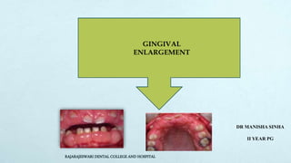
  • 1. GINGIVAL ENLARGEMENT DR MANISHA SINHA II YEAR PG RAJARAJESWARI DENTAL COLLEGE AND HOSPITAL
  • 2. CONTENTS  INTRODUCTION  CLASSIFICATIONS  GRADING  INFLAMMATORY GINGIVAL ENLARGEMENT  DRUG-INDUCED ENLARGEMENT  IDIOPATHIC ENLARGEMENT  ENLARGEMENTS ASS. WITH SYSTEMIC DISEASES  NEOPLASTIC ENLARGEMENT  FALSE ENLARGEMENT  TREATMENT OF GINGIVAL ENLARGEMENT  CONCLUSION
  • 3. INTRODUCTION  Gingival enlargement is the proliferation and intensification of the gingiva which is a prevailing character of the diseased gingival tissues.  Gingival enlargement and gingival overgrowth are terms used interchangeably with hyperplasia, hypertrophy, and fibrosis.  Hyperplasia is an increase in the number of cells in tissues that results in increased tissue volume. Hypertrophy refers to increased tissue size and volume resulting from increased cell size.  Fibrosis refers to a pathologic process in which disrupted wound healing is associated with defective cell proliferation, cell-to-cell interactions, cell-to-matrix interactions, and matrix deposition and with an impaired immune system response.
  • 4. CLASSIFICATION I. Inflammatory enlargement A. Chronic B. Acute II. Drug-induced enlargement III. Enlargements associated with systemic diseases or conditions A. Conditioned enlargement 1. Pregnancy 2. Puberty 3. Vitamin C deficiency 4. Plasma cell gingivitis 5. Nonspecific conditioned enlargement (granuloma pyogenicum) B. Systemic diseases causing gingival enlargement 1. Leukemia 2. Granulomatous disease (Wegener’s granulomatosis, sarcoidosis) IV. Neoplastic enlargement (gingival tumors) A. Benign tumors B. Malignant tumors V. False enlargement
  • 5. Using the criteria of location & distribution, gingival enlargement is designated as follows: LOCALISED GENERALISED MARGINAL PAPILLARY DIFFUSE DISCRETE
  • 6. The degree of gingival enlargement can be scored as follows: ANGELOPOULOS AND GOAZ (1972) None. Not more than 1/3rd of the clinical crown covered. Any part of the middle third of the crown covered. Greater than 2/3rd of the crown covered. 1 0 2 3
  • 7. 0 = No encroachment of the interdental papilla onto the tooth surface 1 = Mild encroachment of the interdental papilla, producing a blunted appearance to papilla tip 2 = Moderate encroachment, involving lateral spread of papilla across buccal tooth surface of less than one quarter of tooth width. 3 = Marked encroachment of papilla, i.e. more than 1/4th tooth width. Loss of normal papilla form Seymour et al 1985
  • 8. Miller and Damm (1992) Horizontal gingival overgrowth index is described as: Grade 0: < 1mm Grade 1: 1 to 2 mm Grade 2: >2mm Vertical gingival overgrowth index is described as: Grade 0: Normal gingival, no alteration Grade 1: Minimal overgrowth, ≤ 2mm, gingiva covering the cervical third or less of the anatomic crown. Grade 2: Moderate overgrowth: 2 to 4 mm, gingival covering the middle third of the anatomic crown. Grade 3: Severe overgrowth: ≥4mm, nodular growth, gingival covering more than two thirds of the dental crown.
  • 9. Bokenkamp A and Bohnhorst B (1994)  Grade 0: No signs of gingival overgrowth  Grade I: Gingival hyperplasia confined to interdental papilla .  Grade II: Hyperplasia of interdental papilla and marginal gingiva  Grade III: Gingival hyperplasia covering at least three-quarters of tooth crowns
  • 10. Eva and Ingles (1999) Grade 0: No overgrowth, firm adaptation of the attached gingiva to the underlying alveolar bone Grade 1: Early overgrowth, as evidenced by an increase in density of the gingiva with marked stippling and granular appearance. The tip of the papilla is rounded and the probing depth is less than or equal to 3mm. Grade 2: Moderate overgrowth, manifested by an increase in the size of the papilla and/ or rolled gingival margins. The contour of the margin is still concave or straight. The probing depth is equal to or less than 6mm and the papilla is somewhat retractable. Grade 3: Marked overgrowth, represented by encroachment of the gingiva onto the clinical crown. Contour of the margin is convex rather than concave. The probing depth is greater than 6mm and the papilla is clearly retractable. Grade 4: Severe overgrowth, characterized by a profound thickening of the gingiva. A large percentage of the clinical crown is covered. The papilla is retractable, the probing depth is greater than 6 mm and the buccolingual dimension is approximately 3 mm
  • 11. Miranda and Brunet index (2001)  described an index in which horizontal measurement of the enlargement is possible. This index is also termed as nodullary papilla index.  In this index the measurement is carried out with the help of a periodontal probe from the enamel surface of the interdental contact point to the outer papillary area. The scores of this index is as mentioned below: Score 0: Papilla thickness < 1 mm Score 1: Papilla thickness 1- 2 mm Score 2: Papilla thickness > 2 mm
  • 12. INFLAMMATORY ENLARGEMENT  Gingival enlargement may result from chronic or acute inflammatory changes; chronic changes are much more common.  In addition, inflammatory enlargements usually are a secondary complication to any of the other types of enlargement, creating a combined gingival enlargement.  In these cases it is important to understand the double etiology and treat them adequately.
  • 13. A) Acute Inflammatory Enlargement  Acute inflammatory enlargement of the gingiva usually is caused by a mechanical, chemical, or physical irritation and can be resolved by removal of the irritant.  Mouth breathing, impacted food items, and poor oral hygiene are usually responsible for acute inflammatory reactions in gingival tissues.  Acute lesions are usually localized to marginal or papillary gingiva.  Acute inflammatory enlargement can lead to the formation of gingival abscess. Gingival abscess is a localized, painful, and rapidly expanding lesion.
  • 14. GINGIVAL ABSCESS  In its early stages, it appears as a red swelling with a smooth, shiny surface. The lesion usually becomes fluctuant and pointed, with a surface orifice and a purulent exudate in 24 to 48 hours  Generally limited to marginal gingiva or interdental papilla.  Etiology- caused by a mechanical, chemical, or physical irritation and can be resolved by removal of the irritant. Mouth breathing, impacted food items, and poor oral hygiene
  • 15. Histopathology:- A purulent focus in the connective tissue, surrounded by a diffuse infiltration of polymorphonuclear leukocytes, edematous tissue, and vascular engorgement. The surface epithelium has varying degrees of intra- and extracellular edema, invasion by leukocytes, and sometimes ulceration. The lesion is confined to the gingiva, and it should be distinguished from periodontal abscess.
  • 16. PERIODONTAL (LATERAL) ABSCESS  It is a localized purulent inflammation in the periodontal tissues.  Also known as Lateral or Parietal abscess.  It involves the supporting periodontal tissues causing enlargement of gingiva.
  • 17.  Periodontal abscess formation may occur in the following ways: --Infection from a periodontal pocket deeply into the supporting periodontal tissues. -- localization of the suppurative inflammatory process Lateral extension of inflammation from the inner surface of a periodontal pocket into the connective tissue of the pocket wall. pocket -- tortuous course around the root, a periodontal abscess may form in the cul - de- sac. Incomplete removal of calculus during treatment of a periodontal pocket. the absence of periodontal disease after trauma to the tooth or perforation of the lateral wall of the root in endodontic therapy
  • 18. B) CHRONIC INFLAMMATORY ENLARGEMENT Etiology:--Prolonged exposure to dental plaque.  Factors that favour plaque accumulation and retention include:- Poor oral hygiene. Irritation by anatomic abnormalities Improper restorative and orthodontic appliances Mouth breathing
  • 19. CLINICAL FEATURES  Originates as Slight ballooning of the interdental papilla and/or the marginal gingiva.  Produces a life preserver-shaped bulge around the involved teeth in early stages.  May be localized or generalized and progresses slowly & painlessly, unless it is complicated by acute infection or trauma.
  • 20.  Occasionally, occurs as a discrete sessile or pedunculated mass resembling a tumor.  May be interproximal or on the marginal or attached gingiva.  Lesions are slow-growing masses and usually painless.  They may undergo spontaneous reduction in size, followed by exacerbation and continued enlargement.  Painful ulceration sometimes occurs in the fold between the mass and the adjacent gingiva
  • 21. Gingival Changes Associated with Mouth Breathing:- o Gingiva appears red and edematous with a diffuse surface shininess of the exposed area. o Maxillary anterior region -common site o Harmful effect is attributed to irritation from surface dehydration. Histopathology  Show the exudative and proliferative features of chronic inflammation.  Lesions that are clinically deep red or bluish red are soft and friable with a smooth, shiny surface, and they bleed easily.  They also have a preponderance of inflammatory cells and fluid, with vascular engorgement, new capillary formation, and associated degenerative changes.  Lesions that are relatively firm, resilient, and pink have a greater fibrotic component with an abundance of fibroblasts and collagen fibers.
  • 23.  Predilection for anterior gingiva  Higher prevalence in children  Onset within 3 months  Change in gingival contour leading to modifi cation of gingival size  Enlargement fi rst observed at the interdental papilla  Change in gingival color  Increased gingival exudate  Bleeding upon provocation  Found in gingiva with or without bone loss but is not associated with attachment loss  Pronounced inflammatory response of gingiva in relation to the plaque present  Reductions in dental plaque can limit the severity of lesion  Must be using phenytoin, cyclosporine A or certain calcium channel blockers; the plasma concentrations to induce the lesion have not been clearly defi ned in humans Characteristics of drug-influenced gingival enlargement (Mariotti 1999)
  • 24. Clinical Features  Growth starts as a painless, bead-like enlargement of the interdental papilla and extends to the facial and lingual gingival margins. Case of phenytoin induced gingival enlargement speech, mastication, tooth eruption, and aesthetic problems. ANTICONVULSANTS, IMMUNOSUPPRESSANTS, AND CALCIUM CHANNEL BLOCKERS
  • 25.  As the condition progresses, the marginal and papillary enlargements unite and may develop into a massive tissue fold covering a considerable portion of the crowns; they may interfere with occlusion.  Spontaneous disappearance occurs within a few months after discontinuation of the drug.  When UNCOMPLICATED BY INFLAMMATION- lesion is mulberry shaped, firm, Pale pink, and resilient minutely lobulated surface and no tendency to bleed.
  • 26.  The presence of the enlargement makes plaque control difficult resulting in a secondary inflammatory process that complicates the gingival overgrowth caused by the drug.  The resultant enlargement then becomes a combination of the increase in size caused by the drug and the complicating inflammation caused by bacteria.  Secondary inflammatory changes add to the size of the lesion caused by the drug and produce a red or bluish red discoloration, obliterate the lobulated surface demarcations, and increase bleeding tendency.  Some investigators believe that inflammation is a prerequisite for development of the enlargement, which therefore could be prevented by plaque removal and fastidious oral hygiene (Ciancio and Yaffe J periodontol 1972)
  • 27. Hassell et al (1982) have hypothesized that in noninflamed gingiva, fibroblasts are less active or even quiescent and do not respond to circulating phenytoin, whereas fibroblasts within inflamed tissue are in an active state as a result of the inflammatory mediators and the endogenous growth factors present. Barclay S et al (J Clin Periodontol 1992) evaluated the incidence and severity of nifedipine-induced gingival overgrowth and concluded that nifedipine therapy results in significant gingival changes, an effect which may be mediated by the drug's action on calcium transport. Thomason JM et al ( J Clin Periodontol 1993) studied the prevalence and severity of cyclosporin and nifedipine induced gingival overgrowth and concluded that patients taking cyclosporin or cyclosporin and nifedipine experience gingival overgrowth and that the severity of the overgrowth is greater in patients taking the combined therapy.
  • 28. Histopathology  Pronounced hyperplasia of the connective tissue and epithelium.  Acanthosis of the epithelium, and elongated rete pegs extend deep into the connective tissue, which exhibits densely arranged collagen bundles with an increase in the number of fibroblasts and new blood vessels.  An abundance of amorphous ground substance as well as marked plasma cell; infiltration has also been reported (Mariani et al 1993).
  • 29. Cyclosporine enlargements -- a more highly vascularized connective tissue with foci of chronic inflammatory cells, particularly plasma cells. “mature” phenytoin enlargements fibroblast/collagen ratio equal to that of normal gingiva from normal individuals, suggesting that at some point in the development of the lesion, fibroblastic proliferation must have been abnormally high Recurring phenytoin enlargements appear as granulation tissue composed of numerous young capillaries and fibroblasts and irregularly arranged collagen fibrils with occasional lymphocytes
  • 30. ANTICONVULSANTS Phenytoin (Dilantin) Ethotoin (Peganone) and mephenytoin (Mesantoin). Succinimides (ethosuximide [Zarontin], Methsuximide [Celontin]) Valproic acid [Depakene]).
  • 31. Prevalence Gingival overgrowth becomes clinically noticeable within 2 to 3 months after initial administration of phenytoin and reaches its maximal severity at 12 to 18 months. (Livingston S 1990)  occurs in 50% of patients receiving the drug, (Taicher S et al1991)  Range is 3% to 84.5%. (Goaz PW,1972; Glickman 1941)  It occurs more often in younger patients .  Its occurrence and severity are not necessarily related to the dosage after a threshold level has been exceeded.
  • 32. Pathogenesis:- Direct stimulating action on gingival fibroblasts or mast-cells with secondary fibroblastic involvement. (Shafer et al 1960) Existence of differential proportions of fibroblast subsets in each individual which exhibit a fibrogenic response to these medications (JISP 2013) Fibroblasts with abnormal susceptibility to drug. fibroblasts from overgrown gingiva in phenytoin treated patients are characterized by elevated levels of proteins, esp. Collagen (J Periodontol 2004)
  • 33. Genetic predisposition is a suspected factor . Hassell et al (1994) hypothesized that gingival enlargement may result from the genetically determined ability or inability of the host to deal effectively with prolonged administration of phenytoin. Other proposed mechanisms include:  the production of inactive fibroblastic collagenase causing a decrease in collagen turnover. Phenytoin may induce a decrease in collagen degradation as a result of the production of an inactive fibroblastic collagenase. (Hassell 1982)  phenytoin-induced folic acid deficiency that can cause degenerative changes in the sulcular epithelium and exacerbate the inflammatory response  phenytoin-induced increase in the synthesis of testosterone metabolites by gingival fibroblasts with resultant overgrowth
  • 34. IMMUNOSUPPRESSANTS potent immunosuppressive agent used to prevent organ transplant rejection and to treat several diseases of autoimmune origin. MOA-not known selectively and reversibly inhibit helperT cells, which play a role in cellular and humoral immune responses. Cyclosporin A (Sandimmune, Neoral) is administered intravenously or by mouth --dosages greater than 500 mg/day have been reported to induce gingival overgrowth.(Daley TD 1986) Occurrence varies from 25% to 70% (Romito GA 2004) CYCLOSPORINE
  • 35.  Gingival enlargement is greater in patients who are medicated with both cyclosporine and calcium channel blockers. (Taylor J 1987)  The microscopic finding of many plasma cells plus the presence of an abundant amorphous extracellular substance has suggested that the enlargement is a hypersensitivity response to the cyclosporine.(Mariani G et al 1993)  In addition to gingival enlargement, cyclosporine induces other major side effects such as –nephrotoxicity,hypertension, hypertrichosis  Another immunosuppressive drug, TACROLIMUS, has been used effectively and is also nephrotoxic, but it results in much less severe hypertension, hypertrichosis, and gingival overgrowth.(Bader G 1988)
  • 36. CALCIUM CHANNEL BLOCKERS They inhibit calcium ion influx across the cell membrane of heart and smooth muscle cells, blocking intracellular mobilization of calcium. This induces direct dilation of the coronary arteries and arterioles, improving oxygen supply to the heart muscle; it also reduces hypertension by dilating the peripheral vasculature. Drugs developed for the treatment of cardiovascular conditions such as hypertension, angina pectoris, coronary artery spasms, and cardiac arrythmias.
  • 37. • DIHYDROPYRIDINE DERIVATIVES (amlodipine [Lotrel, Norvasc], felodipine [Plendil], nicardipine [Cardene], nifedipine [Adalat, Procardia]) • BENZOTHIAZINE DERIVATIVES(diltiazem [Cardizem, Dilacor XR, Tiazac]) • PHENYLALKYLAMINE DERIVATIVES (verapamil [Calan, Isoptin, Verelan, Covera HS]).  Nifedipine, one of the most often used, induces gingival enlargement in 20% of patients. (Lucas RM 1985)
  • 38.  It has been postulated that the drug may stimulate cell proliferation and synthesis indirectly by one of three mechanisms:- 1. By giving rise to the formation of a more potent Metabolic byproduct. 2. By stimulating the production of either interleukin 2 by T cells or metabolites of testosterone by gingival fibroblasts that in turn would promote cellular proliferation and synthesis (Nishikawa S 1991) 3.By causing a calcium dependent inhibitory effect on T cells that would increase gingival susceptibility to bacterial infection through immunosuppression. (Seymour RA 1991)
  • 39.
  • 41.  Idiopathic gingival enlargement is a rare condition of undetermined cause. Etiology  The cause is unknown and thus designated as idiopathic.  Some cases- Hereditary basis but the genetic mechanisms are not well understood.  Mode of inheritance is found to be autosomal recessive in some cases and autosomal dominant in others. (Cocker et al 1974)  Recently a locus for autosomal dominant HGF has been mapped to a region on chromosome 2 (Hart et al. 1998, Xiao et al. 2000)  In some families the gingival enlargement may be linked to impairment of physical development. (Collan Y et al 1978) Gingivomatosis, elephantiasis, idiopathic fibromatosis, hereditary gingival hyperplasia, and congenital familial fibromatosis.
  • 42.  HGF may be an isolated disease entity or part of a syndrome associated with other clinical manifestations, such as hypertrichosis, mental retardation ,epilepsy, hearing loss , growth retardation and abnormalities of extremities.  Studies have suggested that an important pathogenic mechanism may be enhanced production of transforming growth factor (TGF-beta 1) reducing the proteolytic activities of HGF fibroblasts, which again favour the accumulation of extracellular matrix (Coletta et al. 1999).
  • 43. Clinical Features Affects the attached gingiva, as well as the gingival margin and interdental papillae Enlarged gingiva is pink, firm, and almost leathery in consistency characteristic minutely pebbled surface  Severe cases- teeth are almost completely covered & enlargement projects into the oral vestibule.  The jaws appear distorted because of the bulbous enlargement of the gingiva.
  • 45.  Many systemic diseases can develop oral manifestations that may include gingival enlargement.  These diseases and conditions can affect the periodontium by two different mechanisms, as follows:- 1. Magnification of an existing inflammation initiated by dental plaque. This group of diseases (Conditioned Enlargements) includes:  hormonal conditions (e.g., pregnancy and puberty)  nutritional diseases such as vitamin C deficiency  some cases in which the systemic influence is not identified (nonspecific conditioned enlargement). 2.Manifestation of the systemic disease independently of the inflammatory status of the gingiva. This group involves Systemic Diseases Causing Gingival Enlargement and Neoplastic Enlargement (Gingival Tumors)
  • 46. Bacterial plaque is necessary for the initiation of this type of enlargement. However, plaque is not the sole determinant of the nature of the clinical features. The three types of conditioned gingival enlargement are:  Hormonal (pregnancy, puberty),  Nutritional (associated with vitamin C deficiency),  Allergic. Gingival Overgrowth Associated With Systemic Conditions
  • 47.  Pregnancy gingival enlargement may be marginal and generalized or may occur as single or multiple tumor-like masses .  During pregnancy, there is an increase in levels of both progesterone and estrogen, which by the end of the third trimester reach levels 10 and 30 times the levels during the menstrual cycle, respectively. (Amar S 1994)  These hormonal changes induce changes in vascular permeability leading to gingival edema and an increased inflammatory response to dental plaque.  The subgingival microbiota may also undergo changes, including an increase in Prevotella intermedia, Prevotella melaninogenica, and Porphyromonas gingivalis (Kornman KS 1980) 1.Enlargement in Pregnancy A 55 fold increase in the proportion of P intermedia has been demonstrated in pregnant females as compared to non pregnant controls, implying a role for gestational hormones in causing a change in microbial ecology in the gingival pocket. (Jensen et al 1981)
  • 48. MARGINAL GINGIVAL ENLARGEMENT  Marginal gingival enlargement during pregnancy results from the aggravation of previous inflammation,  Incidence reported as 10%and 70%.  The enlargement is usually generalized and tends to be more prominent interproximally than on the facial and lingual surfaces.  The enlarged gingiva is bright red or magenta, soft, and friable and has a smooth, shiny surface.  Bleeding occurs spontaneously or on slight provocation. TUMORLIKE GINGIVAL ENLARGEMENT.  The so-called pregnancy tumor is not a neoplasm; it is an inflammatory response to bacterial plaque and is modified by the patient’s condition.  Usually appears after the third month of pregnancy but may occur earlier.  The reported incidence is 1.8%to 5%.  Appears as a discrete, mushroomlike, flattened spherical mass that protrudes from the gingival margin or more often from the interproximal space and is attached by a sessile or pedunculated base.
  • 49. Histopathology  A central mass of connective tissue, with numerous diffusely arranged, newly formed, and engorged capillaries lined by cuboid endothelial cells and a moderately fibrous stroma.  The stratified squamous epithelium is thickened, with prominent rete pegs and some degree of intracellular and extracellular edema.
  • 50.  It is marginal and interdental  characterized by prominent bulbous interproximal papillae  Often, only the facial gingivae are enlarged, and the lingual surfaces are relatively unaltered.  It is the degree of enlargement and its tendency to recur in the presence of relatively scant plaque deposits that distinguish pubertal gingival enlargement from uncomplicated chronic inflammatory gingival enlargement.  After puberty the enlargement undergoes spontaneous reduction but does not disappear completely until plaque and calculus are removed. 2.Enlargement in Puberty
  • 51.  A longitudinal study of subgingival microbiota of children between ages 11 and 14 and their association with clinical parameters has implicated Capnocytophaga species in the initiation of pubertal gingivitis. (Mombelli A, Lang NP ,1990)  Other studies have reported that hormonal changes coincide with an increase in the proportion of Prevotella intermedia and Prevotella nigrescens. (Fujii H, 1994) The microscopic appearance of gingival enlargement in puberty is chronic inflammation with prominent edema and associated degenerative changes
  • 52.  Acute vitamin C deficiency itself does not cause gingival inflammation, but it does cause hemorrhage, collagen degeneration, and edema of the gingival connective tissue.  These changes modify the response of the gingiva to plaque to the extent that the normal defensive delimiting reaction is inhibited, and the extent of the inflammation is exaggerated, resulting in the massive gingival enlargement seen in scurvy. 3.Enlargement in Vitamin C Deficiency marginal; the gingiva is bluish red, soft, and friable and has a smooth, shiny surface. Hemorrhage, occurring either spontaneously or on slight provocation surface necrosis with pseudomembrane formation
  • 53.  Mild marginal gingival enlargement  Gingiva appears red, friable, and sometimes granular  Lesion is located in the oral aspect of the attached gingiva and therefore differs from plaque-induced gingivitis. 4. Plasma Cell Gingivitis  An associated cheilitis and glossitis have been reported.  Plasma cell gingivitis is thought to be allergic in origin, possibly related to components of chewing gum, dentifrices, or various diet components.  Rare instances-marked inflammatory gingival enlargements with a predominance of plasma cells can appear; these are associated with rapidly progressive periodontitis.
  • 54.  more correctly called telangiectatic granuloma…  frequently ulcerated and the appearance of the fibrin-coated ulcer may resemble purulence.  may occur in all areas of the oral mucosa, but is most frequently found on the marginal gingiva (Makek & Sailer 1985).  may develop rapidly and the size varies considerably.  reddish or bluish, sometimes lobulated, and may be sessile or pedunculated.  Bleeding from the ulcerated lesion is common, but typically it is not painful.  Teeth may become separated due to interdental growth of the lesion. 5.Nonspecific Conditioned Enlargement (Pyogenic Granuloma):
  • 56. Leukemic Infiltration of the Periodontium  Leukemic gingival enlargement can be diffuse or marginal and localized or generalized.  It can appear as a diffuse enlargement of the gingival mucosa, an oversized extension of the marginal gingiva or a discrete tumor-like interproximal mass.  In patients with leukemic enlargement, the gingiva is bluish red, and it has a shiny surface. The consistency is moderately firm, but there is a tendency toward friability and hemorrhage that occur spontaneously or with slight irritation.  True leukemic enlargement often occurs with acute leukemia, but it may also be seen with subacute leukemia. It seldom occurs with chronic leukemia.
  • 57.  Clinically, the gingiva appears bluish red and cyanotic, with a rounding and tenseness of the gingival margin and has a shiny surface.  The consistency is moderately firm, but there is a tendency towards friability and hemorrhage, occurring either spontaneously or on slight irritation. Enlargement may be diffuse or marginal, localized or generalized.
  • 58.  Appear as a diffuse enlargement of the gingival mucosa, an oversized extension of the marginal gingiva, or a discrete tumor like inter-proximal mass.  Acute painful necrotizing ulcerative inflammatory involvement may occur in the crevice formed at the junction of the enlarged gingiva and the contiguous tooth surfaces. Histopathology  Areas of connective tissue infiltrated with a dense mass of immature and proliferating leukocytes  Engorged capillaries, oedematous and degenerated connective tissue, and epithelium with various degrees of leukocytic infiltration and edema are found.  Isolated surface areas of acute necrotizing inflammation with a pseudomembranous meshwork of fibrin, necrotic epithelial cells, polymorphonuclear neutrophils (PMNs), and bacteria are often seen.
  • 59. 2.Granulomatous Diseases WEGENER'S GRANULOMATOSIS:  Rare disease characterized by acute granulomatous necrotizing lesions of the respiratory tract, including nasal and oral defects.  immunologically mediated tissue injury. (Cotran RS 1989)  The initial manifestations of Wegener’s granulomatosis may involve the orofacial region and include: oral mucosal ulceration gingival enlargement abnormal tooth mobility exfoliation of teeth delayed healing response
  • 60.  The granulomatous papillary enlargement is reddish purple and bleeds easily on stimulation. Histopathology Chronic inflammation occurs with scattered giant cells and foci of acute inflammation and microabscesses covered by a thin acanthotic epithelium.
  • 61. SARCOIDOSIS:  granulomatous disease  unknown etiology.  can involve almost any organ, including the gingiva, in which a red, smooth, painless enlargement may appear. Histopathology  Sarcoid granulomas consist of discrete, noncaseating whorls of epithelioid cells and multinucleated, foreign body–type giant cells with peripheral mononuclear cells.
  • 63. arise from the gingival connective tissue or from the periodontal ligament slow-growing, spherical tumors that tend to be firm and nodular but may be soft and vascular. usually pedunculated --giant cell fibroma --peripheral ossifying fibroma FIBROMA Histopathology Bundles of Well-formed collagen fibers with scattering of fibrocytes and a variable vascularity. A) Benign Tumors of the Gingiva
  • 64.  Appear as Solitary, wartlike or "cauliflower"-like protuberances and may be small and discrete or broad, hard elevations with minutely irregular surfaces. Histopathology  Finger like projections of stratified squamous epithelium often hyperkeratotic, with a central core of fibrovascular tissue PAPILLOMA
  • 65.  Arise interdentally or from the gingival margin  sessile or pedunculated  smooth, regularly outlined masses to irregularly shaped, multilobulated protuberances  painless, vary in size  firm or spongy,  pink to deep red or purplish blue.  Local irritation or trauma appears to be important for these lesions to occur.  It has growth potential and may cause separation of teeth due to pressure exerted by the growth PERIPHERAL GIANT CELL GRANULOMA Histopathology Numerous foci of multinuclear giant cells and hemosiderin particles in CT stroma. Overlying epithelium is usually hyperplastic, with ulceration at the base. Bone formation occasionally occurs within the lesion
  • 66.  Leukoplakia of the gingiva varies in appearance from a grayish white, flattened, scaly lesion to a thick, irregularly shaped, keratinous plaque. LEUKOPLAKIA Histopathology •Leukoplakia exhibits hyperkeratosis and acanthosis. •Premalignant and malignant cases have a variable degree of atypical epithelial changes that may be mild, moderate, or severe, depending on the extent of involvement of the epithelial layers. •Inflammatory involvement of the underlying connective tissue is a common associated finding.
  • 67.  involves the marginal & attached gingiva.  occur in the mandibular canine and premolar areas.  painless, but with expansion. GINGIVAL CYST Histopathology:-  A gingival cyst cavity is lined by a thin, flattened epithelium with or without localized areas of thickening.  Less frequently, the following types of epithelium can be found: unkeratinized stratified squamous epithelium, keratinized stratified squamous epithelium, and parakeratinized epithelium with palisading basal cells.
  • 68. B) Malignant Tumors of the Gingiva Carcinoma:-  Squamous cell carcinoma is the most common malignant tumor of the gingiva.  It may be exophytic, presenting as an irregular outgrowth, or ulcerative, which appear as flat, erosive lesions.  It is often symptom free,  locally invasive.
  • 69. Malignant Melanoma Sarcoma Metastasis Rare oral tumor that tends to occur in the hard palate and maxillary gingiva of older persons. • Usually darkly pigmented and is often preceded by localized pigmentation. • May be flat or nodular and is characterized by rapid growth and early metastasis. • Arises from melanoblasts in the gingiva, cheek, or palate. • Infiltration into the underlying bone and metastasis to cervical and axillary lymph nodes are common Fibrosarcoma, lymphosarcoma, and reticulum cell sarcoma of the gingiva are rare • Kaposi’s sarcoma often occurs in the oral cavity of patients with Acquired immunodeficiency syndrome (AIDS), particularly in the palate and the gingiva Tumor metastasis to the gingiva occurs infrequently. • Such metastasis has been reported with various tumors, including adenocarcinoma of the colon, lung carcinoma, melanoma, renal cell carcinoma, hypernephroma, chondrosarcoma, and testicular tumor.
  • 71. Underlying osseous lesions Enlargement of bone subjacent to the gingival area occurs most often in tori and exostoses but it can also occur in Paget’s disease, fibrous dysplasia, cherubism,central giant cell granuloma, ameloblastoma, osteoma, and osteosarcoma. The gingival tissue can appear normal or may have unrelated inflammatory changes. Underlying Dental Tissues During the various stages of eruption, particularly of the primary dentition, the labial gingiva may show a bulbous marginal distortion caused by superimposition of the bulk of the gingiva on the normal prominence of the enamel in the gingival half of the crown. This enlargement has been termed developmental enlargement and often persist until the junctional epithelium has migrated from the enamel to the cementoenamel
  • 73. Treatment of chronic inflammatory enlargement Treated by SCALING AND ROOT PLANING, (provided the size of the enlargement does not interfere with complete removal of deposits from the involved tooth surfaces). When chronic inflammatory gingival enlargements include a significant fibrotic component that does not undergo shrinkage after scaling and root planing or are of such size that they obscure deposits on the tooth surfaces and interfere with access to them. SURGICAL REMOVAL is the treatment of choice GINGIVECTOMY FLAP OPERATION. Used when the enlarged gingiva remains soft and friable even after scaling and root planing Indicated if the gingivectomy incision removes all of the attached, keratinized gingiva, which will create a mucogingival problem Tumorlike inflammatory enlargements are treated by gingivectomy.
  • 74. Treatment Of Drug induced gingival enlargement First, consideration should be given to the possibility of DISCONTINUING THE DRUG OR CHANGING THE MEDICATION. These possibilities should be examined with the patient’s physician. Simple discontinuation of the offending drug is usually not practical, but its SUBSTITUTION WITH ANOTHER MEDICATION might be an option. Second, the clinician should EMPHASIZE PLAQUE CONTROL as the first step in the treatment of drug-induced gingival enlargement Third, in some patients, gingival enlargement persists after careful consideration of the previous approaches. These patients may require SURGERY, either PERIODONTAL FLAP OR GINGIVECTOMY.
  • 75. DRUG SUBSTITUTES PHENYTOIN CARBAMAZEPINE AND VALPROIC ACID, NIFEDIPINE DILTIAZEM OR VERAPAMIL CYCLOSPORINE TACROLIMUS  Also, consideration may be given to the use of another class of ANTIHYPERTENSIVE medications rather than calcium channel blockers, none of which is known to induce gingival enlargement.  Clinical trials have also shown that the substitution of cyclosporine by tacrolimus results in a significant decrease in the severity of gingival enlargement when compared to patients who are kept on cyclosporine therapy.
  • 76.
  • 77.
  • 78.
  • 79.
  • 80. Treatment of Leukemic gingival enlargement The patient’s bleeding and clotting times and platelet count should be checked, and the hematologist should be consulted before periodontal treatment is instituted. After acute symptoms subside, attention is directed to correction of the gingival enlargement. The rationale for therapy is to remove the local irritating factors to control the inflammatory component of the enlargement. The lesion is treated by scaling and root planing performed in stages with topical and local anesthesia and instructing the patient in oral hygiene for plaque control.
  • 81. This portion of the therapy may include, at least initially, the daily use of chlorhexidine mouthwashes. Oral hygiene procedures are extremely important in these patients and should be performed by the nurse if necessary. Progressively deeper scaling is carried out at subsequent visits. Treatments are confined to a small area of the mouth to facilitate the control of bleeding. ANTIBIOTICS are administered systemically the evening before and for 48 hours after each treatment to reduce the risk of infection.
  • 82. Treatment of gingival enlargement in pregnancy  Treatment requires elimination of all local irritants responsible for precipitating the gingival changes in pregnancy.  Marginal and interdental gingival inflammation and enlargement are treated by scaling and root planing.  Treatment of tumorlike gingival enlargements consists of surgical excision and scaling and planing of the tooth surface. The enlargement will recur unless all of the irritants are removed
  • 83. Timing of Treatment and Indications:-  Gingival lesions in pregnancy should be treated as soon as they are detected, although not necessarily by surgical means. Scaling and rootplaning procedures and adequate oral hygiene measures may reduce the size of the enlargement.  Gingival enlargements will shrink after pregnancy but may not completely disappear. After pregnancy, the entire mouth should be reevaluated, a full set of radiographs taken, and the necessary treatment undertaken.  Lesions should be removed surgically during pregnancy only if they interfere with mastication or produce an esthetic disfigurement that the patient wants removed.
  • 84. Treatment of gingival enlargement in puberty  Gingival enlargement in puberty is treated by performing scaling and curettage, removing all sources of irritation, and controlling plaque.  Surgical removal may be required in severe cases.
  • 85. CONCLUSION Inspite of a myriad of etiology, gingival enlargements can often be diagnosed by a careful history, by location or by the clinical presentation. Presence of local irritants (plaque and calculus) could be primary or associated cause of gingival enlargements. Hence, plaque control is an essential aspect of management in all the patients. An excisional/incisional biopsy and/or hematologic/histologic examination may be needed occasionally to correctly diagnose the uncommon cases of gingival enlargement.
  • 86. REFERENCES:-  Newman MG , Takei HH , Klokkevold PR , Carranza FA . Carranza’s Clinical Periodontology, 10th edition  Newman MG , Takei HH , Klokkevold PR , Carranza FA . Carranza’s Clinical Periodontology, 13th edition  Atlas of cosmetic and reconstructive periodontal surgery, Cohen  Marshall R , Bartold M A clinical review of drug induced gingival overgrowth Australian dental journal 1999 ;44:4 219- 232  Seymour RA, Ellis JS, Thomason JM: Risk factors for druginduced gingival overgrowth.J Clin Periodontol 2000; 27: 217-223.  Seymour RA , Thomasan JM Pathogenesis of Drug Induced Gingival Overgrowth- J Clin Periodontol 1996;23:165- 175  Strawberry –like gingival tumor as first sign of Wegener’s Granulomatosis. J Periodontol 2008; 79: 1297-1303  Seymour RA and Heasman PA: Drugs and the periodoniium. J Clin Periodontol 1988: 15: 1-16

Editor's Notes

  1. Although their pathogenetic mechanisms are different, hyperplasia and hypertrophy usually occur simultaneously when cellular involvement in hyperplasia most likely triggers the overgrowth.
  2. ( ACC. TO ETIOLOGICAL FACTORS & PATHOLOGICAL CHANGES)
  3. Loc alizecl: Limited to the gingiva adjacent to a single tooth or group of teeth. • Generalized: Involving the gingiva throughout the mouth. • ,Alar,tiival: Confined to the marginal gingiva. • Papillary: Confined to the interdental papilla. • 1)i1Juse: Involving the marginal and attached gingivae and papillae. • Discrete: An isolated sessile or pedunculated, tumorlike enlargement.
  4. In order to treat and prevent the recurrence of GO, the operator must recognize the extent of the vertical and the horizontal components as well as the severity of GO along with its etiopathogenesis. described an index for measuring the vertical component of gingiva. Three grades based on the enlargement covering the clinical crown were described as:
  5. Seymour RA (1985)8 described a gingival overgrowth index (GOi), in which assessment of gingival hyperplasia is done on plaster study casts that includes both horizontal and vertical overgrowth focusing on the anterior teeth since the overgrowth is more likely to occur in this region. Various authors have suggested that this index allows a three dimensional diagnosis of gingival overgrowth.
  6. enlargement was divided into a vertical and a horizontal component and abbreviated as GOI index. Vertical component is measured from CEJ to the free gingival margin and the horizontal component from the enamel surface at the point of contact to the external margin of the interdental papilla
  7. introduced a new index for measuring gingival overgrowth caused due to drugs. In this index for standardization, the buccal and lingual papillae were scored separately. The criteria by which scores were divided are as mentioned below:
  8. In some cases, gingival enlargement is a direct outcome of gingivitis without any complicating factors or involvement of systemic conditions. When a patient with gingival enlargement is seen, the initial assessment is made by careful visual examination of abnormalities of gingival contours, texture, and color, which are compared with normal standards. Visual inspection is accompanied by a detailed medical history to exclude potential systemic factors and conditions. Dental irregularities, dysfunctional habits, and oral care eficiency should be considered in the evaluation, and clinical measurements should be recorded.
  9. The main etiologic factor for acute inlammatory gingival enlargement is trauma. Traumatic lesions occur when a foreign substance (e.g., toothbrush bristle) is forcefully embedded in the gingiva and complicated by resident microbes. Trauma-induced injuries result in a chronic process characterized by granulation tissue formation and ibrosis.
  10. Adjacent teeth are sensitive to percussion. >> Acute inflammatory gingival enlargement results from bacteria carried deep into the tissues when foreign substances such as a toothbrush bristle, a piece of apple core, or a lobster shell fragment is forcefully embedded into the gingiva. (Lesion is confined to gingiva).
  11. 1. Extension of infection from a periodontal pocket deeply into the supporting periodontal tissues and localization of the suppurative inflammatory process along the lateral aspect of the root. 2. Lateral extension of inflammation from the inner surface of a periodontal pocket into the connective tissue of the pocket wall. Localization of the abscess results when drainage into the pocket space is impaired . 3. In a pocket that describes a tortuous course around the root, a periodontal abscess may form in the cul -de- sac, the deep end of which is shut off from the surface. 4. Incomplete removal of calculus during treatment of a periodontal pocket. In this instance, the gingival wall shrinks, occluding the pocket orifice, and a periodontal abscess occurs in the sealed-off portion of the pocket. 5. A periodontal abscess may occur in the absence of periodontal disease after trauma to the tooth or perforation of the lateral wall of the root in endodontic therapy.
  12. Gingival enlargement may result from chronic or acute inflammatory changes; chronic changes are much more common. In addition, inflammatory enlargements usually are a secondary complication to any of the other types of enlargement, creating a combined gingival enlargement
  13. Gingival enlargement is a well-known consequence of the administration of some……….. and may create Three drugs associated with DIGO are phenytoin, nifedipine, and cyclosporine.
  14. •It occurs in areas in which teeth are present, not in edentulous spaces, and the enlargement disappears in areas from which teeth are extracted. •Hyperplasia of the mucosa in edentulous mouths has been reported but is rare •The enlargement is chronic and slowly increases in size •When surgically removed, it recurs. Drug-induced enlargement may occur in mouths with little or no plaque and may be absent in mouths with abundant deposits
  15. The first drug-induced gingival enlargements reported were those produced by Other hydantoins known to induce gingival enlargement are Other anticonvulsants that have the same side effect are the (Hallmon et al 1999)
  16. susceptibility or resistance to pharmacologically induced gingival overgrowth may be governed by the …. only few subsets of patients on phenytoin develop enlargement. These individuals have ,, Tissue culture experiments by Shafer in 1960 indicate that phenytoin stimulates proliferation of fibroblast-like cells and epithelium
  17. In conclusion, the pathogenesis of gingival enlargement induced by phenytoin is not known, but some evidence links it to a direct effect on specific, genetically predetermined subpopulations of fibroblasts, inactivation of collagenase, and plaque-induced inflammation
  18. Cyclosporine-induced gingival enlargement is more vascularized than phenytoin enlargement. Affects children more frequently, and its magnitude appears to be related more to the plasma concentration than to the patient’s periodontal status.
  19. Schematic diagram to illustrate the potential multifactorial features and interactions involved in the pathogenesis of drug-induced gingival overgrowth. Despite their pharmacological diversity, the three major drugs causing gingival overgrowth, namely; anticonvulsants, calcium channel blockers, and immunosuppressants; have similar mechanism of action at the cellular level, where they inhibit intracellular calcium ion influx. The action of these drugs on calcium and sodium ion flux may prove to be the key in understanding why three dissimilar drugs have a common side effect upon a secondary target tissue, such as gingival connective tissue. An appraisal of the various investigations into the pathogenesis of drug-induced gingival overgrowth supports the hypothesis that it is multifactorial [Figure 1].[3,4,19] Plaque scores and gingival inflammation appear to exacerbate the expression of drug-induced gingival overgrowth, irrespective of the initiating drug. The severity of gingival enlargement in patients taking medications correlates well with poor plaque control and is commensurate with the degree of plaque-induced inflammation. The importance of plaque as a cofactor in the etiology of drug-associated gingival enlargement has been recognized in the most recent classification system for periodontal diseases. In this classification, “drug-induced gingival enlargements” are categorized as plaque-induced gingival diseases modified by medications.[
  20. It has been designated by terms like… The enlargement usually begins with the eruption of the primary or secondary dentition and may regress after extraction, suggesting that the teeth (or the plaque attached to them) may be initiating factors. The presence of bacterial plaque is a complicating factor.
  21. The facial and lingual surfaces of the mandible and maxilla are generally affected, but the involvement may be limited to either jaw.
  22. •Conditioned enlargement occurs when the systemic condition of the patient exaggerates or distorts the usual gingival response to dental plaque. The specific manner in which the clinical picture of conditioned gingival enlargement differs from that of chronic gingivitis depends on the nature of the modifying systemic influence
  23. Tumour gingival enlargement It tends to expand laterally, and pressure from the tongue and the cheek perpetuates its lattened appearance. It is dusky red or magenta; it has a smooth, glistening surface that often exhibits numerous deep red, pinpoint markings.
  24. Generally dusky red or magenta, it has a smooth, glistening surface that often exhibits numerous deep-red, pinpoint markings. Superficial lesion and usually does not invade the underlying bone. Consistency varies; the mass is usually semifirm, but it may have various degrees of softness and friability. Usually painless unless its size and shape foster accumulation of debris under its margin or interfere with occlusion, in which case, painful ulceration may occur
  25. Enlargement of the gingiva is sometimes seen during puberty and occurs in both male and female adolescents and appears in areas of plaque accumulation. The size of the gingival enlargement greatly exceeds that usually seen in association with comparable local factors. --the mechanical action of the tongue and the excursion of food prevent a heavy accumulation of local irritants on the lingual surface. Gingival enlargement during puberty has all the clinical features generally associated with chronic inflammatory gingival disease.
  26. Enlargement of the gingiva is generally included in classic descriptions of scurvy.
  27. Consists of a mild marginal gingival enlargement that extends to the attached gingiva. Gingiva appears red, friable, and sometimes granular and bleeds easily; usually it does not induce a loss of attachment. Lesion is located in the oral aspect of the attached gingiva and therefore differs from plaque-induced gingivitis
  28. tumorlike gingival enlargement that may be conceived as an exaggerated reaction to minor trauma without it having been possible to demonstrate a definite infectious organism ….since the lesion is highly vascular and usually is not purulent as the term pyogenic suggests.
  29. Leukemic infiltration causing localized gingival swelling of the interdental papillae between the maxillary CI and LI
  30. Patients with leukemia may also have a simple chronic inflammation without the involvement of leukemic cells and may present with the same clinical and microscopic features seen in patients without the systemic disease. Most cases reveal features of both simple chronic inflammation and leukemic infiltrate.
  31. Cause is unknown but it is considered as an … Renal lesions develop, and acute necrotizing vasculitis affects the blood vessels.
  32. It starts in individuals in their twenties or thirties, predominantly affects blacks
  33. Hard fibromas of the gingiva are rare; most of the lesions diagnosed clinically as “fibromas” are inflammatory enlargements. •Also called giant cell fibroma contains multinucleated fibroblasts. •In another variant of fibroma, mineralised tissue (bone, cementum like material, dystrophic calcification) may be found and is called peripheral ossifying fibroma.
  34. Benign proliferations of surface epithelium associated with the human papilloma virus (HPV). •Viral subtypes HPV-6 and HPV-11 have been found in most cases of oral papillomas.
  35. Giant cell lesions of the gingiva arise interdentally or from the gingival margin, occur most frequently on the labial surface, and may be sessile or pedunculated. •They vary in appearance from smooth, regularly outlined masses to irregularly shaped, multilobulated protuberances with surface indentations . •Ulceration of the margin is occasionally seen. •The lesions are painless, vary in size, and may cover several teeth. •They may be firm or spongy, and the color varies from pink to deep red or purplish blue. •Local irritation or trauma appears to be important for these lesions to occur.
  36. Leukoplakia is a strictly clinical term defined by the World Health Organization as a white patch or plaque that does not rub off and cannot be diagnosed as any other disease. The cause of leukoplakia remains obscure, although it is associated with the use of tobacco (smoke or smokeless). Other probable factors are Candida albicans, HPV-16 and HPV-18, and trauma. 
  37. Gingival cysts develop from odontogenic epithelium or from surface or sulcular epithelium traumatically implanted in the area. Other benign tumors or masses have also been described as rare or infrequent findings in the gingiva. These include: Nevus Myoblastoma Ameloblastoma Hemangioma Neurilemoma Neurofibroma Mucoceles
  38. Oral cancer accounts for less than 3% of all malignant tumors in the body. The gingiva is not a frequent site of oral malignancy (6% of oral cancers). (Krolls SO,1976) Symptom free…often going unnoticed until complicated by inflammatory changes that may mask the neoplasm but cause pain; sometimes it becomes evident after tooth extraction. Locally invasive…. involving the underlying bone and periodontal ligament of adjoining teeth and the adjacent mucosa
  39. 1…..False enlargements are not true enlargements of the gingival tissues but may appear as such as a result of increases in size of the underlying osseous or dental tissues. The gingiva usually presents with no abnormal clinical features except the massive increase in size of the area. 2…. Developmental gingival enlargements are physiologic and usually present no problems. However, when such enlargement is complicated by marginal inflammation, the composite picture gives the impression of extensive gingival enlargement Treatment to alleviate the marginal inflammation, rather than resection of the enlargement, is sufficient in these patients.
  40. Chronic enlargement of the gingiva due to gingivitis is reversible and can be resolved by removal of the etiologic factors, including the bioilm, and correction of environmental factors. In severe form s of inlammatory enlargement, surgical approaches may be required.
  41. Treatment of drug-induced gingival enlargement should be based on the medication being used and the clinical features of the case. 1,,,,,,If any drug substitution is attempted, it is important to allow for a 6- to I2-month period to elapse between discontinuation of the offending drug and the possible resolution of gingival enlargement before a decision to implement surgical treatment is made. 2,,,,,plaque decrease the degree of gingival enlargement and improves overall gingival health. The presence of drug-induced enlargement is associated with pseudopocket formation, frequently with abundant plaque accumulation, which may lead to development of periodontitis; meticulous plaque control therefore helps maintain attachment levels. Also, adequate plaque control may aid in preventing the recurrence of gingival enlargement in surgically treated cases.
  42. Decision tree for treatment of drug-induced gingival enlargement
  43. Gingivectomy technique. A, Enlarged gingival tissue with pocketing. B, Horizontal bone loss. C, Use of pocket markers to establish bleeding points for incisions. D, Correct and Incorrect placement of pocket markers and how inclusions are affected, resulting in bone exposure and possible removal of all attached gingiva. E, Continuous Incision on the buccal aspect. Note how Incisions follow the outline of bleeding points F, Discontinuous incision. G, Palatal Incision. Note that the incisal papilla (ip) is outlined or avolded in this area. H, Continuous follow the outline of bleeding points. F, Discontinuous incision. G, Patatal Incision. Note that the incisal papilla (lP) is outlined or avolded in this area. H, Continuous incision extending from the tuberosity area onto the buccal aspect of the teeth. I, Continuous Incision on the palatal surface. : 1 = correct marking with a beveied inclusion to the base of the pocket; 2 = Incorrect shallow marking, resulting in incision above the base of the pocket; and 3 = Incorrect deep Incision
  44. Continued. J, Periodontal knife angulated at 45 degree, following the continuous Incision outline. K, Interproximal knife used to separate and detach tissue buc-collngually. L, Proper angulation of an Interpoximal knife to permit sort tissue coverage. M, Incision. 1 = correct Incision beveled above bone to the base of the pocket; 2 = Incorrect Incision: there is no bevel and the Incision is too deep, resultting in bone exposure; 3 = Incorrect shallow incision, resulting in failure to remove the pocket; and 4 = incomplete incision because of failure to carry the Incision to the tooth, resulting In ragged, torn tissue. N, Removal of excised rissue with a hoe or heavy scalers. O, Scalers and curets are now used to remove residual granulation tissue (1) and subgingival plaque and calculus (2). P and Q, Gingivopiasty is now completed using tissue nippers and diamond stones to establish a thin, even-flowing gingival architecture that has a scalloped outline rising interproximally to a conlcal shape. R, Final healed tissue.
  45. Gingivectomy and gingivoplasty procedures. A, Before treatment. B, Bleeding points show marked pocket. Probe shows 4 to 5 mm pockets. C, Initial Incision with a periodontal knife angled at 45 degree. D, A no. 15 scalpel blade used for the initial incision. E, Orban knife used to release interdental tissue, F, Heavy scalers used to remove incised tissue. G, Tissue removed. Note the ledge of beveled tissue. H, Sclssors used for reduction of the ledge and gingivoplasty. I, Small diamonds are used to blend the tissue, especially Interproximally on bulky tissue J, tissue nippers may be used for gingivoplasty. Note how tissue has been thinned and blended (K). L, Healed tissue 6 months later.
  46. Severe cases may require removal during the second trimester; however, removal of the GO lesions without establishment of an optimal oral hygiene regimen ensures recurrence of gingival enlargement. Although spontaneous reduction in the size of gingival enlargement typically follows the termination of pregnancy, complete elimination of the residual inlammatory lesion and GO requires removal of all plaque deposits, elimination of factors that favor its accumulation, and in some ibrotic cases, surgical intervention. Treatment of pyogenic granuloma consists of the removal of the lesions and the elimination of irritating local factors. The recurrence rate of pyogenic granuloma is about 15%.
  47. Inspite of a myriad of etiology, gingival enlargements can often be diagnosed by a careful history (e.g., drug influenced or hormonal influenced gingival enlargement), by location (e.g., mouth-breathing enlargement around anterior teeth) or by the clinical presentation (e.g., strawberry gingivitis). Presence of local irritants (plaque and calculus) could be primary or associated cause of gingival enlargements. Hence, plaque control is an essential aspect of management in all the patients. An excisional/incisional biopsy and/or hematologic/histologic examination may be needed occasionally to correctly diagnose the uncommon cases of gingival enlargement. The clinician should have an open mind and consider all possibilities before coming to the final diagnosis of the condition at hand.