Dr. NABAJYOTI HAZARIKA
CTVS DEPTT. (GMCH)
DEFINITION
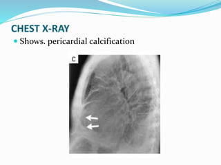
Chronic constrictive pericarditis is a chronic
inflammatory process that involves both fibrous and
serous layers of the pericardium, and that leads to
pericardial thickening and compression (constriction) of
the ventricles. The resultant impairment in diastolic
filling reduces cardiac function.
This disorder results when the healing of an acute
fibrinous or serofibrinous pericarditis or the resorption
of a chronic pericardial effusion is followed by
obliteration of the pericardial cavity with the
formation of granulation tissue, which latter gradually
contracts and forms a firm scar, which may be calcified,
encasing the heart and interfering with filling of the
ventricles.
Pericardial fluid is generally absent or of normal
volume.
Thickened wall of pericardial sac, may be 3-20mm
thick, versus 1-2 mm thickness for normal pericardium.
CAUSES
Idiopathic (most common in developed nations)
Irradiation
Postsurgical
Infectious (most common in developing nations)
Neoplastic
Autoimmune (connective tissue) disorders
Uremia
Post-trauma
Sarcoid
Methysergide therapy
Implantable defibrillator patches
 The end result is dense fibrosis, often calcification,
and adhesions of the parietal and visceral
pericardium. Scarring is usually more or less
symmetric and impedes filling of all heart
chambers. In a subset of patients, the process
develops relatively rapidly and is reversible. This
variant is seen most commonly after cardiac
surgery.
PATHOPHYSIOLOGY
The pathophysiologic consequence of pericardial scarring is
markedly restricted filling of the heart.[This results in
elevation and equilibration of filling pressures in all
chambers and the systemic and pulmonary veins.
Impairs cardiac filling only in late diastole.
Early diastolic filling of RV occurs breifly in constrictive
pericarditis untill the ventricle suddenly reaches the rigid
constraint of the pericardium, producing the “square root”
sign in the LV and RV diastolic filling pressure waveforms.
CVP tracing has a prominent y descent that corresponds to
the initial dip of the “square root” sign. This y descent
results from exaggerated diastolic collapse of the normal
venous pressure as rapid atrial filling occurs.
Systemic venous congestion results in hepatic
congestion, peripheral edema, ascites and
sometimes anasarca, and cardiac cirrhosis.
 Reduced cardiac output is a consequence of
impaired ventricular filling and causes fatigue,
muscle wasting, and weight loss.
 In “absolute” constriction, contractile function is
preserved, although ejection fraction can be
reduced because of reduced preload. The
myocardium is occasionally involved in the chronic
inflammation and fibrosis, leading to true
contractile dysfunction that can at times be quite
severe and predicts a poor response to
pericardiectomy.
Representation of transvalvular and central venous flow velocities
in constrictive pericarditis. During inspiration, the decrease in left
ventricular filling results in a leftward septal shift, allowing
augmented flow into the right ventricle. The opposite occurs
during expiration
CLINICAL PRESENTATION
 The usual presentation consists of signs and
symptoms of right-sided heart failure. At a
relatively early stage, these include lower extremity
edema, vague abdominal complaints, and passive
hepatic congestion.
 Later on hepatic congestion worsens and can
progress to ascites, anasarca, and jaundice due to
cardiac cirrhosis.
 Elevated pulmonary venous pressures, leading to
exertional dyspnea, cough, and orthopnea, may
also appear with progressive disease.
 Atrial fibrillation and tricuspid regurgitation
 In end-stage constrictive pericarditis, the effects of
a chronically low cardiac output are prominent,
including severe fatigue, muscle wasting, and
cachexia.
 Other findings include recurrent pleural effusions
and syncope.
 Can be mistaken for any cause of right-sided heart
failure as well as end-stage primary hepatic
disease.
PHYSICAL EXAMINATION
 Markedly elevated jugular venous pressure with a prominent, rapidly
collapsing y descent.
 Kussmaul sign.
 In cases with extensive calcification and adhesion of the heart to
adjacent structures, the position of the cardiac point of maximal
impulse may fail to change with changes in body position.
 Pericardial knock in around 50% cases.
 Widening of second heart sound splitting.
 Prominent S3 gallop may be present.
 Secondary tricuspid regurgitation with its characteristic murmur.
 Abdominal examination reveals hepatomegaly, often with palpable
venous pulsations, with or without ascites. Other signs of hepatic
congestion or cardiac cirrhosis include jaundice, spider angiomas,
and palmar erythema.
 Lower extremity edema.
 Inspiratory increase in right atrial pressure
(Kussmaul’s sign)
"A" wave: atrial contraction (ABSENT
in atrial fibrillation)
"C" wave: ventricular contraction
(tricuspid bulges).
"X" descent: atrial relaxation
"V" wave: atrial venous filling
(occurs at same of time of
ventricular contraction)
"Y" descent: ventricular filling
(tricuspid opens)
CHEST X-RAY
 Shows. pericardial calcification
ECG with low voltage
ECHOCARDIOGRAPHY
 Pericardial thickening,
 Abrupt displacement of the interventricular septum
during early diastole (septal “bounce”).
 Premature pulmonic valve opening as a result of
elevated right ventricular early diastolic pressure may
also be observed.
 Exaggerated septal shifting during respiration is often
present.
 Transesophageal echocardiography is superior to
transthoracic echocardiography for measuring
pericardial thickness and has an excellent correlation
with CT.
CT SCAN
CT showing dialated SVC relative to AA and DA
 CT with thickened pericardium
CARDIAC CATHETERIZATION
 CVP greater than 14 mm hg
 Near equalization of CVP, PA diastolic pressure,
and capillary wedge pressures.
 Decreased cardiac output.
 “square root” sign in RV and LV pressure tracings
 Prominent y descent
 Accentuated by 500ml volume change
Right sided pressure tracings showing near
equalization of pressures
 Simultaneous RV and LV tracings with early
diastolic pressure dip (characteristic “square
root” sign)
CONSTRICTION RESTRICTION
Prominent y descent in
venous pressure
Present Variable
Paradoxical pulse 1/3rd of cases Absent
Pericardial knock Present Absent
Equal right- and left-sided
filling pressures
Present Left at least 3-5 mm Hg > right
Filling pressures >25 mm Hg Rare Common
Pulmonary artery systolic
pressure >60 mm Hg
No Common
“Square root” sign Present Variable
Respiratory variation in left-
and right-sided pressures or
flows
Exaggerated Normal
Ventricular wall thickness Normal Usually increased
Atrial size Possible left atrial enlargement Biatrial enlargement
Septal bounce Present Absent
Tissue Doppler E′ velocity Increased Reduced
Pericardial thickness Increased Normal
MANAGEMENT
 With the exception of patients with transient constriction,
surgical pericardiectomy is the definitive treatment.
 Patients with major comorbidities or severe debilitation
and radiation-induced disease are relatively
contraindicated for pericardiectomy.
 Medical management with diuretics and salt restriction.
Because sinus tachycardia is a compensatory mechanism,
beta-adrenergic blockers and calcium antagonists that
slow the heart rate should be avoided. In patients with
atrial fibrillation and a rapid ventricular response, digoxin
is recommended as initial treatment to slow the
ventricular rate before resorting to beta blockers or
calcium antagonists.
PERICARDIECTOMY
 Usually done through a median sternotomy.
 Possible through bilateral thoracotomies, and left
anterolateral thoracotomy has been used to achieve
effective pericardiectomy.
 For sternotomy a midline incision is made from just
inferior to the suprasternal notch to the inferior extent of
the xiphoid process.
 Sternum is divided with a sternal saw and opened with a
sternal retractor.
 Relatively thin area of anterior pericardium is incised and a
plane between the epicardium and the pericardium is
established using sharp dissection.
 Pericardium is opened in the midline from the level of the
diaphragm to the base of the great vessels.
 LV should be freed first, to avoid RV distension.
 Ant. Pericardium is freed 1-2 cm anterior to both phrenic
nerves.
 Dense epicardial scar can be removed with grid pattern of
crossing lines 1-2 cm apart.
 Once the pericardium is freed from the epicardium, it is
excised using low electrocautery to achieve hemostasis.
 Anterior pericardiectomy described above provides
maximal hemodynamic benefit in relieving right
ventricular constriction and usually can be performed
without cardiopulmonary bypass.
 Resection of the diaphragmatic pericardium, atrial
pericardium and posterior pericardiectomy remains
unclear benefit.
 Intraoperative findings of pericardial
thickening.
OUTCOME
 Pericardiectomy provides excellent symptomatic
improvement, with 85% of late survivors being
NYHA class I and the remainder being class II.
 Operative mortality 14%, primarily as a result of
low cardiac output in 70% of deaths.
 The operative mortality rate varies with NYHA
class- 1% for class I-II
10% for class III
46% for class IV
TUBERCULOUS CONSTRICTIVE PERICARDITIS
 Pericardiectomy should be performed early and as radically as
possible.
 A combination of chemotherapy and surgery yields gratifying
results.
 Tuberculous pericarditis affects 1% to 2% of all patients with
TBC by direct extension from the mediastinal lymph nodes
and, occasionally, by hematogenous spread or by contiguous
spread from the myocardium.
 Triple-drug antituberculous therapy is to be administered for
a minimum of 9 months.
 In patients in whom pericardial effusion persists or recurs
despite the use of anti-TBC drugs, 3 months of corticosteroid
therapy may be a useful adjunct.
APPENDIX
 Kussmaul's Sign
 Kussmaul’s Sign In Effusive
Constrictive Pericarditis
Chronic constrictive pericarditis

Chronic constrictive pericarditis

  • 1.
  • 2.
    DEFINITION Chronic constrictive pericarditisis a chronic inflammatory process that involves both fibrous and serous layers of the pericardium, and that leads to pericardial thickening and compression (constriction) of the ventricles. The resultant impairment in diastolic filling reduces cardiac function.
  • 3.
    This disorder resultswhen the healing of an acute fibrinous or serofibrinous pericarditis or the resorption of a chronic pericardial effusion is followed by obliteration of the pericardial cavity with the formation of granulation tissue, which latter gradually contracts and forms a firm scar, which may be calcified, encasing the heart and interfering with filling of the ventricles. Pericardial fluid is generally absent or of normal volume. Thickened wall of pericardial sac, may be 3-20mm thick, versus 1-2 mm thickness for normal pericardium.
  • 4.
    CAUSES Idiopathic (most commonin developed nations) Irradiation Postsurgical Infectious (most common in developing nations) Neoplastic Autoimmune (connective tissue) disorders Uremia Post-trauma Sarcoid Methysergide therapy Implantable defibrillator patches
  • 5.
     The endresult is dense fibrosis, often calcification, and adhesions of the parietal and visceral pericardium. Scarring is usually more or less symmetric and impedes filling of all heart chambers. In a subset of patients, the process develops relatively rapidly and is reversible. This variant is seen most commonly after cardiac surgery.
  • 6.
    PATHOPHYSIOLOGY The pathophysiologic consequenceof pericardial scarring is markedly restricted filling of the heart.[This results in elevation and equilibration of filling pressures in all chambers and the systemic and pulmonary veins. Impairs cardiac filling only in late diastole. Early diastolic filling of RV occurs breifly in constrictive pericarditis untill the ventricle suddenly reaches the rigid constraint of the pericardium, producing the “square root” sign in the LV and RV diastolic filling pressure waveforms. CVP tracing has a prominent y descent that corresponds to the initial dip of the “square root” sign. This y descent results from exaggerated diastolic collapse of the normal venous pressure as rapid atrial filling occurs.
  • 7.
    Systemic venous congestionresults in hepatic congestion, peripheral edema, ascites and sometimes anasarca, and cardiac cirrhosis.  Reduced cardiac output is a consequence of impaired ventricular filling and causes fatigue, muscle wasting, and weight loss.  In “absolute” constriction, contractile function is preserved, although ejection fraction can be reduced because of reduced preload. The myocardium is occasionally involved in the chronic inflammation and fibrosis, leading to true contractile dysfunction that can at times be quite severe and predicts a poor response to pericardiectomy.
  • 8.
    Representation of transvalvularand central venous flow velocities in constrictive pericarditis. During inspiration, the decrease in left ventricular filling results in a leftward septal shift, allowing augmented flow into the right ventricle. The opposite occurs during expiration
  • 9.
    CLINICAL PRESENTATION  Theusual presentation consists of signs and symptoms of right-sided heart failure. At a relatively early stage, these include lower extremity edema, vague abdominal complaints, and passive hepatic congestion.  Later on hepatic congestion worsens and can progress to ascites, anasarca, and jaundice due to cardiac cirrhosis.  Elevated pulmonary venous pressures, leading to exertional dyspnea, cough, and orthopnea, may also appear with progressive disease.
  • 10.
     Atrial fibrillationand tricuspid regurgitation  In end-stage constrictive pericarditis, the effects of a chronically low cardiac output are prominent, including severe fatigue, muscle wasting, and cachexia.  Other findings include recurrent pleural effusions and syncope.  Can be mistaken for any cause of right-sided heart failure as well as end-stage primary hepatic disease.
  • 11.
    PHYSICAL EXAMINATION  Markedlyelevated jugular venous pressure with a prominent, rapidly collapsing y descent.  Kussmaul sign.  In cases with extensive calcification and adhesion of the heart to adjacent structures, the position of the cardiac point of maximal impulse may fail to change with changes in body position.  Pericardial knock in around 50% cases.  Widening of second heart sound splitting.  Prominent S3 gallop may be present.  Secondary tricuspid regurgitation with its characteristic murmur.  Abdominal examination reveals hepatomegaly, often with palpable venous pulsations, with or without ascites. Other signs of hepatic congestion or cardiac cirrhosis include jaundice, spider angiomas, and palmar erythema.  Lower extremity edema.
  • 12.
     Inspiratory increasein right atrial pressure (Kussmaul’s sign)
  • 14.
    "A" wave: atrialcontraction (ABSENT in atrial fibrillation) "C" wave: ventricular contraction (tricuspid bulges). "X" descent: atrial relaxation "V" wave: atrial venous filling (occurs at same of time of ventricular contraction) "Y" descent: ventricular filling (tricuspid opens)
  • 15.
    CHEST X-RAY  Shows.pericardial calcification
  • 16.
    ECG with lowvoltage
  • 17.
    ECHOCARDIOGRAPHY  Pericardial thickening, Abrupt displacement of the interventricular septum during early diastole (septal “bounce”).  Premature pulmonic valve opening as a result of elevated right ventricular early diastolic pressure may also be observed.  Exaggerated septal shifting during respiration is often present.  Transesophageal echocardiography is superior to transthoracic echocardiography for measuring pericardial thickness and has an excellent correlation with CT.
  • 18.
    CT SCAN CT showingdialated SVC relative to AA and DA
  • 19.
     CT withthickened pericardium
  • 20.
    CARDIAC CATHETERIZATION  CVPgreater than 14 mm hg  Near equalization of CVP, PA diastolic pressure, and capillary wedge pressures.  Decreased cardiac output.  “square root” sign in RV and LV pressure tracings  Prominent y descent  Accentuated by 500ml volume change
  • 21.
    Right sided pressuretracings showing near equalization of pressures
  • 22.
     Simultaneous RVand LV tracings with early diastolic pressure dip (characteristic “square root” sign)
  • 23.
    CONSTRICTION RESTRICTION Prominent ydescent in venous pressure Present Variable Paradoxical pulse 1/3rd of cases Absent Pericardial knock Present Absent Equal right- and left-sided filling pressures Present Left at least 3-5 mm Hg > right Filling pressures >25 mm Hg Rare Common Pulmonary artery systolic pressure >60 mm Hg No Common “Square root” sign Present Variable Respiratory variation in left- and right-sided pressures or flows Exaggerated Normal Ventricular wall thickness Normal Usually increased Atrial size Possible left atrial enlargement Biatrial enlargement Septal bounce Present Absent Tissue Doppler E′ velocity Increased Reduced Pericardial thickness Increased Normal
  • 24.
    MANAGEMENT  With theexception of patients with transient constriction, surgical pericardiectomy is the definitive treatment.  Patients with major comorbidities or severe debilitation and radiation-induced disease are relatively contraindicated for pericardiectomy.  Medical management with diuretics and salt restriction. Because sinus tachycardia is a compensatory mechanism, beta-adrenergic blockers and calcium antagonists that slow the heart rate should be avoided. In patients with atrial fibrillation and a rapid ventricular response, digoxin is recommended as initial treatment to slow the ventricular rate before resorting to beta blockers or calcium antagonists.
  • 25.
    PERICARDIECTOMY  Usually donethrough a median sternotomy.  Possible through bilateral thoracotomies, and left anterolateral thoracotomy has been used to achieve effective pericardiectomy.  For sternotomy a midline incision is made from just inferior to the suprasternal notch to the inferior extent of the xiphoid process.  Sternum is divided with a sternal saw and opened with a sternal retractor.  Relatively thin area of anterior pericardium is incised and a plane between the epicardium and the pericardium is established using sharp dissection.  Pericardium is opened in the midline from the level of the diaphragm to the base of the great vessels.
  • 26.
     LV shouldbe freed first, to avoid RV distension.  Ant. Pericardium is freed 1-2 cm anterior to both phrenic nerves.  Dense epicardial scar can be removed with grid pattern of crossing lines 1-2 cm apart.  Once the pericardium is freed from the epicardium, it is excised using low electrocautery to achieve hemostasis.  Anterior pericardiectomy described above provides maximal hemodynamic benefit in relieving right ventricular constriction and usually can be performed without cardiopulmonary bypass.  Resection of the diaphragmatic pericardium, atrial pericardium and posterior pericardiectomy remains unclear benefit.
  • 27.
     Intraoperative findingsof pericardial thickening.
  • 28.
    OUTCOME  Pericardiectomy providesexcellent symptomatic improvement, with 85% of late survivors being NYHA class I and the remainder being class II.  Operative mortality 14%, primarily as a result of low cardiac output in 70% of deaths.  The operative mortality rate varies with NYHA class- 1% for class I-II 10% for class III 46% for class IV
  • 29.
    TUBERCULOUS CONSTRICTIVE PERICARDITIS Pericardiectomy should be performed early and as radically as possible.  A combination of chemotherapy and surgery yields gratifying results.  Tuberculous pericarditis affects 1% to 2% of all patients with TBC by direct extension from the mediastinal lymph nodes and, occasionally, by hematogenous spread or by contiguous spread from the myocardium.  Triple-drug antituberculous therapy is to be administered for a minimum of 9 months.  In patients in whom pericardial effusion persists or recurs despite the use of anti-TBC drugs, 3 months of corticosteroid therapy may be a useful adjunct.
  • 30.
    APPENDIX  Kussmaul's Sign Kussmaul’s Sign In Effusive Constrictive Pericarditis