SlideShare a Scribd company logo
1 of 14
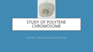
STUDY OF POLYTENE
CHROMOSOME
POLYTENE CHROMOSOMES IN DROSOPHILA
INTRODUCTION
• Polytene chromosomes are over-sized chromosomes which have
developed from standard chromosomes and are commonly found in the
salivary glands of DROSOPHILA MELANOGASTER Specialized cells
undergo repeated rounds of DNA REPLICATION without CELL DIVISION,
to increase CELL volume, forming a giant polytene chromosome.
Polytene chromosomes form when multiple rounds of replication
produce many sister chromatids that remain synapsed together.
HISTORY
• Polytene chromosomes were originally observed in the larval SALIVARY
GLANDS of CHIROMOMUS midges by Balbiani in 1881, but the
hereditary nature of these structures was not confirmed until they were
studied in DROSOPHILA MELANGOSTER in the early 1930s by EMIL
HEITZ and HANS BAUER. They are known to occur in secretory tissues of
other DIPTERAN insects such as the MALPHIGIAN TUBULES of Sciara and
also in protists, plants, mammals, or in cells from other INSECTS. Some
of the largest polytene chromosomes described thus far occur in larval
salivary gland cells of the Chironomid genus Axarus.
FUNCTIONS
• In addition to increasing the volume of the cells' nuclei and causing cell
expansion, polytene cells may also have a metabolic advantage as
multiple copies of genes permits a high level of gene expression.
In Drosophila melanogaster, for example, the chromosomes of the larval
salivary glands undergo many rounds of endoreduplication, to produce
large amounts of glue before pupation. Another example, within the
organism itself is the tandem duplication of various polytene bands
located near the centromere of the X chromosome which results in the
Bar phenotype of kidney-shaped eyes.
STRUCTURE
• Polytene chromosomes have characteristic light and dark banding
patterns that can be used to identify chromosomal rearrangements and
deletions. Dark banding frequently corresponds to inactive chromatin,
whereas light banding is usually found at areas with higher
transcriptional activity. The banding patterns of the polytene
chromosomes of Drosophila melanogaster were sketched in 1935
by Calvin B. Bridges.
STRUCTURE CONTINUED..
• The banding patterns of the chromosomes are especially helpful in
research, as they provide an excellent visualization of transcriptionally
active chromatin and general chromatin structure. For example, the
polytene chromosomes in Drosophila have been used to support the
theory of genomic equivalence, which states that all of the cells in the
body maintain the same genome. Polytene chromosomes are about
200 micrometer in length. The chromonema of these chromosomes
divide but do not separate. Therefore, they remain together to become
large in size.
PROCEDURE
• The success of these experiments is largely determined by the
developmental stage of the larvae.
• If they are too small, their salivary glands will not be sufficiently
developed to yield good chromosomes.
• Choose the largest larvae that you can find that is still moving actively.
• Do not choose the larvae that is noticebly darker than the rest.
• Darkening of the cuticle is a sign that pupation is imminent.
PREPARING SALIVARY SQUASHES
• Place a drop of 0.7% saline on a clean slide.
• Transfer an appropriate larva to the slide and place it on the stage of a dissecting
microscope.
• Using the enhanced detail afforded by the microscope, firmly grasp the posterior end
of the larva with forceps and use a dissecting needle to pierce through the head.
• Using a continuous motion pull the needle away from the body.
• The salivary glands are recognised by the foll. features:
• They are paired and each is identical in shape and size.
• They have a glistening, translucent appearance.
• Each gland should have an opaque fat body associated with it.
• While continuing to observe under the microscope, separate the salivary
glands from any extraneous material, such as the fat bodies or parts of
the digestive tract.
• Once cleaned, remove the used saline and debris from the slide. Add
fresh saline and allow the glands to soak for ten minutes.
• Carefully place 1-2 drops of acetocarmine or aceto-orcein stain on each
of the regions.
• Wait for a couple of minutes and proceed to the next step.
• Crushing glands to expose cells: Place a cover slip carefully in each of
the regions of the slide where salivary glands were placed.
• Crush each cover slip against the slide with the help of your thumb.
• If the crushed region appears too thick, flatten it further by tapping the
coverslip multiple times with the back of a dissecting needle
• Allow the exposed cells to absorb the stain for 5-10 minutes.
IDENTIFYING THE POLYTENE
CHROMOSOME UNDER A MICROSCOPE
• Place the slide under the microscope .
• Locate the squashed regions under 4x and 10x objective magnification
to see where to focus.
• Once those are determined change the objective to 40x to locate
individual cells.
• If certain fibrous projections appear, switch to 100x.
• Light and dark bands are observed as shown.
• Hence the procedure comes to an end.
REFERENCES
• https://en.wikipedia.org/wiki/Polytene_chromosome
• https://www.ncbi.nlm.nih.gov/pmc/articles/PMC2821744/
• http://msg.ucsf.edu/sedat/polytene_chrom.html

More Related Content

What's hot

Cytotaxonomy by Ishtiyaq sir.pptx
Cytotaxonomy by Ishtiyaq sir.pptxCytotaxonomy by Ishtiyaq sir.pptx
Cytotaxonomy by Ishtiyaq sir.pptxIshtiyaqMir3
 
Sex determination XX-XY
Sex determination XX-XYSex determination XX-XY
Sex determination XX-XYAsr Cv
 
Hardy – weinberg law
 Hardy – weinberg law Hardy – weinberg law
Hardy – weinberg lawShabnamkhan113
 
Polytene chromosomes
Polytene chromosomesPolytene chromosomes
Polytene chromosomespooja singh
 
07 mendelian genetics and humans
07 mendelian genetics and humans07 mendelian genetics and humans
07 mendelian genetics and humansmrtangextrahelp
 
Eukaryotic chromosomal organization
Eukaryotic chromosomal organizationEukaryotic chromosomal organization
Eukaryotic chromosomal organizationPraveen Garg
 
RNA- Structure and their types
RNA- Structure and their typesRNA- Structure and their types
RNA- Structure and their typesHEENA KAUSAR
 
Cytogenetic analysis
Cytogenetic analysisCytogenetic analysis
Cytogenetic analysisIkram Ullah
 
Fundamentals of Fluorescence in situ Hybridization
Fundamentals of Fluorescence in situ Hybridization Fundamentals of Fluorescence in situ Hybridization
Fundamentals of Fluorescence in situ Hybridization Amartya Pradhan
 
Fluorescence in situ hybridization
Fluorescence in situ hybridizationFluorescence in situ hybridization
Fluorescence in situ hybridizationTukaramBorkar
 
Karyotyping
KaryotypingKaryotyping
Karyotypingwildask
 
Role of kinetochore in cell division
Role of kinetochore in cell divisionRole of kinetochore in cell division
Role of kinetochore in cell divisionBINAYAKJENAMANI
 
X chromosome inactivation
X chromosome inactivationX chromosome inactivation
X chromosome inactivationIkram Ullah
 

What's hot (20)

KARYOTYPING
KARYOTYPINGKARYOTYPING
KARYOTYPING
 
Cytotaxonomy by Ishtiyaq sir.pptx
Cytotaxonomy by Ishtiyaq sir.pptxCytotaxonomy by Ishtiyaq sir.pptx
Cytotaxonomy by Ishtiyaq sir.pptx
 
Sex determination XX-XY
Sex determination XX-XYSex determination XX-XY
Sex determination XX-XY
 
aCGH in PGD/PGS
aCGH in PGD/PGSaCGH in PGD/PGS
aCGH in PGD/PGS
 
Hardy – weinberg law
 Hardy – weinberg law Hardy – weinberg law
Hardy – weinberg law
 
Polytene chromosomes
Polytene chromosomesPolytene chromosomes
Polytene chromosomes
 
07 mendelian genetics and humans
07 mendelian genetics and humans07 mendelian genetics and humans
07 mendelian genetics and humans
 
Genetic heterogeneity |Genetics|Animal Genetics|Plant Genetics|
Genetic heterogeneity |Genetics|Animal Genetics|Plant Genetics|Genetic heterogeneity |Genetics|Animal Genetics|Plant Genetics|
Genetic heterogeneity |Genetics|Animal Genetics|Plant Genetics|
 
Vntr marker
Vntr markerVntr marker
Vntr marker
 
Eukaryotic chromosomal organization
Eukaryotic chromosomal organizationEukaryotic chromosomal organization
Eukaryotic chromosomal organization
 
LAMPBRUSH CHROMOSOMES
LAMPBRUSH CHROMOSOMESLAMPBRUSH CHROMOSOMES
LAMPBRUSH CHROMOSOMES
 
RNA- Structure and their types
RNA- Structure and their typesRNA- Structure and their types
RNA- Structure and their types
 
Pedigree analysis
Pedigree analysisPedigree analysis
Pedigree analysis
 
Cytogenetic analysis
Cytogenetic analysisCytogenetic analysis
Cytogenetic analysis
 
Fundamentals of Fluorescence in situ Hybridization
Fundamentals of Fluorescence in situ Hybridization Fundamentals of Fluorescence in situ Hybridization
Fundamentals of Fluorescence in situ Hybridization
 
Fluorescence in situ hybridization
Fluorescence in situ hybridizationFluorescence in situ hybridization
Fluorescence in situ hybridization
 
Development of chick
Development of chickDevelopment of chick
Development of chick
 
Karyotyping
KaryotypingKaryotyping
Karyotyping
 
Role of kinetochore in cell division
Role of kinetochore in cell divisionRole of kinetochore in cell division
Role of kinetochore in cell division
 
X chromosome inactivation
X chromosome inactivationX chromosome inactivation
X chromosome inactivation
 

Similar to Lab Study of Polytene Chromosomes.

Polytene chromosomes
Polytene chromosomesPolytene chromosomes
Polytene chromosomesAasim Faiz
 
EARLY MAMMALIAN DEVELOPMENT
EARLY MAMMALIAN DEVELOPMENTEARLY MAMMALIAN DEVELOPMENT
EARLY MAMMALIAN DEVELOPMENTAhmad Raza
 
Dog vaginal cytology by Dr.mehdi moradi
Dog vaginal cytology by Dr.mehdi moradiDog vaginal cytology by Dr.mehdi moradi
Dog vaginal cytology by Dr.mehdi moradimehdi moradi
 
Fundamental unit of life
Fundamental unit of lifeFundamental unit of life
Fundamental unit of lifeKrishnaDhaked
 
Development in drosophila
Development in drosophilaDevelopment in drosophila
Development in drosophilaKAUSHAL SAHU
 
Method for observing polytene chromosomes in Drosophila Larvae
Method for observing polytene chromosomes in Drosophila LarvaeMethod for observing polytene chromosomes in Drosophila Larvae
Method for observing polytene chromosomes in Drosophila LarvaeUddeep Chaudhury
 
Salivary glands dina+sahar audio 2
Salivary glands dina+sahar audio 2Salivary glands dina+sahar audio 2
Salivary glands dina+sahar audio 2dina hassouna
 
Megasporogenesis by Anurag yadav
Megasporogenesis  by Anurag yadavMegasporogenesis  by Anurag yadav
Megasporogenesis by Anurag yadavAnuragsinghYadav3
 
Ch.1 Cell Structures and Functions
Ch.1 Cell Structures and FunctionsCh.1 Cell Structures and Functions
Ch.1 Cell Structures and FunctionsCDA-PamelaOrtiz
 
PSILOTUM : structure, morphology, anatomy, reproduction , life cycle etc.
PSILOTUM : structure, morphology, anatomy,  reproduction , life cycle etc.PSILOTUM : structure, morphology, anatomy,  reproduction , life cycle etc.
PSILOTUM : structure, morphology, anatomy, reproduction , life cycle etc.Cherry
 
Barr Body Staining
Barr Body StainingBarr Body Staining
Barr Body StainingAmna Jalil
 
Oral epithelium and Lamina Propria
Oral epithelium and Lamina PropriaOral epithelium and Lamina Propria
Oral epithelium and Lamina PropriaSayana Nazrine
 
Oral mucosa part 1
Oral mucosa part 1Oral mucosa part 1
Oral mucosa part 1samah khaled
 

Similar to Lab Study of Polytene Chromosomes. (20)

Presentation on Polytene Chromosome
Presentation on Polytene ChromosomePresentation on Polytene Chromosome
Presentation on Polytene Chromosome
 
Polytene chromosomes
Polytene chromosomesPolytene chromosomes
Polytene chromosomes
 
EARLY MAMMALIAN DEVELOPMENT
EARLY MAMMALIAN DEVELOPMENTEARLY MAMMALIAN DEVELOPMENT
EARLY MAMMALIAN DEVELOPMENT
 
Dog vaginal cytology by Dr.mehdi moradi
Dog vaginal cytology by Dr.mehdi moradiDog vaginal cytology by Dr.mehdi moradi
Dog vaginal cytology by Dr.mehdi moradi
 
Fundamental unit of life
Fundamental unit of lifeFundamental unit of life
Fundamental unit of life
 
Development in drosophila
Development in drosophilaDevelopment in drosophila
Development in drosophila
 
Giant chromosomes
Giant chromosomesGiant chromosomes
Giant chromosomes
 
Method for observing polytene chromosomes in Drosophila Larvae
Method for observing polytene chromosomes in Drosophila LarvaeMethod for observing polytene chromosomes in Drosophila Larvae
Method for observing polytene chromosomes in Drosophila Larvae
 
Salivary glands dina+sahar audio 2
Salivary glands dina+sahar audio 2Salivary glands dina+sahar audio 2
Salivary glands dina+sahar audio 2
 
Megasporogenesis by Anurag yadav
Megasporogenesis  by Anurag yadavMegasporogenesis  by Anurag yadav
Megasporogenesis by Anurag yadav
 
Ch.1 Cell Structures and Functions
Ch.1 Cell Structures and FunctionsCh.1 Cell Structures and Functions
Ch.1 Cell Structures and Functions
 
Giant chromosomes
Giant chromosomesGiant chromosomes
Giant chromosomes
 
Protozoan disease
Protozoan diseaseProtozoan disease
Protozoan disease
 
Chap 5 Cleavage.pptx
Chap 5 Cleavage.pptxChap 5 Cleavage.pptx
Chap 5 Cleavage.pptx
 
Desarrollo embrionario
Desarrollo embrionarioDesarrollo embrionario
Desarrollo embrionario
 
PSILOTUM : structure, morphology, anatomy, reproduction , life cycle etc.
PSILOTUM : structure, morphology, anatomy,  reproduction , life cycle etc.PSILOTUM : structure, morphology, anatomy,  reproduction , life cycle etc.
PSILOTUM : structure, morphology, anatomy, reproduction , life cycle etc.
 
Barr Body Staining
Barr Body StainingBarr Body Staining
Barr Body Staining
 
Oral epithelium and Lamina Propria
Oral epithelium and Lamina PropriaOral epithelium and Lamina Propria
Oral epithelium and Lamina Propria
 
Dental Pulp
Dental Pulp Dental Pulp
Dental Pulp
 
Oral mucosa part 1
Oral mucosa part 1Oral mucosa part 1
Oral mucosa part 1
 

Recently uploaded

GBSN - Biochemistry (Unit 3) Metabolism
GBSN - Biochemistry (Unit 3) MetabolismGBSN - Biochemistry (Unit 3) Metabolism
GBSN - Biochemistry (Unit 3) MetabolismAreesha Ahmad
 
Porella : features, morphology, anatomy, reproduction etc.
Porella : features, morphology, anatomy, reproduction etc.Porella : features, morphology, anatomy, reproduction etc.
Porella : features, morphology, anatomy, reproduction etc.Cherry
 
COMPOSTING : types of compost, merits and demerits
COMPOSTING : types of compost, merits and demeritsCOMPOSTING : types of compost, merits and demerits
COMPOSTING : types of compost, merits and demeritsCherry
 
Terpineol and it's characterization pptx
Terpineol and it's characterization pptxTerpineol and it's characterization pptx
Terpineol and it's characterization pptxMuhammadRazzaq31
 
X-rays from a Central “Exhaust Vent” of the Galactic Center Chimney
X-rays from a Central “Exhaust Vent” of the Galactic Center ChimneyX-rays from a Central “Exhaust Vent” of the Galactic Center Chimney
X-rays from a Central “Exhaust Vent” of the Galactic Center ChimneySérgio Sacani
 
Role of AI in seed science Predictive modelling and Beyond.pptx
Role of AI in seed science  Predictive modelling and  Beyond.pptxRole of AI in seed science  Predictive modelling and  Beyond.pptx
Role of AI in seed science Predictive modelling and Beyond.pptxArvind Kumar
 
Understanding Partial Differential Equations: Types and Solution Methods
Understanding Partial Differential Equations: Types and Solution MethodsUnderstanding Partial Differential Equations: Types and Solution Methods
Understanding Partial Differential Equations: Types and Solution Methodsimroshankoirala
 
development of diagnostic enzyme assay to detect leuser virus
development of diagnostic enzyme assay to detect leuser virusdevelopment of diagnostic enzyme assay to detect leuser virus
development of diagnostic enzyme assay to detect leuser virusNazaninKarimi6
 
LUNULARIA -features, morphology, anatomy ,reproduction etc.
LUNULARIA -features, morphology, anatomy ,reproduction etc.LUNULARIA -features, morphology, anatomy ,reproduction etc.
LUNULARIA -features, morphology, anatomy ,reproduction etc.Cherry
 
Lipids: types, structure and important functions.
Lipids: types, structure and important functions.Lipids: types, structure and important functions.
Lipids: types, structure and important functions.Cherry
 
Taphonomy and Quality of the Fossil Record
Taphonomy and Quality of the  Fossil RecordTaphonomy and Quality of the  Fossil Record
Taphonomy and Quality of the Fossil RecordSangram Sahoo
 
Cyathodium bryophyte: morphology, anatomy, reproduction etc.
Cyathodium bryophyte: morphology, anatomy, reproduction etc.Cyathodium bryophyte: morphology, anatomy, reproduction etc.
Cyathodium bryophyte: morphology, anatomy, reproduction etc.Cherry
 
Daily Lesson Log in Science 9 Fourth Quarter Physics
Daily Lesson Log in Science 9 Fourth Quarter PhysicsDaily Lesson Log in Science 9 Fourth Quarter Physics
Daily Lesson Log in Science 9 Fourth Quarter PhysicsWILSONROMA4
 
Fourth quarter science 9-Kinetic-and-Potential-Energy.pptx
Fourth quarter science 9-Kinetic-and-Potential-Energy.pptxFourth quarter science 9-Kinetic-and-Potential-Energy.pptx
Fourth quarter science 9-Kinetic-and-Potential-Energy.pptxrosenapiri1
 
Energy is the beat of life irrespective of the domains. ATP- the energy curre...
Energy is the beat of life irrespective of the domains. ATP- the energy curre...Energy is the beat of life irrespective of the domains. ATP- the energy curre...
Energy is the beat of life irrespective of the domains. ATP- the energy curre...Nistarini College, Purulia (W.B) India
 
Pteris : features, anatomy, morphology and lifecycle
Pteris : features, anatomy, morphology and lifecyclePteris : features, anatomy, morphology and lifecycle
Pteris : features, anatomy, morphology and lifecycleCherry
 
(May 9, 2024) Enhanced Ultrafast Vector Flow Imaging (VFI) Using Multi-Angle ...
(May 9, 2024) Enhanced Ultrafast Vector Flow Imaging (VFI) Using Multi-Angle ...(May 9, 2024) Enhanced Ultrafast Vector Flow Imaging (VFI) Using Multi-Angle ...
(May 9, 2024) Enhanced Ultrafast Vector Flow Imaging (VFI) Using Multi-Angle ...Scintica Instrumentation
 
Concept of gene and Complementation test.pdf
Concept of gene and Complementation test.pdfConcept of gene and Complementation test.pdf
Concept of gene and Complementation test.pdfCherry
 
Thyroid Physiology_Dr.E. Muralinath_ Associate Professor
Thyroid Physiology_Dr.E. Muralinath_ Associate ProfessorThyroid Physiology_Dr.E. Muralinath_ Associate Professor
Thyroid Physiology_Dr.E. Muralinath_ Associate Professormuralinath2
 
Reboulia: features, anatomy, morphology etc.
Reboulia: features, anatomy, morphology etc.Reboulia: features, anatomy, morphology etc.
Reboulia: features, anatomy, morphology etc.Cherry
 

Recently uploaded (20)

GBSN - Biochemistry (Unit 3) Metabolism
GBSN - Biochemistry (Unit 3) MetabolismGBSN - Biochemistry (Unit 3) Metabolism
GBSN - Biochemistry (Unit 3) Metabolism
 
Porella : features, morphology, anatomy, reproduction etc.
Porella : features, morphology, anatomy, reproduction etc.Porella : features, morphology, anatomy, reproduction etc.
Porella : features, morphology, anatomy, reproduction etc.
 
COMPOSTING : types of compost, merits and demerits
COMPOSTING : types of compost, merits and demeritsCOMPOSTING : types of compost, merits and demerits
COMPOSTING : types of compost, merits and demerits
 
Terpineol and it's characterization pptx
Terpineol and it's characterization pptxTerpineol and it's characterization pptx
Terpineol and it's characterization pptx
 
X-rays from a Central “Exhaust Vent” of the Galactic Center Chimney
X-rays from a Central “Exhaust Vent” of the Galactic Center ChimneyX-rays from a Central “Exhaust Vent” of the Galactic Center Chimney
X-rays from a Central “Exhaust Vent” of the Galactic Center Chimney
 
Role of AI in seed science Predictive modelling and Beyond.pptx
Role of AI in seed science  Predictive modelling and  Beyond.pptxRole of AI in seed science  Predictive modelling and  Beyond.pptx
Role of AI in seed science Predictive modelling and Beyond.pptx
 
Understanding Partial Differential Equations: Types and Solution Methods
Understanding Partial Differential Equations: Types and Solution MethodsUnderstanding Partial Differential Equations: Types and Solution Methods
Understanding Partial Differential Equations: Types and Solution Methods
 
development of diagnostic enzyme assay to detect leuser virus
development of diagnostic enzyme assay to detect leuser virusdevelopment of diagnostic enzyme assay to detect leuser virus
development of diagnostic enzyme assay to detect leuser virus
 
LUNULARIA -features, morphology, anatomy ,reproduction etc.
LUNULARIA -features, morphology, anatomy ,reproduction etc.LUNULARIA -features, morphology, anatomy ,reproduction etc.
LUNULARIA -features, morphology, anatomy ,reproduction etc.
 
Lipids: types, structure and important functions.
Lipids: types, structure and important functions.Lipids: types, structure and important functions.
Lipids: types, structure and important functions.
 
Taphonomy and Quality of the Fossil Record
Taphonomy and Quality of the  Fossil RecordTaphonomy and Quality of the  Fossil Record
Taphonomy and Quality of the Fossil Record
 
Cyathodium bryophyte: morphology, anatomy, reproduction etc.
Cyathodium bryophyte: morphology, anatomy, reproduction etc.Cyathodium bryophyte: morphology, anatomy, reproduction etc.
Cyathodium bryophyte: morphology, anatomy, reproduction etc.
 
Daily Lesson Log in Science 9 Fourth Quarter Physics
Daily Lesson Log in Science 9 Fourth Quarter PhysicsDaily Lesson Log in Science 9 Fourth Quarter Physics
Daily Lesson Log in Science 9 Fourth Quarter Physics
 
Fourth quarter science 9-Kinetic-and-Potential-Energy.pptx
Fourth quarter science 9-Kinetic-and-Potential-Energy.pptxFourth quarter science 9-Kinetic-and-Potential-Energy.pptx
Fourth quarter science 9-Kinetic-and-Potential-Energy.pptx
 
Energy is the beat of life irrespective of the domains. ATP- the energy curre...
Energy is the beat of life irrespective of the domains. ATP- the energy curre...Energy is the beat of life irrespective of the domains. ATP- the energy curre...
Energy is the beat of life irrespective of the domains. ATP- the energy curre...
 
Pteris : features, anatomy, morphology and lifecycle
Pteris : features, anatomy, morphology and lifecyclePteris : features, anatomy, morphology and lifecycle
Pteris : features, anatomy, morphology and lifecycle
 
(May 9, 2024) Enhanced Ultrafast Vector Flow Imaging (VFI) Using Multi-Angle ...
(May 9, 2024) Enhanced Ultrafast Vector Flow Imaging (VFI) Using Multi-Angle ...(May 9, 2024) Enhanced Ultrafast Vector Flow Imaging (VFI) Using Multi-Angle ...
(May 9, 2024) Enhanced Ultrafast Vector Flow Imaging (VFI) Using Multi-Angle ...
 
Concept of gene and Complementation test.pdf
Concept of gene and Complementation test.pdfConcept of gene and Complementation test.pdf
Concept of gene and Complementation test.pdf
 
Thyroid Physiology_Dr.E. Muralinath_ Associate Professor
Thyroid Physiology_Dr.E. Muralinath_ Associate ProfessorThyroid Physiology_Dr.E. Muralinath_ Associate Professor
Thyroid Physiology_Dr.E. Muralinath_ Associate Professor
 
Reboulia: features, anatomy, morphology etc.
Reboulia: features, anatomy, morphology etc.Reboulia: features, anatomy, morphology etc.
Reboulia: features, anatomy, morphology etc.
 

Lab Study of Polytene Chromosomes.

  • 1. STUDY OF POLYTENE CHROMOSOME POLYTENE CHROMOSOMES IN DROSOPHILA
  • 2. INTRODUCTION • Polytene chromosomes are over-sized chromosomes which have developed from standard chromosomes and are commonly found in the salivary glands of DROSOPHILA MELANOGASTER Specialized cells undergo repeated rounds of DNA REPLICATION without CELL DIVISION, to increase CELL volume, forming a giant polytene chromosome. Polytene chromosomes form when multiple rounds of replication produce many sister chromatids that remain synapsed together.
  • 3. HISTORY • Polytene chromosomes were originally observed in the larval SALIVARY GLANDS of CHIROMOMUS midges by Balbiani in 1881, but the hereditary nature of these structures was not confirmed until they were studied in DROSOPHILA MELANGOSTER in the early 1930s by EMIL HEITZ and HANS BAUER. They are known to occur in secretory tissues of other DIPTERAN insects such as the MALPHIGIAN TUBULES of Sciara and also in protists, plants, mammals, or in cells from other INSECTS. Some of the largest polytene chromosomes described thus far occur in larval salivary gland cells of the Chironomid genus Axarus.
  • 4. FUNCTIONS • In addition to increasing the volume of the cells' nuclei and causing cell expansion, polytene cells may also have a metabolic advantage as multiple copies of genes permits a high level of gene expression. In Drosophila melanogaster, for example, the chromosomes of the larval salivary glands undergo many rounds of endoreduplication, to produce large amounts of glue before pupation. Another example, within the organism itself is the tandem duplication of various polytene bands located near the centromere of the X chromosome which results in the Bar phenotype of kidney-shaped eyes.
  • 5. STRUCTURE • Polytene chromosomes have characteristic light and dark banding patterns that can be used to identify chromosomal rearrangements and deletions. Dark banding frequently corresponds to inactive chromatin, whereas light banding is usually found at areas with higher transcriptional activity. The banding patterns of the polytene chromosomes of Drosophila melanogaster were sketched in 1935 by Calvin B. Bridges.
  • 6.
  • 7.
  • 8. STRUCTURE CONTINUED.. • The banding patterns of the chromosomes are especially helpful in research, as they provide an excellent visualization of transcriptionally active chromatin and general chromatin structure. For example, the polytene chromosomes in Drosophila have been used to support the theory of genomic equivalence, which states that all of the cells in the body maintain the same genome. Polytene chromosomes are about 200 micrometer in length. The chromonema of these chromosomes divide but do not separate. Therefore, they remain together to become large in size.
  • 9. PROCEDURE • The success of these experiments is largely determined by the developmental stage of the larvae. • If they are too small, their salivary glands will not be sufficiently developed to yield good chromosomes. • Choose the largest larvae that you can find that is still moving actively. • Do not choose the larvae that is noticebly darker than the rest. • Darkening of the cuticle is a sign that pupation is imminent.
  • 10. PREPARING SALIVARY SQUASHES • Place a drop of 0.7% saline on a clean slide. • Transfer an appropriate larva to the slide and place it on the stage of a dissecting microscope. • Using the enhanced detail afforded by the microscope, firmly grasp the posterior end of the larva with forceps and use a dissecting needle to pierce through the head. • Using a continuous motion pull the needle away from the body. • The salivary glands are recognised by the foll. features: • They are paired and each is identical in shape and size. • They have a glistening, translucent appearance. • Each gland should have an opaque fat body associated with it.
  • 11. • While continuing to observe under the microscope, separate the salivary glands from any extraneous material, such as the fat bodies or parts of the digestive tract. • Once cleaned, remove the used saline and debris from the slide. Add fresh saline and allow the glands to soak for ten minutes. • Carefully place 1-2 drops of acetocarmine or aceto-orcein stain on each of the regions. • Wait for a couple of minutes and proceed to the next step.
  • 12. • Crushing glands to expose cells: Place a cover slip carefully in each of the regions of the slide where salivary glands were placed. • Crush each cover slip against the slide with the help of your thumb. • If the crushed region appears too thick, flatten it further by tapping the coverslip multiple times with the back of a dissecting needle • Allow the exposed cells to absorb the stain for 5-10 minutes.
  • 13. IDENTIFYING THE POLYTENE CHROMOSOME UNDER A MICROSCOPE • Place the slide under the microscope . • Locate the squashed regions under 4x and 10x objective magnification to see where to focus. • Once those are determined change the objective to 40x to locate individual cells. • If certain fibrous projections appear, switch to 100x. • Light and dark bands are observed as shown. • Hence the procedure comes to an end.