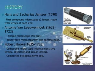
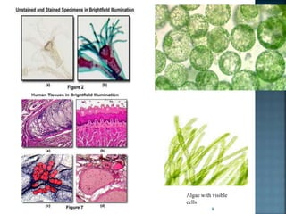
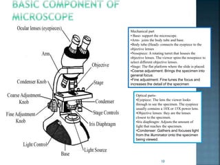
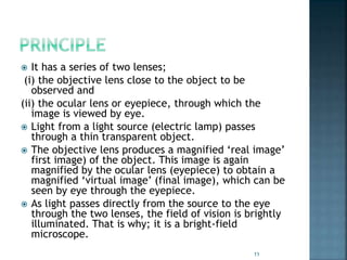
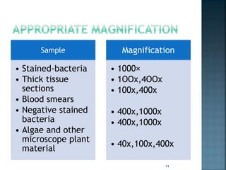
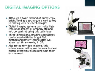
1
By
KAUSHAL KUMAR SAHU
Assistant Professor (Ad Hoc)
Department of Biotechnology
Govt. Digvijay Autonomous P. G. College
Raj-Nandgaon ( C. G. )
 Introduction
 History
 Basic Component of Microscope
 Light Microscopy
 Types of Light Microscopy
 What Are Bright Microscopy
 Principle of Bright Microscope
 Advantage
 Disadvantage
 Application
 Conclusion
 Reference
2
 Originated from Greek word
Micros=small, scopien= to watch
 Generally human eye feels difficulty in seeing an
object smaller then 1mm; therefore the microscope
is used to observe smaller then this size.
 Microscopy plays a critical role in a majority of life
sciences.
 Microscopes have contributed significantly in the
fields of cell biology and histology where great
discoveries have been made over the years.
 The discovery of blood cells in the human body
paved the way for advanced studies in cell biology
3
 Hans and Zacharias Jansser (1590)
First compound microscope (2 lenses),tube
with lenses at each end.
 Antonie Van Leeuwenhoek (1632-
1723)
Simple microscope (1lenses)
Discovered microorganism and spermatozoa.
 Robert Hooke(1635-1703)
Compound microscope improvement(two
lenses, objective and ocular lenses).
Coined the biological term cell.
4
Depending on number of lenses
 Simple microscope- uses a single lens to magnify an
object and cannot reach high magnification.
 Compound microscope-use two sets of lenses - an
objective lens and an eyepiece - to produce images
Depending on number of eyepiece
 Monocular microscope- microscopes have one
eyepiece.
 Binocular microscope- microscopes have two
eyepieces and reduce eye strain.
Depending on source
 Light microscope
 Electron microscope
5
 Light microscope are simplest of all microscope.
 A light microscope uses focused light and lenses to
magnify a specimen, usually a cell.
 Most cells (bacterial or otherwise) are both small
and transparent, and so light can easily pass through
them.
 A light microscope is much like a telescope, except
that instead of the object being very large and very
far away; it is very small and very close to the lens.
 Great importance in the study of microorganisms and
biomolecules.
6
 Bright-field microscope- color-
stained, high –contrast sample.
 Dark-field microscope- find
structure and tiny sample in dark
background.
 Phase-contrast- low-contrast,
transparent sample.
 Differential interference
contrast- low-contrast sample,
for surface observation.
Bright-field
microscope
Dark-field
microscope
Phase-
contrast
7
 Dark sample on a bright background.
 In bright-field microscopy, the sample illumination is
transmitted white light and contrast in sample is
caused by absorbance of some of the transmitted
light in dense areas of the sample and this contrast
allows us to see the specimen.
 High-contrast sample.
 Staining is often required to increase contrast.
 Bright-field illumination is useful for samples which
have an intrinsic color, for example chloroplasts in
plant cells.
8
Algae with visible
cells
9
10
Mechanical part
• Base- support the microscope.
•Arm- joins the body tube and base.
•Body tube (Head)- connects the eyepiece to the
objective lenses
•Nosepiece: A rotating turret that houses the
objective lenses. The viewer spins the nosepiece to
select different objective lenses.
•Stage: The flat platform where the slide is placed.
•Coarse adjustment: Brings the specimen into
general focus.
•Fine adjustment: Fine tunes the focus and
increases the detail of the specimen
Optical parts-
•Eyepiece: The lens the viewer looks
through to see the specimen. The eyepiece
usually contains a 10X or 15X power lens.
•Objective lenses: they are the lenses
closest to the specimen.
•Iris diaphragm: Adjusts the amount of
light that reaches the specimen.
•Condenser: Gathers and focuses light
from the illuminator onto the specimen
being viewed.
 It has a series of two lenses;
(i) the objective lens close to the object to be
observed and
(ii) the ocular lens or eyepiece, through which the
image is viewed by eye.
 Light from a light source (electric lamp) passes
through a thin transparent object.
 The objective lens produces a magnified ‘real image’
first image) of the object. This image is again
magnified by the ocular lens (eyepiece) to obtain a
magnified ‘virtual image’ (final image), which can be
seen by eye through the eyepiece.
 As light passes directly from the source to the eye
through the two lenses, the field of vision is brightly
illuminated. That is why; it is a bright-field
microscope.
11
12
13
Sample
• Stained-bacteria
• Thick tissue
sections
• Blood smears
• Negative stained
bacteria
• Algae and other
microscope plant
material
Magnification
• 1000×
• 1OOx,4OOx
• 100x,400x
• 400x,1000x
• 400x,1000x
• 40x,100x,400x
14
 Although a basic method of microscopy,
bright field as a technique is well suited
to mating with new technologies.
 Digital imaging systems can make high
resolution images of properly stained
microorganisms using this technique.
 Three-dimensional imaging accessories
can be used with the bright field
method and newer technologies will
allow real time viewing in 3D.
 Also suited to video imaging, this
enhancement will allow the user to view
motile organisms interacting with their
environment.
15
 Bright field microscopy is very simple to use
with fewer adjustments needed to be made
to view specimens.
 Some specimens can be viewed without
staining and the optics used in the bright
field technique don’t alter the color of the
specimen.
 It is adaptable with new technology and
optional pieces of equipment can be
implemented with bright field illumination to
give versatility in the tasks it can perform.
16
 Very low contrast of most biological samples.
 The practical limit to magnification with a
light microscope is around 1300X. Although
higher magnifications are possible, it
becomes increasingly difficult to maintain
image clarity as the magnification increases.
 Samples that are naturally colorless and
transparent cannot be seen well, e.g. many
types of mammalian cells. These samples
often have to be stained before viewing.
17
 Widely use in pathology to view fixed tissue
sections or cell films/smears.
 Important for hematology, microbiology, TB and
malaria testing. Microscope magnifies the blood
samples, so, the doctor can see the malaria
parasites attacking the red blood cells.
 Used in bacteriology, biology and medicine to
examine minute objects such as bacteria, other
unicellular organisms and plant and animal cells
and tissue.
 Advances in flurochrome stains and monoclonal
antibody techniques caused growth in use of
analysis and cell biology.
18
 In conclusion, it is safe to say that microscopes have
played a central part in life sciences.
 This has positively contributed to the enhancement
of quality of life since a lot of discoveries directly
contributed to the development of drugs and cures
used in the treatment of diseases and conditions that
were previously misunderstood or not well
understood.
 A cell is the single unit of life, and to understand and
study it, the microscope is necessary.
 The discovery of cells and genes were major
milestones in the medical sciences and were a great
influence to the development of new effective cures
and a reduction of mortality cases among
populations.
19
 Cell and Molecular Biology,6th Edition- Gerald
Karp
 Internet
 www.ruf.rice.edu/~bioslabs/methods/microscop
y/microscopy.htm
 https://www.microscopemaster.com/life-
sciences.html
 http://www.open.edu/openlearn/science-
maths-technology/science/biology/introduction-
microscopy/content-section-7
 http://www.ruf.rice.edu/~bioslabs/methods/mi
croscopy/microscopy.html
20

Bright field microscopy, Principle and applications

  • 1.
    1 By KAUSHAL KUMAR SAHU AssistantProfessor (Ad Hoc) Department of Biotechnology Govt. Digvijay Autonomous P. G. College Raj-Nandgaon ( C. G. )
  • 2.
     Introduction  History Basic Component of Microscope  Light Microscopy  Types of Light Microscopy  What Are Bright Microscopy  Principle of Bright Microscope  Advantage  Disadvantage  Application  Conclusion  Reference 2
  • 3.
     Originated fromGreek word Micros=small, scopien= to watch  Generally human eye feels difficulty in seeing an object smaller then 1mm; therefore the microscope is used to observe smaller then this size.  Microscopy plays a critical role in a majority of life sciences.  Microscopes have contributed significantly in the fields of cell biology and histology where great discoveries have been made over the years.  The discovery of blood cells in the human body paved the way for advanced studies in cell biology 3
  • 4.
     Hans andZacharias Jansser (1590) First compound microscope (2 lenses),tube with lenses at each end.  Antonie Van Leeuwenhoek (1632- 1723) Simple microscope (1lenses) Discovered microorganism and spermatozoa.  Robert Hooke(1635-1703) Compound microscope improvement(two lenses, objective and ocular lenses). Coined the biological term cell. 4
  • 5.
    Depending on numberof lenses  Simple microscope- uses a single lens to magnify an object and cannot reach high magnification.  Compound microscope-use two sets of lenses - an objective lens and an eyepiece - to produce images Depending on number of eyepiece  Monocular microscope- microscopes have one eyepiece.  Binocular microscope- microscopes have two eyepieces and reduce eye strain. Depending on source  Light microscope  Electron microscope 5
  • 6.
     Light microscopeare simplest of all microscope.  A light microscope uses focused light and lenses to magnify a specimen, usually a cell.  Most cells (bacterial or otherwise) are both small and transparent, and so light can easily pass through them.  A light microscope is much like a telescope, except that instead of the object being very large and very far away; it is very small and very close to the lens.  Great importance in the study of microorganisms and biomolecules. 6
  • 7.
     Bright-field microscope-color- stained, high –contrast sample.  Dark-field microscope- find structure and tiny sample in dark background.  Phase-contrast- low-contrast, transparent sample.  Differential interference contrast- low-contrast sample, for surface observation. Bright-field microscope Dark-field microscope Phase- contrast 7
  • 8.
     Dark sampleon a bright background.  In bright-field microscopy, the sample illumination is transmitted white light and contrast in sample is caused by absorbance of some of the transmitted light in dense areas of the sample and this contrast allows us to see the specimen.  High-contrast sample.  Staining is often required to increase contrast.  Bright-field illumination is useful for samples which have an intrinsic color, for example chloroplasts in plant cells. 8
  • 9.
  • 10.
    10 Mechanical part • Base-support the microscope. •Arm- joins the body tube and base. •Body tube (Head)- connects the eyepiece to the objective lenses •Nosepiece: A rotating turret that houses the objective lenses. The viewer spins the nosepiece to select different objective lenses. •Stage: The flat platform where the slide is placed. •Coarse adjustment: Brings the specimen into general focus. •Fine adjustment: Fine tunes the focus and increases the detail of the specimen Optical parts- •Eyepiece: The lens the viewer looks through to see the specimen. The eyepiece usually contains a 10X or 15X power lens. •Objective lenses: they are the lenses closest to the specimen. •Iris diaphragm: Adjusts the amount of light that reaches the specimen. •Condenser: Gathers and focuses light from the illuminator onto the specimen being viewed.
  • 11.
     It hasa series of two lenses; (i) the objective lens close to the object to be observed and (ii) the ocular lens or eyepiece, through which the image is viewed by eye.  Light from a light source (electric lamp) passes through a thin transparent object.  The objective lens produces a magnified ‘real image’ first image) of the object. This image is again magnified by the ocular lens (eyepiece) to obtain a magnified ‘virtual image’ (final image), which can be seen by eye through the eyepiece.  As light passes directly from the source to the eye through the two lenses, the field of vision is brightly illuminated. That is why; it is a bright-field microscope. 11
  • 12.
  • 13.
  • 14.
    Sample • Stained-bacteria • Thicktissue sections • Blood smears • Negative stained bacteria • Algae and other microscope plant material Magnification • 1000× • 1OOx,4OOx • 100x,400x • 400x,1000x • 400x,1000x • 40x,100x,400x 14
  • 15.
     Although abasic method of microscopy, bright field as a technique is well suited to mating with new technologies.  Digital imaging systems can make high resolution images of properly stained microorganisms using this technique.  Three-dimensional imaging accessories can be used with the bright field method and newer technologies will allow real time viewing in 3D.  Also suited to video imaging, this enhancement will allow the user to view motile organisms interacting with their environment. 15
  • 16.
     Bright fieldmicroscopy is very simple to use with fewer adjustments needed to be made to view specimens.  Some specimens can be viewed without staining and the optics used in the bright field technique don’t alter the color of the specimen.  It is adaptable with new technology and optional pieces of equipment can be implemented with bright field illumination to give versatility in the tasks it can perform. 16
  • 17.
     Very lowcontrast of most biological samples.  The practical limit to magnification with a light microscope is around 1300X. Although higher magnifications are possible, it becomes increasingly difficult to maintain image clarity as the magnification increases.  Samples that are naturally colorless and transparent cannot be seen well, e.g. many types of mammalian cells. These samples often have to be stained before viewing. 17
  • 18.
     Widely usein pathology to view fixed tissue sections or cell films/smears.  Important for hematology, microbiology, TB and malaria testing. Microscope magnifies the blood samples, so, the doctor can see the malaria parasites attacking the red blood cells.  Used in bacteriology, biology and medicine to examine minute objects such as bacteria, other unicellular organisms and plant and animal cells and tissue.  Advances in flurochrome stains and monoclonal antibody techniques caused growth in use of analysis and cell biology. 18
  • 19.
     In conclusion,it is safe to say that microscopes have played a central part in life sciences.  This has positively contributed to the enhancement of quality of life since a lot of discoveries directly contributed to the development of drugs and cures used in the treatment of diseases and conditions that were previously misunderstood or not well understood.  A cell is the single unit of life, and to understand and study it, the microscope is necessary.  The discovery of cells and genes were major milestones in the medical sciences and were a great influence to the development of new effective cures and a reduction of mortality cases among populations. 19
  • 20.
     Cell andMolecular Biology,6th Edition- Gerald Karp  Internet  www.ruf.rice.edu/~bioslabs/methods/microscop y/microscopy.htm  https://www.microscopemaster.com/life- sciences.html  http://www.open.edu/openlearn/science- maths-technology/science/biology/introduction- microscopy/content-section-7  http://www.ruf.rice.edu/~bioslabs/methods/mi croscopy/microscopy.html 20